International Journal of Virology and Molecular Biology

p-ISSN: 2163-2219    e-ISSN: 2163-2227

2026;  15(7): 157-162

doi:10.5923/j.ijvmb.20261507.02

Received: Sep. 25; Accepted: Oct. 12, 2026; Published: Oct. 20, 2026

 

Age-Related Features of Serum Aminotransferases (ALT, AST) and the AST/ALT Ratio in Patients with Pulmonary Tuberculosis

Dildora B. Muhammadjonova1, Shohista O. Rustamova2

1Trainee Researcher, Department of Biochemistry, Namangan State University, Namangan, Uzbekistan

2PhD, Associate Professor, Department of Anatomy and Physiology, Namangan State University, Namangan, Uzbekistan

Correspondence to: Dildora B. Muhammadjonova, Trainee Researcher, Department of Biochemistry, Namangan State University, Namangan, Uzbekistan.

Email:

Copyright © 2026 The Author(s). Published by Scientific & Academic Publishing.

This work is licensed under the Creative Commons Attribution International License (CC BY).
http://creativecommons.org/licenses/by/4.0/

Abstract

Pulmonary tuberculosis (TB) remains a major infectious disease, and the hepatotoxicity of first-line anti-TB drugs requires regular monitoring of liver function. This study aimed to compare alanine aminotransferase (ALT), aspartate aminotransferase (AST) and the AST/ALT (De Ritis) ratio between patients with pulmonary TB and healthy individuals across age groups. In this cross-sectional study (2023-2024), 20 patients treated for active pulmonary TB and 11 healthy volunteers were examined. ALT and AST were measured by the IFCC-recommended kinetic UV method, and groups were compared with the Mann-Whitney U and Kruskal-Wallis tests. In all patients, ALT (16-40 U/L) and AST (13-32 U/L) were within reference limits; nevertheless, the AST/ALT ratio was 0.85 ± 0.20 in patients versus 0.24 in healthy individuals (≈3.6-fold; p < 0.001), mainly because of lower ALT and relatively higher AST. The ratio was 3.2-4.0 times higher in patients in every age group, but differences between age groups within the TB group were not significant (p = 0.70). Even when aminotransferases are within reference limits, the De Ritis ratio is markedly shifted in pulmonary TB and may serve as an additional marker for liver monitoring during anti-TB therapy.

Keywords: Pulmonary tuberculosis, Alanine aminotransferase, Aspartate aminotransferase, De Ritis ratio, Hepatotoxicity

Cite this paper: Dildora B. Muhammadjonova, Shohista O. Rustamova, Age-Related Features of Serum Aminotransferases (ALT, AST) and the AST/ALT Ratio in Patients with Pulmonary Tuberculosis, International Journal of Virology and Molecular Biology, Vol. 15 No. 7, 2026, pp. 157-162. doi: 10.5923/j.ijvmb.20261507.02.

1. Introduction

Pulmonary tuberculosis (TB) is still one of the most widespread and dangerous infectious diseases in the world. According to the World Health Organization (WHO), 10.6 million people fell ill with TB and 1.3 million died from it in 2022 [1]. Although comprehensive TB control measures are being implemented in Uzbekistan, TB incidence remains an important public health problem [2].
First-line anti-TB drugs (isoniazid, rifampicin, pyrazinamide and ethambutol) are highly effective, but their hepatotoxicity is one of the main adverse effects of treatment. Drug-induced liver injury may lead to interruption of therapy or a change of regimen, and its frequency varies widely depending on the population, drug regimen and risk factors [3-5]. Therefore, regular monitoring of serum aminotransferase activity is an important condition for the safety of TB treatment [4]. The main conclusions on the hepatotoxicity of anti-TB drugs were summarised in the American Thoracic Society (ATS) statement, which describes the mechanisms of liver injury caused by isoniazid, rifampicin and pyrazinamide and the procedure for monitoring [4]. Tostmann et al. reviewed the risk factors for hepatotoxicity, including older age, sex, alcohol use, viral hepatitis and acetylator phenotype [3]; Yew and Leung analysed the formation of toxic metabolites such as acetylhydrazine during isoniazid metabolism [12]; and Ramappa and Aithal discussed the mechanisms of hepatotoxicity and approaches to its management [5].
ALT is located mainly in the cytoplasm of hepatocytes and is relatively specific for the liver, whereas AST is present in both the cytoplasm and mitochondria and is also found in the heart, skeletal muscle and erythrocytes [6,7]. Pratt and Kaplan proposed an algorithm for evaluating abnormal liver enzyme results in asymptomatic patients [7], and Giannini et al. systematically described the clinical interpretation of enzyme alterations [6]. The AST/ALT ratio (De Ritis index) was proposed in 1957 for the diagnosis of viral hepatitis [8] and is still used to assess the type of liver injury. In healthy adults the ratio is usually close to 1; a marked increase is associated with alcoholic liver disease, fibrosis and cirrhosis [9]. Botros and Sikaris confirmed its diagnostic and prognostic value and recommended measuring aminotransferases with reagents supplemented with pyridoxal-5′-phosphate (P5P) [9]; the IFCC primary reference procedures for ALT and AST also include P5P in the reaction mixture [10,11]. Importantly, both enzymes require P5P (the active form of vitamin B6) as a coenzyme: in B6 deficiency, especially when P5P is not added to the reagent, ALT activity is underestimated more than AST activity and the ratio increases artificially [9-11]. Vitamin B6 deficiency is common in TB patients treated with isoniazid, so this factor must be considered when interpreting the results.
Clinical studies have shown that hepatitis developed in 18.9% of TB patients during treatment in Taiwan, with slow NAT2 acetylator status and viral hepatitis load being independent risk factors [13]. The frequency and outcomes of drug-induced liver injury have been studied in a large TB centre in the United Kingdom [14] and risk factors for hepatotoxicity in the Indian population [15]; global progress and challenges in multidrug-resistant TB have also been analysed [16]. The clinical practice guidelines of the Asian Pacific Association for the Study of the Liver (APASL) [17] and the European Association for the Study of the Liver (EASL) [18] define monitoring criteria for drug-induced liver injury, and the national clinical guidelines of Uzbekistan also require monitoring of liver function during TB treatment [19].
However, the literature shows several gaps: (1) the influence of age on ALT, AST and the De Ritis index in patients with pulmonary TB has not been sufficiently studied; (2) comparative age-stratified data for the population of Uzbekistan are limited; and (3) the significance of a shifted De Ritis index when enzyme activities remain within reference limits has received little attention. We hypothesised that pulmonary TB alters aminotransferase activity and the De Ritis index and that this alteration differs between age groups.
The aim of this study was to compare ALT and AST activities and the AST/ALT ratio between patients with pulmonary TB and healthy volunteers across age groups. The objectives were: (1) to determine ALT, AST and the AST/ALT ratio by age group in the healthy and TB groups; (2) to assess the differences between groups statistically; (3) to classify TB patients by AST/ALT ratio ranges; and (4) to substantiate the relevance of the findings for liver monitoring during TB treatment.

2. Materials and Methods

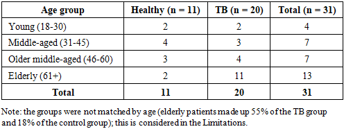
Participants. A total of 31 individuals were enrolled: 20 patients with confirmed active pulmonary TB receiving treatment (main group) and 11 volunteers found to be healthy on clinical and laboratory examination (control group). The diagnosis was based on clinical signs, radiological findings, detection of acid-fast bacilli (AFB+) in sputum and the national clinical guidelines [19]. At the time of examination, patients were receiving standard anti-TB therapy in accordance with the national guidelines [19]. Sex distribution: TB group-14 men (70%) and 6 women (30%); control group-2 men (18.2%) and 9 women (81.8%). The age distribution of participants is shown in Table 1.
Table 1. Distribution of study participants by age group
     
Exclusion criteria: HBsAg or anti-HCV positivity, HIV infection, known chronic liver disease, alcohol abuse, use of hepatotoxic drugs other than anti-TB drugs, and pregnancy.
Biochemical analysis. In the morning after an overnight fast (at least 8 h), 5 mL of blood was collected from the cubital vein of each participant. Samples were allowed to clot for 30 min at room temperature and centrifuged at 3000 rpm for 10 min. Serum was analysed immediately or stored at −20 °C. ALT and AST activities were determined at 37 °C by the kinetic UV method according to the IFCC recommendations [10, 11] on a Mindray BS-200 automated biochemistry analyser (China); the reagent did not contain added P5P. Activity was expressed in U/L. Laboratory reference limits were 7-56 U/L for ALT and 5-40 U/L for AST. The AST/ALT ratio was calculated for each participant individually.
Grouping by AST/ALT ratio. For descriptive purposes, TB patients were divided into four ratio ranges: < 0.4; 0.4-< 0.7; 0.7-< 1.0; and ≥ 1.0. These ranges were used descriptively and are not independent diagnostic criteria.
Statistical analysis. Data were processed in Microsoft Excel 2019 and GraphPad Prism 9.0. Quantitative variables are presented as mean and standard deviation (M ± SD) and as median and interquartile range (Me [Q1-Q3]). Because the samples were small and normality could not be assumed, non-parametric tests were used: the Mann-Whitney U test for comparison of two groups and the Kruskal-Wallis test for comparison of the four age groups within TB patients. Differences were considered statistically significant at p < 0.05. Separate statistical tests were not performed for subgroups with n ≤ 2.

3. Result and Discussion

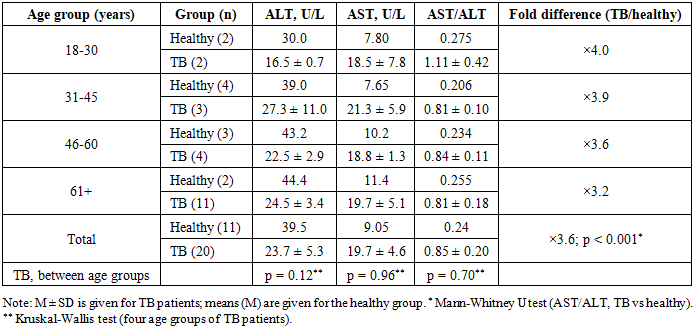
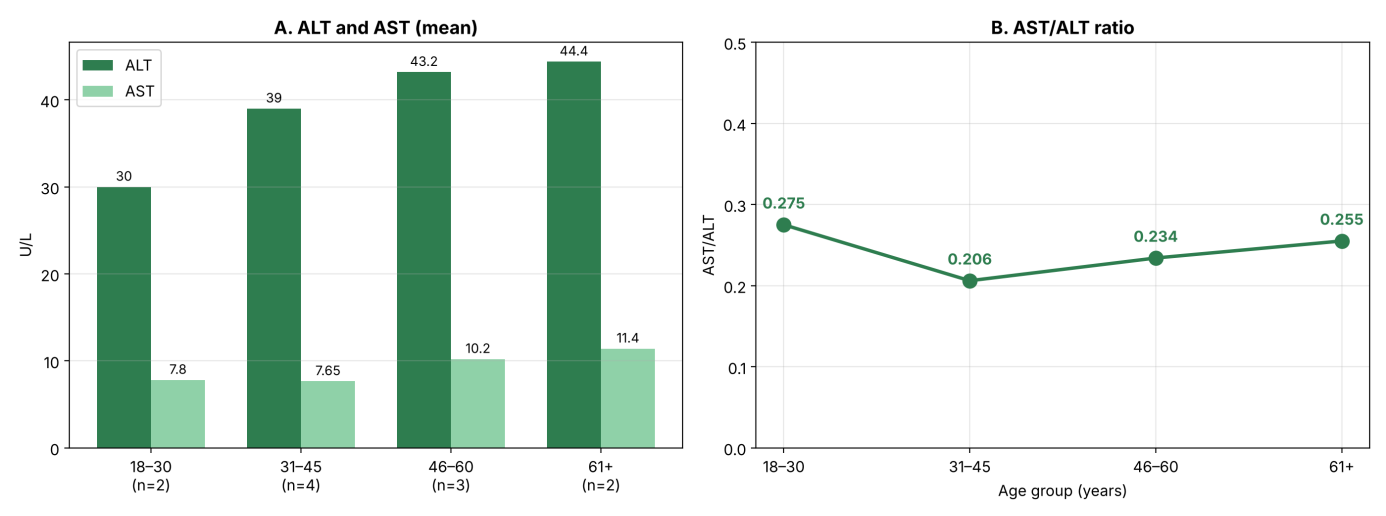
In healthy volunteers, ALT activity across age groups ranged from 30.0 to 44.4 U/L and AST from 7.65 to 11.4 U/L; the AST/ALT ratio ranged from 0.206 to 0.275, with an overall mean of 0.24. In no healthy individual did the ratio exceed 0.44 (Table 2, Figure 1).
Table 2. ALT, AST and the AST/ALT ratio in healthy individuals and TB patients by age group
     
Figure 1. Healthy volunteers (n = 11) by age group: A-mean ALT and AST activity; B-AST/ALT ratio
In TB patients, all individual ALT (16-40 U/L) and AST (13-32 U/L) values were within the laboratory reference limits; that is, no patient showed aminotransferase elevation above the upper limit (cytolytic syndrome). At the same time, mean ALT was lower in patients than in healthy individuals (23.7 ± 5.3 vs 39.5 U/L), while AST was higher (19.7 ± 4.6 vs 9.1 U/L). As a result, the AST/ALT ratio in TB patients was 0.85 ± 0.20 (Me 0.82 [0.73-0.87]; range 0.56-1.41), approximately 3.6 times higher than in healthy individuals. Because the ratio in every TB patient (≥ 0.56) exceeded that in every healthy individual (≤ 0.44), the groups were completely separated (Mann-Whitney U = 0; p < 0.001) (Table 2).
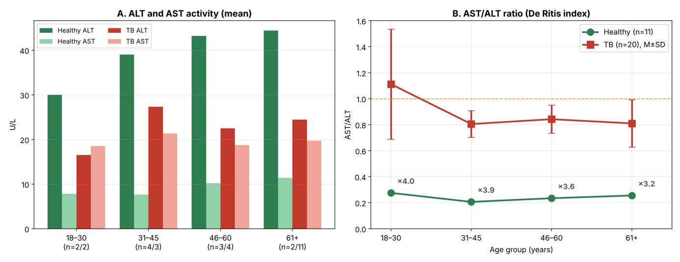
Across age groups, the ratio in TB patients was 3.2 (61+ years) to 4.0 (18-30 years) times higher than in healthy individuals (Figure 2). The highest mean ratio was observed in the 18-30-year group (1.11), but this group included only two patients with widely differing values (0.81 and 1.41). In the remaining groups the ratio was about 0.81-0.84. Within the TB group, no significant differences between age groups were found for the AST/ALT ratio (p = 0.70), ALT (p = 0.12) or AST (p = 0.96).
Figure 2. Healthy individuals (n = 11) and TB patients (n = 20) by age group: A-mean ALT and AST activity (n = healthy/TB); B-AST/ALT ratio (M ± SD for TB); numbers show the fold difference between the TB and healthy ratios
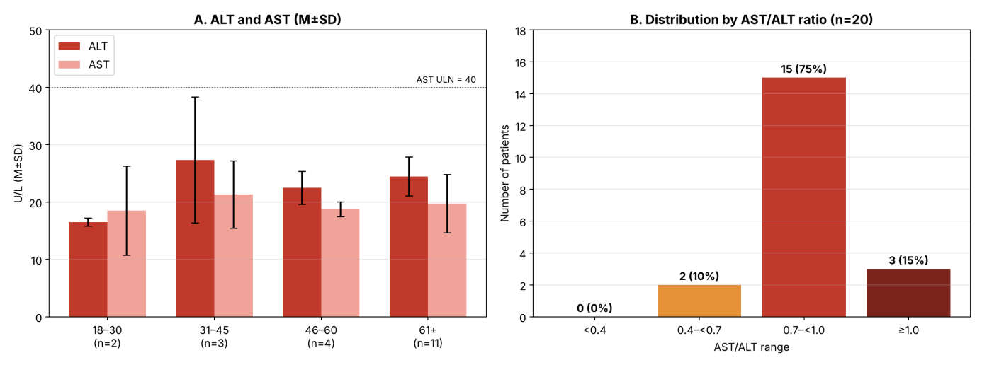
By AST/ALT ratio range, TB patients were distributed as follows (Figure 3): ≥ 1.0-3 patients (15%; one each in the 18-30, 46-60 and 61+ groups); 0.7-< 1.0-15 patients (75%); 0.4-< 0.7-2 patients (10%); no patient had a ratio below 0.4. The 11 elderly (61+) patients formed the largest part of the TB group, and the highest AST value (32 U/L) was also recorded in this group.
Figure 3. Patients with pulmonary TB (n = 20): A-ALT and AST activity by age group (M ± SD; dotted line-AST upper reference limit, 40 U/L); B-distribution of patients by AST/ALT ratio range
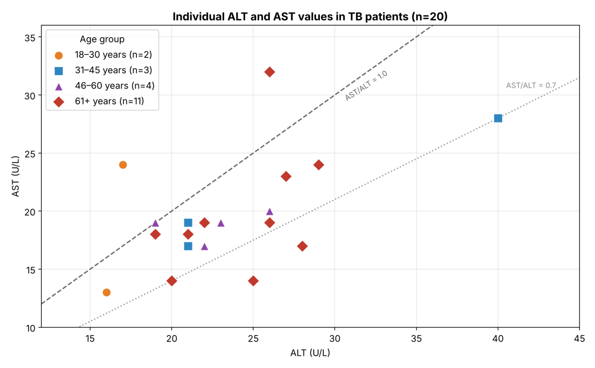
Analysis of individual values (Figure 4) showed that the increase in the ratio was associated with low ALT values: in patients with a ratio ≥ 1.0, ALT was 17-26 U/L, whereas AST did not exceed 19-32 U/L. The patient with the highest ratio (1.41), aged 18-30 years, had ALT 17 U/L and AST 24 U/L.
Figure 4. Individual ALT and AST values in TB patients (n = 20; two patients had identical values-ALT 26, AST 19 U/L-so their points overlap). Dashed line-AST/ALT = 1.0; dotted line-AST/ALT = 0.7
Thus, the De Ritis index was significantly higher in patients with pulmonary TB than in healthy individuals (0.85 vs 0.24; p < 0.001). However, this change was not accompanied by a classical cytolytic syndrome: in no patient did ALT or AST exceed the reference limit. Therefore, there are insufficient grounds to interpret the observed shift as "hepatic cytolysis" or "mitochondrial injury"; such a conclusion would require elevated absolute enzyme activities and additional markers (bilirubin, GGT, alkaline phosphatase, clinical signs) [6,7,18].
The main reason for the increased ratio was low ALT activity in TB patients, which may be explained by several factors. First, isoniazid disturbs vitamin B6 metabolism, and malnutrition is common in TB patients; in P5P deficiency, ALT activity decreases more than AST activity and the ratio increases artificially [9,10]. Second, chronic inflammation and a general catabolic state may reduce hepatic ALT synthesis. Third, the relatively higher AST may also originate from extrahepatic sources (skeletal muscle, erythrocytes), since AST is not liver-specific [6]. The hepatotoxic effect of anti-TB drugs may also contribute to the change in the ratio [3-5,12]; however, the cross-sectional design does not allow these factors to be separated.
The highest mean ratio by age was observed in the 18-30-year group, but this group included only two patients, and the result depends mainly on the value of a single patient (1.41). No significant differences between age groups were found within the TB group (p = 0.70), so the apparent age-related trend can be regarded only as a preliminary observation. The fact that the ratio increased in the same direction in all age groups (3.2-4.0-fold) indicates that the change is related primarily to the disease and its treatment.
Compared with the literature, Wang et al. reported clinically significant hepatitis in 18.9% of patients during anti-TB treatment [13]. In our small sample, no patient had enzyme levels above the reference limits, which may be explained by differences in sample size, treatment stage and population characteristics. The ATS, APASL and EASL guidelines recommend regular monitoring of liver function during TB treatment [4,17,18]; our results suggest that monitoring the AST/ALT ratio as an additional indicator may be useful even when enzyme activities are within reference limits.
Limitations. This study has several limitations. First, the sample was small, particularly in the younger age subgroups (n = 2-3), which limits statistical power for age comparisons. Second, the groups were not matched by age (55% vs 18% elderly) or sex (70% vs 18.2% men), which may have influenced enzyme activities. Third, the cross-sectional design and the lack of data on treatment duration, vitamin B6 status and acetylator phenotype do not allow causal conclusions. Fourth, enzymes were measured without P5P supplementation, and other liver markers (bilirubin, GGT, alkaline phosphatase) were not assessed. Finally, the AST/ALT ratio in the control group (0.24) was considerably lower than values usually reported for healthy adults (about 1) [9], so the fold differences should be interpreted with caution and verified in a larger, matched control group.

4. Conclusions

1. In healthy volunteers (n = 11), the mean AST/ALT ratio was 0.24 and did not exceed 0.44 in any individual.
2. In patients with pulmonary TB (n = 20), despite ALT and AST being within reference limits, the AST/ALT ratio was 0.85 ± 0.20 (0.56-1.41), approximately 3.6 times higher than in healthy individuals (p < 0.001). This shift was mainly due to low ALT activity.
3. The ratio was ≥ 1.0 in 15% (3/20) of TB patients, 0.7-< 1.0 in 75% (15/20) and 0.4-< 0.7 in 10% (2/20); no patient had aminotransferase levels above the reference limits.
4. During TB treatment, it is advisable to consider the AST/ALT ratio together with the absolute aminotransferase values when monitoring liver status and to perform regular monitoring in accordance with national and international guidelines [4,18,19].

References

[1]  World Health Organization (2023), Global Tuberculosis Report 2023, World Health Organization, Geneva. ISBN 978-92-4-008385-1.
[2]  Ministry of Health of the Republic of Uzbekistan (2024), Report on the state of tuberculosis control in Uzbekistan in 2023, Ministry of Health, Tashkent, 48 p. (in Uzbek).
[3]  Tostmann A., Boeree M.J., Aarnoutse R.E., de Lange W.C., van der Ven A.J., Dekhuijzen P.N.R. (2008), Antituberculosis drug-induced hepatotoxicity: concise up-to-date review, Journal of Gastroenterology and Hepatology, 23(2), 192-202. https://doi.org/10.1111/j.1440-1746.2007.05207.x.
[4]  Saukkonen J.J., Cohn D.L., Jasmer R.M., Schenker S., Jereb J.A., Nolan C.M., et al. (2006), An official ATS statement: hepatotoxicity of antituberculosis therapy, American Journal of Respiratory and Critical Care Medicine, 174(8), 935-952. https://doi.org/10.1164/rccm.200510-1666ST.
[5]  Ramappa V., Aithal G.P. (2013), Hepatotoxicity related to anti-tuberculosis drugs: mechanisms and management, Journal of Clinical and Experimental Hepatology, 3(1), 37-49. https://doi.org/10.1016/j.jceh.2012.12.001.
[6]  Giannini E.G., Testa R., Savarino V. (2005), Liver enzyme alteration: a guide for clinicians, Canadian Medical Association Journal, 172(3), 367-379. https://doi.org/10.1503/cmaj.1040752.
[7]  Pratt D.S., Kaplan M.M. (2000), Evaluation of abnormal liver-enzyme results in asymptomatic patients, New England Journal of Medicine, 342(17), 1266-1271. https://doi.org/10.1056/NEJM200004273421707.
[8]  De Ritis F., Coltorti M., Giusti G. (1957), An enzymic test for the diagnosis of viral hepatitis: the transaminase serum activities, Clinica Chimica Acta, 2(1), 70-74. https://doi.org/10.1016/0009-8981(57)90027-X.
[9]  Botros M., Sikaris K.A. (2013), The De Ritis ratio: the test of time, Clinical Biochemist Reviews, 34(3), 117-130. PMID: 24353357; PMCID: PMC3866949.
[10]  Schumann G., Bonora R., Ceriotti F., Férard G., Ferrero C.A., Franck P.F.H., et al. (2002), IFCC primary reference procedures for the measurement of catalytic activity concentrations of enzymes at 37 °C. Part 4. Reference procedure for the measurement of catalytic concentration of alanine aminotransferase, Clinical Chemistry and Laboratory Medicine, 40(7), 718-724. https://doi.org/10.1515/CCLM.2002.124.
[11]  Schumann G., Bonora R., Ceriotti F., Férard G., Ferrero C.A., Franck P.F.H., et al. (2002), IFCC primary reference procedures for the measurement of catalytic activity concentrations of enzymes at 37 °C. Part 5. Reference procedure for the measurement of catalytic concentration of aspartate aminotransferase, Clinical Chemistry and Laboratory Medicine, 40(7), 725-733. https://doi.org/10.1515/CCLM.2002.125.
[12]  Yew W.W., Leung C.C. (2006), Antituberculosis drugs and hepatotoxicity, Respirology, 11(6), 699-707. https://doi.org/10.1111/j.1440-1843.2006.00941.x.
[13]  Wang J.Y., Liu C.H., Hu F.C., Chang H.C., Liu J.L., Chen J.M., et al. (2011), Risk factors of hepatitis during anti-tuberculous treatment and implications of hepatitis virus load, Journal of Infection, 62(6), 448-455. https://doi.org/10.1016/j.jinf.2011.04.005.
[14]  Abbara A., Chitty S., Roe J.K., et al. (2017), Drug-induced liver injury from antituberculous treatment: a retrospective study from a large TB centre in the UK, BMC Infectious Diseases, 17, 231. https://doi.org/10.1186/s12879-017-2330-z.
[15]  Singla R., Sharma S.K., Mohan A., et al. (2010), Evaluation of risk factors for antituberculosis treatment induced hepatotoxicity, Indian Journal of Medical Research, 132, 81-86.
[16]  Falzon D., Mirzayev F., Wares F., Baena I.G., Zignol M., Linh N., et al. (2015), Multidrug-resistant tuberculosis around the world: what progress has been made?, European Respiratory Journal, 45(1), 150-160. https://doi.org/10.1183/09031936.00101814.
[17]  Devarbhavi H., Aithal G.P., Treeprasertsuk S., et al. (2021), Drug-induced liver injury: Asia Pacific Association for Study of Liver consensus guidelines, Hepatology International, 15(2), 258-282. https://doi.org/10.1007/s12072-021-10144-3.
[18]  European Association for the Study of the Liver (2019), EASL Clinical Practice Guidelines: Drug-induced liver injury, Journal of Hepatology, 70(6), 1222-1261. https://doi.org/10.1016/j.jhep.2019.02.014.
[19]  Ministry of Health of the Republic of Uzbekistan (2022), Clinical guidelines on pulmonary tuberculosis, Ministry of Health, Tashkent, 124 p. (in Uzbek).