
-
Paper Information
- Next Paper
- Previous Paper
- Paper Submission
-
Journal Information
- About This Journal
- Editorial Board
- Current Issue
- Archive
- Author Guidelines
- Contact Us
International Journal of Virology and Molecular Biology
p-ISSN: 2163-2219 e-ISSN: 2163-2227
2026; 15(6): 142-145
doi:10.5923/j.ijvmb.20261506.02
Received: Aug. 18, 2026; Accepted: Sep. 16, 2026; Published: Sep. 22, 2026

Immunogistochemical Characteristics of Neoangiogenesis Processes in Regional Lymph Nodes in Alcoholic Pancreatitis
 Abstract
 Reference
 Full-Text PDF
 Full-text HTML
Musaeva Yulduz Alpersovna1, Latibjonov Azizbek Erkinjon o‘g‘li2
1Associate Professor of Tashkent State Medical University, Department of Neurology and Medical Psychology, Doctor of Medicine
2Independent Researcher of Fergana Medical Institute of Public Health, Department of Trauma and Orthopedics
Correspondence to: Musaeva Yulduz Alpersovna, Associate Professor of Tashkent State Medical University, Department of Neurology and Medical Psychology, Doctor of Medicine.
| Email: |  |
Copyright © 2026 The Author(s). Published by Scientific & Academic Publishing.
This work is licensed under the Creative Commons Attribution International License (CC BY).
http://creativecommons.org/licenses/by/4.0/

In alcoholic pancreatitis, numerous foci of neoangiogenesis and abundant formation of microcirculatory plexuses were detected in simple morphological studies. This, in turn, was used to assess the endothelial proliferative index in lymph nodes using the CD34 marker. In immunohistochemical (IHC) examination of regional lymph nodes and pancreas in alcoholic pancreatitis, each of the above markers is expressed in a specific way, reflecting the processes of inflammation, activation of immune cells, and blood vessel remodeling.
Keywords: Immunohistochemical examination, Lymph nodes, Alcoholic pancreatitis, Morphology
Cite this paper: Musaeva Yulduz Alpersovna, Latibjonov Azizbek Erkinjon o‘g‘li, Immunogistochemical Characteristics of Neoangiogenesis Processes in Regional Lymph Nodes in Alcoholic Pancreatitis, International Journal of Virology and Molecular Biology, Vol. 15 No. 6, 2026, pp. 142-145. doi: 10.5923/j.ijvmb.20261506.02.
Article Outline
1. Relevance of the Topic
- In the world, pancreatitis is the third most common acute abdominal disease. An average of 960 million people worldwide are diagnosed with pancreatitis, of which 36.6% (262 million) are alcoholic pancreatitis patients between the ages of 25 and 40 [1,2]. In the United States, more than 25 million people aged 25 to 45 suffer from this disease [3]. In Europe, an average of 66.8 million people suffer from alcoholic pancreatitis [4]. In the Russian Federation, an average of 52.3 million people aged 25 to 55 suffer from this disease [5]. In the CIS countries, a total of more than 12 million people are registered with this indicator. Every year, an average of 12-30 million people die from various forms of alcoholic pancreatitis [6,7,8]. However, no source provides information on the morphological features of regional lymph nodes in acute and chronic alcoholic pancreatitis, however, it is important to develop morphofunctional parameters of regional lymph nodes in acute pancreatitis with edematous and necrotic changes and important determinants of the prevention of severe complications [9,10,12].Research objective: To study the immunohistochemical features of regional lymph nodes in alcoholic pancreatitis.
2. Materials and Methods
- Morphological examination methods: hematoxylin eosin, histochemical methods Van Gieson, Alcian blue, clinical anamnestic and statistical analysis methods are used. Appropriate immunomarkers were obtained for immunohistochemical examinations. Materials of regional lymph nodes of the pancreas were obtained during autopsy of patients who died from chronic and acute pancreatitis.
3. Results and Discussion
- Analysis of the level of expression of the CD34 (T-lymphocyte center) marker in alcoholic pancreatitis and its assessment criteria. CD34 is a membrane protein, an intercellular adhesion molecule, which plays an important role in the early stages of hematopoiesis. CD34 ensures the binding of stem cells to the extracellular matrix of the bone marrow or directly to stromal cells.It serves as a protein scaffold for binding specific glycans that allow the stem cells to bind to lectins produced by the stroma or other components of the bone marrow. In addition, it provides carbohydrate ligands for highly glycosylated CD34 selectins. Mainly when assessing the level of neoangiogenesis after vascular endothelial damage, intussusceptive angiogenesis (the vessel wall is one, the endothelium is divided into 2, the vascular channel is divided into 2 or continues with the doubling of small-caliber vessels), as a result of a sharp increase in vascular stromal structures in the tissue, which allows for the early assessment of sclerosis and cirrhosis.In our study, it was revealed that in lymph nodes examined with the CD34 marker, a large number of small vessels with a glomeruloid lattice shape were detected on surfaces of 32,000 μm2, 64,000 μm2, a large number of lymphoid follicles, paracortical areas. These changes indicate that in alcoholic pancreatitis, cytokines and other inflammatory factors are produced in large quantities, bone marrow, and hematogenous cell proliferation is increased.Therefore, The CD34 marker is the most important and widely used marker of endothelial and hematopoietic stem cells in immunohistochemical (IHC) studies (it stains the endothelium of small blood vessels, especially capillaries and venules, with high sensitivity).Against the background of inflammation, hypoxia, and compensatory processes observed in regional lymph nodes in alcoholic pancreatitis, the following important immunomorphological changes are detected by CD34 expression. Activation of angiogenesis, expansion of the microcirculatory network, and formation of a vascular stroma in the lymph node.
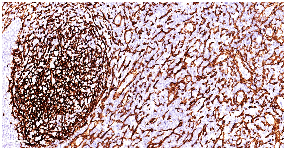 | Figure 1. Sample-1. Mesenteric lymph node in alcoholic pancreatitis. High positive expression of the CD34 marker, tortuous vascular trajectory are detected. A tangle consisting of a vascular skeleton is detected in the traces of lymphoid follicles. Staining Dab chromogen. Size 20x10 |
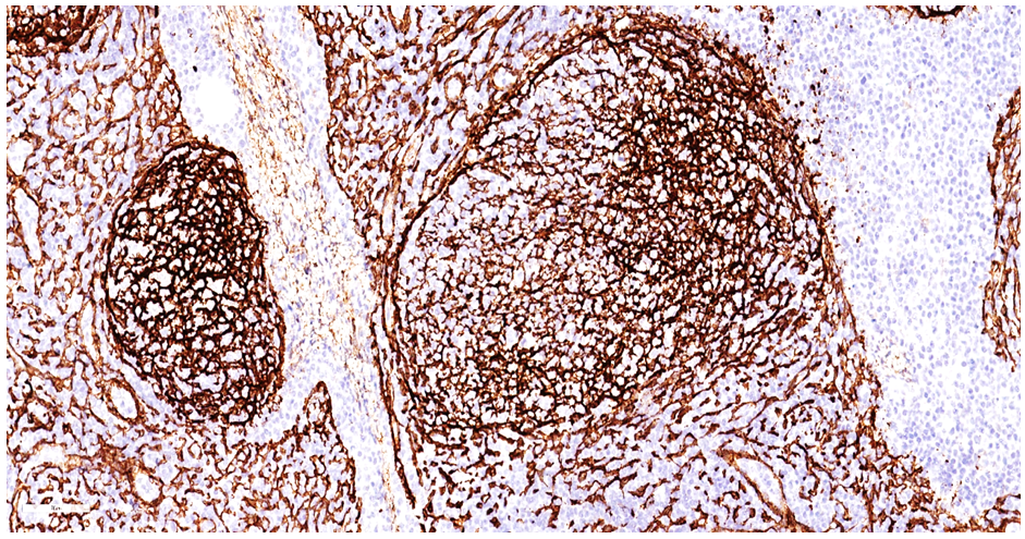 | Figure 2. Sample-2. Mesenteric lymph node in alcoholic pancreatitis. High positive expression of the CD34 marker, tortuous vascular trajectory are detected. A tangle consisting of a vascular skeleton is detected in the traces of lymphoid follicles. Staining Dab chromogen. Size 20x10 |
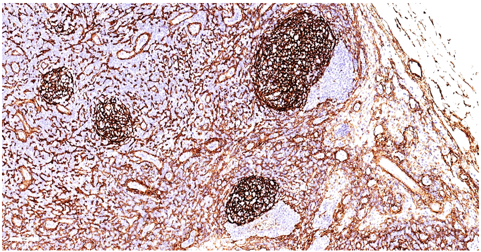 | Figure 3. Sample-3. Mesenteric lymph node in alcoholic pancreatitis. High positive expression of the CD34 marker, tortuous vascular trajectory are detected. A tangle consisting of a vascular skeleton is detected in the traces of lymphoid follicles. Staining Dab chromogen. Size 10x10 |
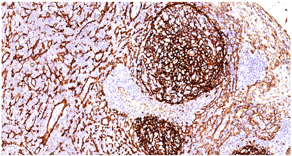 | Figure 4. Sample-4. Mesenteric lymph node in alcoholic pancreatitis. High positive expression of the CD34 marker, tortuous vascular trajectory are detected. A tangle consisting of a vascular skeleton is detected in the traces of lymphoid follicles. Staining Dab chromogen. Size 20x10 |
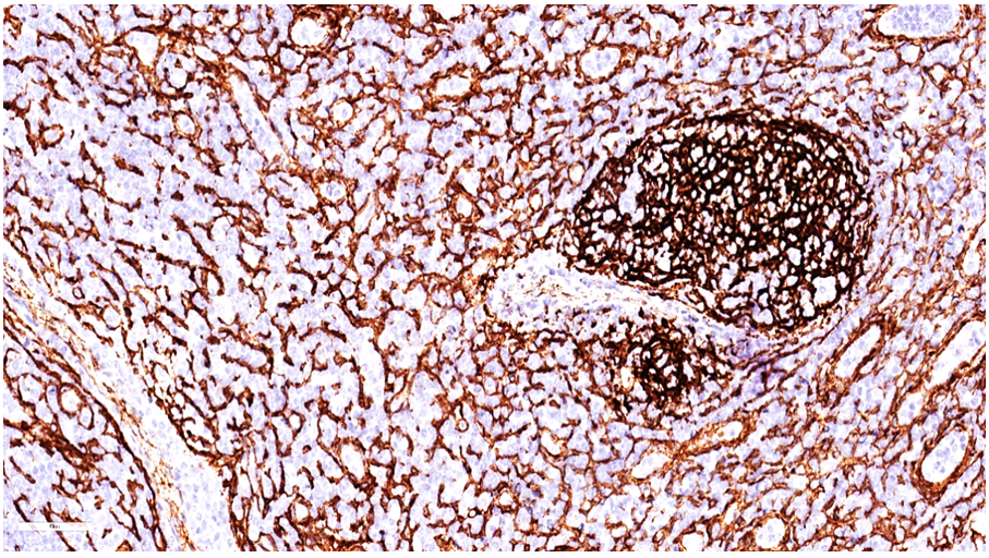 | Figure 5. Sample-5. Mesenteric lymph node in alcoholic pancreatitis. High positive expression of the CD34 marker, tortuous vascular trajectory are detected. A tangle consisting of a vascular skeleton is detected in the traces of lymphoid follicles. Staining Dab chromogen. Size 20x10 |
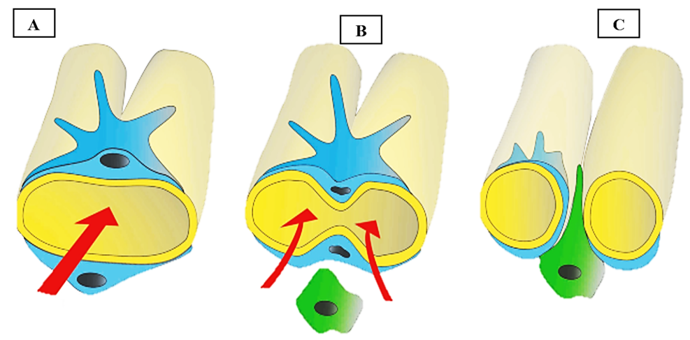 | Figure 6. The process of intussusceptive angiogenesis of blood vessels under the influence of the CD 34 marker. In the blood vessel, endothelial proliferation initially leads to the division of the lumen into two and the subsequent formation of two separate vascular lumens. |
Authors’ Contribution
- Conceptualization, Y.A.M. and A.E.L.; methodology, Y.A.M.; validation, Y.A.M. and A.E.L.; formal analysis, A.E.L.; investigation, A.E.L.; resources, Y.A.M.; data curation, A.E.L.; writing—original draft preparation, A.E.L.; writing—review and editing, Y.A.M.; visualization, A.E.L.; supervision, Y.A.M.; project administration, Y.A.M. All authors have read and agreed to the published version of the manuscript.
Funding Source
- This research received no external funding.
Ethics Approval
- The study was conducted in accordance with the Declaration of Helsinki and was approved by the Institutional Ethics Committee of Fergana Medical Institute of Public Health (protocol code K29, approval date 08.26.2025). Only archival, de-identified post-mortem material was used.
Consent for Publication
- Patient consent was waived because the study used only de-identified archival autopsy material and no individual can be identified from the data or images presented.
Data Availability Statement
- The histological slides, digital micrographs and anonymised grading datasets that support the findings of this study are available from the corresponding author upon reasonable request. The data are not publicly archived because of institutional restrictions on post-mortem material.
ACKNOWLEDGEMENTS
- The authors thank the staff of the pathology laboratories of Tashkent State Medical University and the Fergana Medical Institute of Public Health for technical assistance with tissue processing and staining.
Conflict of Interest
- The authors declare no conflicts of interest. No funders had any role in the design of the study; in the collection, analyses or interpretation of data; in the writing of the manuscript; or in the decision to publish the results.