
-
Paper Information
- Previous Paper
- Paper Submission
-
Journal Information
- About This Journal
- Editorial Board
- Current Issue
- Archive
- Author Guidelines
- Contact Us
International Journal of Virology and Molecular Biology
p-ISSN: 2163-2219 e-ISSN: 2163-2227
2026; 15(5): 118-123
doi:10.5923/j.ijvmb.20261505.03
Received: Aug. 10, 2026; Accepted: Sep. 3, 2026; Published: Sep. 10, 2026

Effect of Low-Dose Gamma Irradiation on Lipolytic Activity and Pepsinogen Level in Rat Salivary Glands and Blood Serum, and Their Correction with Bioflavonoids
 Abstract
 Reference
 Full-Text PDF
 Full-text HTML
Mirzabekov Ismoiljon Adxamjon o'g'li
Department of Human Physiology and Life Safety, Andijan State Medical Institute, Andijan, Uzbekistan
Correspondence to: Mirzabekov Ismoiljon Adxamjon o'g'li, Department of Human Physiology and Life Safety, Andijan State Medical Institute, Andijan, Uzbekistan.
| Email: |  |
Copyright © 2026 The Author(s). Published by Scientific & Academic Publishing.
This work is licensed under the Creative Commons Attribution International License (CC BY).
http://creativecommons.org/licenses/by/4.0/

This study investigated the effects of 1 and 2 Gray (Gy) gamma irradiation on lipolytic activity and pepsinogen levels in the parotid salivary glands and blood serum of rats, as well as their correction with the bioflavonoids glabridin, pulicarin, and rutin. Rats were irradiated using a Terabalt-80 apparatus with a 60Co source at a dose rate of 0.86-0.85 Gy/min, with total absorbed doses of 1 and 2 Gy, and lipolytic activity and pepsinogen levels were examined on days 1, 3, 10, 20, 30, and 60 post-irradiation and after bioflavonoid administration. The results demonstrated that increasing radiation doses led to a decrease in lipolytic activity and pepsinogen levels, while bioflavonoids exerted a protective effect on these parameters. Rutin exhibited the highest efficacy, followed by pulicarin and glabridin. Bioflavonoids are recommended as promising natural agents for the correction of radiation-induced damage.
Keywords: Gamma irradiation, Salivary glands, Lipolytic activity, Pepsinogen, Bioflavonoids, Rutin, Pulicarin, Glabridin, Radioprotector
Cite this paper: Mirzabekov Ismoiljon Adxamjon o'g'li, Effect of Low-Dose Gamma Irradiation on Lipolytic Activity and Pepsinogen Level in Rat Salivary Glands and Blood Serum, and Their Correction with Bioflavonoids, International Journal of Virology and Molecular Biology, Vol. 15 No. 5, 2026, pp. 118-123. doi: 10.5923/j.ijvmb.20261505.03.
Article Outline
1. Introduction
- Ionizing radiation, particularly gamma irradiation, is a powerful stress factor that induces profound biochemical and functional alterations in living tissues. In radiotherapy for head and neck tumors, the salivary glands (parotid and submandibular) are among the most radiosensitive organs. Modern research indicates that gamma irradiation causes destruction and apoptosis of acinar cells in salivary glands [1], [2].Salivary glands perform not only exocrine but also recretory and incretory functions. They have the ability to extract various metabolites and enzymes from the blood (recretion) and re-secrete them into saliva. The enzyme homeostasis in salivary gland secretions and blood — particularly the lipase (lipolytic activity) system involved in lipid metabolism — is highly sensitive to radiation [3].According to the literature, the effect of gamma irradiation is dose-dependent. At low doses (0.5–2 Gy), short-term compensatory activation or mild functional changes in enzyme secretion are observed. At higher doses (4 Gy and above), severe hyposalivation (dryness), disruption of cell membrane permeability, and damage to recretory pathways occur [4]. Consequently, lipolytic activity in saliva decreases, while pathological increases in plasma lipolytic activity may occur due to enzyme leakage into the blood (hyperfermentemia).The damaging effects of gamma radiation on tissues occur through two main mechanisms: direct (breakage of biomolecules and DNA strands) and indirect (formation of free radicals due to water radiolysis).Contemporary studies confirm that the primary factor in salivary gland injury is nitro-oxidative stress. Radiation exposure leads to a sharp increase in reactive oxygen species (ROS) in cells, which promotes lipid peroxidation (LPO) in lysosomal and cell membranes. ROS oxidize sulfhydryl (SH) groups in the active centers of enzymes, including lipase, thereby reducing their catalytic activity, and disrupt mitochondrial ultrastructure, ATP synthesis, and calcium signaling pathways.Currently, due to the high toxicity of synthetic radioprotectors in medicine and radiobiology, there is growing interest in bioflavonoids derived from natural sources. Bioflavonoids (e.g., quercetin, rutin, hesperidin, curcumin, and epigallocatechin-3-gallate) have low toxicity and can be used long-term for prophylactic purposes [5]. Literature analysis shows that the protective mechanisms of bioflavonoids manifest in various ways [6].Flavonoids neutralize ROS and free radicals through their phenolic rings, thereby reducing LPO and malondialdehyde (MDA) levels. They enhance Nrf-2 (nuclear factor erythroid 2–related factor 2) signaling, which coordinates the activity of intracellular superoxide dismutase, catalase, and glutathione systems. They maintain the integrity of acinar cell membranes and excretory duct membranes in the salivary glands, ensuring normal recretion of lipase and protecting enzymatic homeostasis between blood and saliva. Flavonoids also suppress the synthesis of pro-inflammatory cytokines (TNF-α, IL-1β) and inflammatory mediators, and block caspase-3 activity, preventing cell death [7]. The dynamic changes in lipolytic activity in the parotid salivary gland and blood under 1 and 2 Gy gamma irradiation, and the effects of flavonoids on these parameters, have not yet been studied.The aim of this work was to study the effect of low-dose gamma irradiation on lipolytic activity and pepsinogen levels in rat salivary gland homogenates and blood serum, and to evaluate their correction with bioflavonoids. Rats were irradiated using a Terabalt-80 apparatus with a 60Co source at a dose rate of 0.86–0.85 Gy/min, with total absorbed doses of 1 and 2 Gy, and lipolytic activity and pepsinogen levels were examined on days 1, 3, 10, 20, 30, and 60 post-irradiation and after bioflavonoid administration.
2. Materials and Methods
2.1. Animals and Experimental Design
- A chronic experiment was conducted on rats. Adult male outbred white rats weighing 175–200 g were used. The animals were kept under standard vivarium conditions and fed a carbohydrate-protein mixed diet with free access to water. The rats were divided into five groups: I — control (healthy); II — irradiated with 1 or 2 Gy gamma radiation; III — irradiated (1 or 2 Gy) + glabridin; IV — irradiated (1 or 2 Gy) + pulicarin; V — irradiated (1 or 2 Gy) + rutin.The control group received no treatment. Rats in groups II–V were irradiated locally (head and neck area) using a Terabalt-80 apparatus with a 60Co source. The irradiation field was 20×20 cm, focus distance 80 cm, dose rate 0.86–0.85 Gy/min, and absorbed doses were 1 and 2 Gy. Flavonoids (glabridin, pulicarin, rutin) were administered at 50 mg/kg via gastric tube once daily, starting 5 days before irradiation and continuing after irradiation. Irradiated rats were kept in standard cages and decapitated on days 1, 3, 10, 20, 30, and 60 post-irradiation.
2.2. Determination of Lipolytic Activity
- Lipolytic activity was determined by the Tietz method, based on titration of free fatty acids released during tributyrin lipolysis with 0.05 N NaOH [8], [9]. Lipase activity was expressed in Tietz units, calculated as the difference in alkali consumption between test and control tubes. The assay procedure was as follows: 4 ml of emulsion (containing 1.5 ml TRIS-hydroxymethylaminomethane buffer, 1.0 ml 0.1% CaCl2, 2 ml sodium cholate mixture, 0.25 ml tributyrin, and 0.25 ml distilled water) was added to each test and control tube. Then, 1 ml of test material (salivary gland homogenate or blood serum) was added. The control tube received 3 ml of 96% ethanol immediately to stop the reaction. The test tube was incubated for 2 hours at 37°C in a water bath, after which the reaction was stopped by adding 3 ml of 96% ethanol. After stopping the reaction, 3–4 drops of thymolphthalein were added, and titration with 0.05 N NaOH was performed until a stable blue color appeared. One unit of lipase activity was defined as the amount of alkali consumed to reach a faint pink endpoint.
2.3. Determination of Pepsinogen Levels
- Pepsinogen levels in salivary gland homogenates were determined using a modified Hirschowitz tyrosine method [10], [11]. In our modification, the incubation time was extended to 24 hours, and dried plasma was used as the protein substrate instead of hemoglobin. For analysis, 0.5 ml of test material (salivary gland homogenate or blood plasma) was mixed with 2.5 ml of 2% plasma solution in 0.05 N HCl. The mixture was incubated at 37°C for 24 hours. A parallel control tube containing 2.5 ml of 2% plasma solution in 0.05 N HCl was also incubated. After incubation, 0.5 ml of test material was added to the control tube. Then, 2.5 ml of 0.5 N trichloroacetic acid (TCA) was added to both tubes, and the mixture was filtered through thick filter paper. To 1 ml of filtrate, 2.0 ml of 0.5 N NaOH and 0.6 ml of Folin–Ciocalteu reagent (diluted 1:2 with water) were added. The resulting solution was colorimetried against water using a red filter for 3–20 minutes. The extinction of the control tube was subtracted from that of the test tube. The difference was converted to arbitrary activity units using a calibration curve prepared with tyrosine. One unit of pepsinogen activity was defined as the amount of enzyme that released 1 µg of tyrosine from protein substrate at 37°C over 24 hours.
2.4. Statistical Analysis
- Statistical processing of the obtained results and plotting of figures were performed using OriginPro 8.6 (Microsoft, USA) computer software. Experimental data were statistically processed according to the Student–Fisher method and subjected to correlation analysis. Differences between the values obtained from the control, experimental, and experimental + test substance groups were calculated using the t-test, where P<0.05 and P<0.01 indicate statistical significance.
3. Results and Discussion
- Salivary gland secretions contain lipase primarily via the recretory pathway. We studied the effect of different doses of gamma irradiation on lipolytic activity in rat salivary glands and blood.
3.1. Lipolytic Activity in Salivary Glands
- To evaluate the dose-dependent effect of ionizing radiation and the radioprotective potential of natural polyphenols, we examined the dynamics of lipolytic activity in the parotid salivary gland from day 1 to day 60 under 1 Gy and 2 Gy gamma irradiation (Figure 1 and Figure 2). The results showed that increasing radiation dose significantly deepened enzyme suppression, and the protective effect of flavonoids was proportionally dose-dependent.
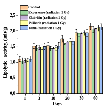 | Figure 1. Effect of 1 Gy gamma irradiation on lipolytic activity (U/g) in the parotid salivary gland. (All cases: significance level P<0.05; n=6) |
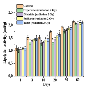 | Figure 2. Effect of 2 Gy gamma irradiation on lipolytic activity (U/g) in the parotid salivary gland. (All cases: significance level P<0.05; n=6) |
3.2. Lipolytic Activity in Blood
- We simultaneously studied lipolytic activity in the blood of irradiated rats (Figure 3 and Figure 4). The results showed that low-dose gamma irradiation affects the secretory and incretory processes of the glands similarly. Lipolytic activity in salivary gland homogenates showed no significant changes after 1–2 Gy irradiation; however, at 2 Gy, blood lipolytic activity remained at control levels on days 3 and 10.
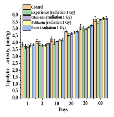 | Figure 3. Effect of 1 Gy gamma irradiation on lipolytic activity (U/g) in blood. Ordinate — enzyme activity (U/g), abscissa — experimental days. (All cases: significance level P<0.05; n=6) |
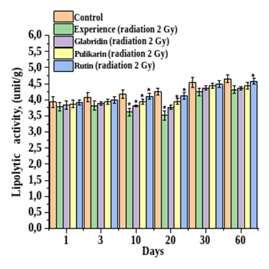 | Figure 4. Effect of 2 Gy gamma irradiation on lipolytic activity (U/g) in blood. Ordinate — enzyme activity (U/g), abscissa — experimental days. (All cases: significance level P<0.05; n=6) |
|
3.3. Pepsinogen Levels in Salivary Gland Homogenates
- The effect of low-dose (1 and 2 Gy) gamma irradiation on pepsinogen content in rat salivary gland homogenates is shown in Figure 5 and Figure 6. A clear dose-dependent change was observed. At 1 Gy, pepsinogen levels in all salivary glands did not change significantly and remained at control levels (Figure 5). However, at 2 Gy, notable changes occurred.
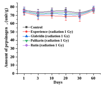 | Figure 5. Effect of 1 Gy gamma irradiation on pepsinogen levels (U/g) in the parotid salivary gland. (All cases: significance level P<0.05; n=6) |
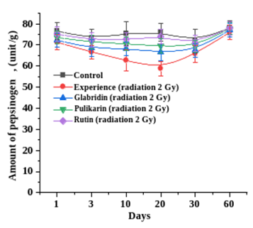 | Figure 6. Effect of 2 Gy gamma irradiation on pepsinogen levels (U/g) in the parotid salivary gland. (All cases: significance level P<0.05; n=6) |
3.4. Pepsinogen Levels in Blood
- The dose-dependent effect of gamma irradiation on blood pepsinogen levels is shown in Figure 7 and Figure 8. At 1 Gy, blood pepsinogen levels did not change and remained at control levels.
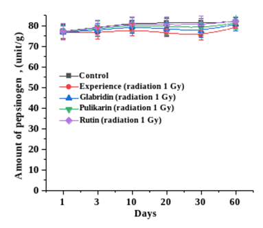 | Figure 7. Effect of 1 Gy gamma irradiation on pepsinogen levels (U/g) in blood. (All cases: significance level P<0.05; n=6) |
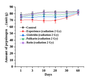 | Figure 8. Effect of 2 Gy gamma irradiation on pepsinogen levels (U/g) in blood. (All cases: significance level P<0.05; n=6) |
3.5. Correlation Between Blood and Salivary Gland Pepsinogen Levels
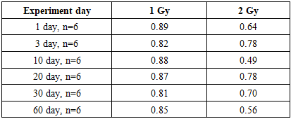
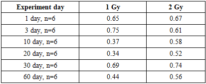
- To determine the relationship between pepsinogen levels in blood and salivary glands, we calculated correlation coefficients (Table 2). The data show that with increasing radiation dose, the correlation between blood and parotid salivary gland pepsinogen levels becomes stronger. For example, at 1 Gy, correlation coefficients ranged from 0.34 to 0.75; at 2 Gy, from 0.52 to 0.74. These results indicate that as radiation dose increases, blood pepsinogen levels and its excretion via the parotid salivary gland decrease in the same direction.
|
4. Conclusions
- Gamma irradiation exerts a negative effect on the recretory function of rat salivary glands, lipolytic homeostasis in the blood–saliva system, and pepsinogen activity. The administration of natural bioflavonoids before or after irradiation demonstrates highly effective radioprotective effects by reducing oxidative stress, protecting membranes, and stabilizing enzyme activity. Among the tested compounds, the radioprotective efficacy follows the order Rutin > Pulicarin > Glabridin, which is directly related to their chemical structure, the number and position of hydroxyl groups, and their free radical scavenging potential. This substantiates the significant theoretical and practical potential of such research for medicine, and rutin and pulicarin in particular hold high promise as natural radioprotective agents for ensuring the metabolic stability of salivary glands.