International Journal of Virology and Molecular Biology

p-ISSN: 2163-2219    e-ISSN: 2163-2227

2026;  15(4): 99-107

doi:10.5923/j.ijvmb.20261504.03

Received: Jan. 16, 2026; Accepted: Feb. 12, 2026; Published: Jul. 30, 2026

 

Morphological and Molecular Characterization of Selected Soil Microalgae Isolated from Rice Fields in the Surkhandarya Region, Uzbekistan

Abdullaeva G. K.1, Tukhtaboeva Y. A.2, Kamalova G. G.2

1Namangan State University, Uzbekistan

2KIUT Namangan Branch, Uzbekistan

Correspondence to: Abdullaeva G. K., Namangan State University, Uzbekistan.

Email:

Copyright © 2026 The Author(s). Published by Scientific & Academic Publishing.

This work is licensed under the Creative Commons Attribution International License (CC BY).
http://creativecommons.org/licenses/by/4.0/

Abstract

Scientific interest in the ecological and functional roles of soil microalgae in rice paddy agroecosystems has increased considerably since the mid-20th century, evolving from basic morphological descriptions to advanced applications in agricultural biotechnology. The present study aimed to characterize the diversity of soil microalgae in rice fields located in the Termez and Muzrabat districts of the Surkhandarya region, Uzbekistan. A polyphasic approach integrating traditional morphological observations with molecular genetic analysis based on the internal transcribed spacer 2 (ITS2) rDNA marker was employed. Four microalgal strains were successfully isolated and identified. The identified strains exhibited specific adaptations to local soil conditions and may serve as valuable bioindicators for assessing the agroecological status and salinity of rice-growing soils in the region. The findings provide new information on the taxonomic diversity of indigenous soil microalgae and establish a basis for future studies on their potential application in sustainable rice cultivation and soil reclamation.

Keywords: Microalgae, Soil biodiversity, ITS2 rDNA, Soil algae, Salinity, Oryza sativa L., Bioindicators

Cite this paper: Abdullaeva G. K., Tukhtaboeva Y. A., Kamalova G. G., Morphological and Molecular Characterization of Selected Soil Microalgae Isolated from Rice Fields in the Surkhandarya Region, Uzbekistan, International Journal of Virology and Molecular Biology, Vol. 15 No. 4, 2026, pp. 99-107. doi: 10.5923/j.ijvmb.20261504.03.

1. Introduction

The optimization of agricultural productivity under conditions of climate change, soil degradation, and increasing salinization represents one of the major challenges facing modern agriculture. In Uzbekistan, the Surkhandarya region, particularly the Muzrabat district within the Amu Darya River Basin, is characterized by an arid climate and widespread soil salinity, which substantially limit crop productivity. Rice (Oryza sativa L.) is one of the country's strategically important crops, and improving its cultivation under saline conditions has become increasingly important for sustainable agricultural development.
Soil microalgae constitute an essential component of terrestrial ecosystems and play important roles in soil formation, nutrient cycling, and the maintenance of soil fertility. Recent studies have emphasized that integrating classical morphology with molecular approaches has significantly improved the accuracy of microalgal taxonomy and revealed a greater diversity than previously recognized [6,7,15,18,20]. Furthermore, agricultural soils represent important reservoirs of indigenous microalgal diversity with considerable ecological and potential biotechnological value [17,21,22].
The algoflora of Central Asia has been investigated for several decades, and classical taxonomic studies established the foundation for regional phycological research [1,10,13]. More recently, molecular investigations conducted in Uzbekistan have considerably expanded our understanding of soil microalgal diversity and demonstrated the effectiveness of combining morphological observations with molecular analyses for species identification [18,19]. Nevertheless, information regarding the diversity of cultivable soil microalgae associated with rice agroecosystems in the Surkhandarya region remains extremely limited.
Accordingly, the aim of the present study was to investigate the diversity of cultivable soil microalgae isolated from rice fields in the Surkhandarya region using a polyphasic approach integrating morphological characterization and ITS2 rDNA sequence analysis. The results expand current knowledge of the taxonomic diversity of indigenous microalgae associated with rice agroecosystems in Uzbekistan and provide a scientific basis for future ecological and biotechnological studies of these microorganisms.
Microalgae inhabiting arid and eroded soils possess numerous adaptive characteristics, including small cell size and the production of extracellular polysaccharides, mycosporine-like amino acids, and secondary carotenoids. These adaptations enable their survival under severe environmental conditions and enhance their ecological significance. Therefore, the indigenous strains identified in this study may represent promising candidates for future investigations aimed at developing functional microalgal consortia for the restoration and sustainable management of degraded agricultural ecosystems [18].

2. Materials and Methods

The study was conducted in the Surkhandarya region, the southernmost administrative region of the Republic of Uzbekistan. This area is characterized by unique agroecological conditions and is located within the Amu Darya River Basin. The Amu Darya River plays a crucial role in maintaining the hydrological balance of local rice-growing ecosystems by regulating irrigation water availability and influencing soil salinity under arid climatic conditions.
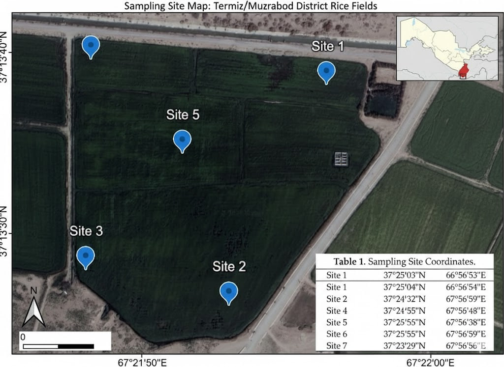
The Termez district is located in the southernmost part of Uzbekistan. The region is dominated by alluvial landforms formed within the Amu Darya River valley, where long-term irrigated agriculture has shaped the prevailing soil characteristics. The dominant soil types are alluvial-meadow and meadow-serozem soils, which possess relatively high agricultural potential within irrigated river terraces. Arid and typical serozem soils occur in the surrounding steppe landscapes, whereas poorly drained depressions are characterized by saline (solonchak) soils and other low-fertility soil types (Fig. 1 and 2).
According to regional soil surveys, the humus content of the arable layer generally ranges from 1.0 to 1.5%. Soil fertility is primarily limited by carbonate and gypsum accumulation, together with varying degrees of secondary soil salinity.
The study area experiences a sharply continental arid climate, with a mean annual air temperature of 19–20°C and annual precipitation ranging from 150 to 170 mm, most of which falls between November and April. During summer, maximum air temperatures frequently reach 40–42°C.
Soil samples were collected systematically from rice fields following standard soil sampling procedures. Samples containing visible colonies of cyanobacteria and microalgae were carefully transferred into sterile sampling bags and transported to the laboratory for further analyses [19]. The sampling sites were located at an altitude of 300–302 m above sea level (37°13′31″N, 67°21′54″E).
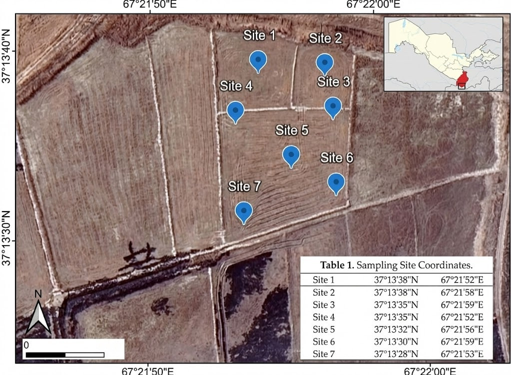
Figure 1. High-resolution satellite mapping of sampling sites in the Termez district
The Muzrabat district is characterized by proluvial-deluvial, alluvial, and aeolian deposits typical of desert-steppe environments. Serozem soils predominate throughout the district, whereas saline soils (solonchaks) and takyrs are commonly found in lowland areas. Irrigated agricultural lands are particularly susceptible to secondary salinization, making soil reclamation an important component of sustainable land management.
The average annual temperature is approximately 19–20°C, with summer temperatures reaching 37–42°C. Annual precipitation ranges from 110 to 120 mm, occurring mainly between November and April. The sampling sites were located at an altitude of 288 m above sea level (37°25′01″N, 66°56′54″E) (Fig. 2).
Figure 2. High-resolution satellite mapping of sampling sites in the Muzrabat district
Isolation and cultivation of microalgal strains. Microalgal strains were isolated from soil samples using the enrichment culture technique followed by inoculation onto BG-11 agar medium. Axenic cultures were obtained through repeated streak plating and the isolation of individual colonies using a sterile Pasteur pipette [15].
The isolates were cultivated on solid BG-11 medium supplemented with nitrogen (pH 7.0, 1.4% agar) in a controlled-environment chamber maintained at 23–25°C, with a light intensity of 60–75 μmol photons m⁻² s⁻¹ under a 12 h light/12 h dark photoperiod.
The isolated strains were deposited in the All-Russian Collection of Microorganisms (VKM) under accession numbers VKM Al-507, VKM Al-508, VKM Al-509, and VKM Al-510.
Light Microscopy
Morphological characteristics and life-cycle features of the isolated microalgal strains were examined using a Leica DM750 light microscope (Germany). Images were captured using a Leica Flexacam C3 digital color camera (Germany). The observation period ranged from 1 to 12 weeks.
Morphological identification was performed using standard taxonomic criteria, including thallus organization, cell morphology, cell size, chloroplast number and morphology, presence of pyrenoids, mucilaginous sheath characteristics, reproductive structures, and other diagnostic features.
Morphometric analyses were conducted using Leica Application Suite X software. Measurements were obtained from 100 cells per strain.
Taxonomic identification was performed using published taxonomic keys and molecular studies [2–4,11,12,14,16]. Nomenclature and taxonomy followed the AlgaeBase classification system (Guiry & Guiry, 2025).
DNA Extraction, PCR Amplification, and Sequencing
Total genomic DNA was extracted using the DNeasy Plant Mini Kit (Qiagen, USA) according to the manufacturer's instructions.
PCR amplification was performed using ScreenMix-HS Master Mix (Evrogen, Russia). Amplification of the ITS2 rDNA region was carried out using the primers described by Johnson et al. (2007). The primer sequences and PCR conditions are presented in Table 1.
Table 1. Primers and conditions for ITS2 amplification
     
The detection of target PCR products was performed by electrophoresis in a 1% agarose gel. For the subsequent purification of amplicons from the gel, the Cleanup Standard kit (Evrogen, Russia) was utilized. Sequencing was conducted by the commercial company Evrogen (Russia).
Molecular Phylogenetic Analysis. For the molecular identification of the isolated microalgal strains, ITS2 nucleotide sequences were compared with reference sequences available in the NCBI GenBank database using the BLASTn algorithm. Reference sequences were selected based on sequence similarity, sequence quality (absence of ambiguous nucleotides), sequence length, and the availability of authentic or type strains.
The phylogenetic dataset for Bracteacoccus comprised 51 ITS2 sequences, including Neochloris aquatica UTEX B138 as the outgroup. The dataset for strain VKM Al-508 included 57 sequences, with Vitreochlamys aulata SAG 69.72 used as the outgroup. The dataset for Chlorella vulgaris strains VKM Al-509 and VKM Al-510 consisted of 40 sequences, using Chlorella sp. ACSSI 342 as the outgroup.
Multiple sequence alignment was performed using ClustalW implemented in BioEdit. The optimal nucleotide substitution model was selected using the IQ-TREE web server according to the Bayesian Information Criterion (BIC). Maximum Likelihood (ML) phylogenetic trees were reconstructed using IQ-TREE, and branch support was evaluated with the Ultrafast Bootstrap method (1000 replicates) together with the SH-aLRT test. Phylogenetic trees were visualized using FigTree version 1.3.1.
Pairwise genetic distances were calculated in MEGA 11 as the percentage of nucleotide differences between aligned ITS2 sequences.
The newly generated ITS2 sequences were deposited in the NCBI GenBank database under accession numbers PV432759, PV432760, PV432761, and PV432762.

3. Results and Discussion

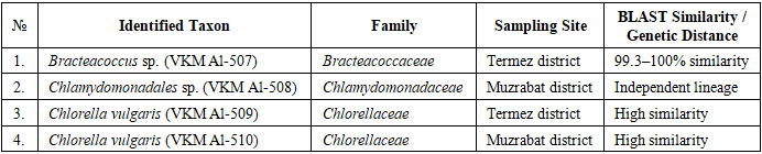
The polyphasic investigation of soil microalgae isolated from rice field agroecosystems in the Termez and Muzrabat districts resulted in the identification of four taxonomically distinct microalgal strains. Using an integrative approach that combined detailed morphological characterization with ITS2 rDNA sequence analysis, the isolates were successfully identified and taxonomically characterized. The strains, designated VKM Al-507 to VKM Al-510, represent important members of the microalgal communities inhabiting the investigated rice field soils. Their taxonomic affiliations, sampling locations, and molecular identification data are summarized in Table 2.
Table 2. Taxonomic identification and molecular-genetic characteristics of microalgae strains isolated from rice field agroecosystems in the Surkhandarya region
     
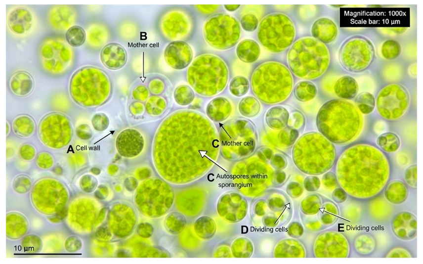
Strain Bracteacoccus sp. VKM Al-507 The strain, exhibiting Bracteacoccus-like morphology, had the following morphological description (Fig. 3).
Figure 3. Morphology of Bracteacoccus sp. strain VKM Al-507. A – cell wall, B – mother cell, C – autospores within sporangium, D – dividing cells, E – dividing cells. Magnification: 1000x. Scale bar = 10 μm
Cells are solitary or occur in irregular aggregates, spherical in shape, and measure 5–28 μm in diameter. The cell wall is relatively robust and does not undergo marked thickening during cell maturation. Young cells contain 2–4 chloroplasts, whereas mature cells possess numerous pyrenoid-free chloroplasts. Orange lipid droplets serve as the primary storage products. Asexual reproduction occurs through the formation of autospores, with each mother cell typically producing four or more autospores [18]. The autospores frequently mature within the sporangial wall and may remain aggregated following its rupture. Zoospore formation was not observed under the culture conditions used in this study.
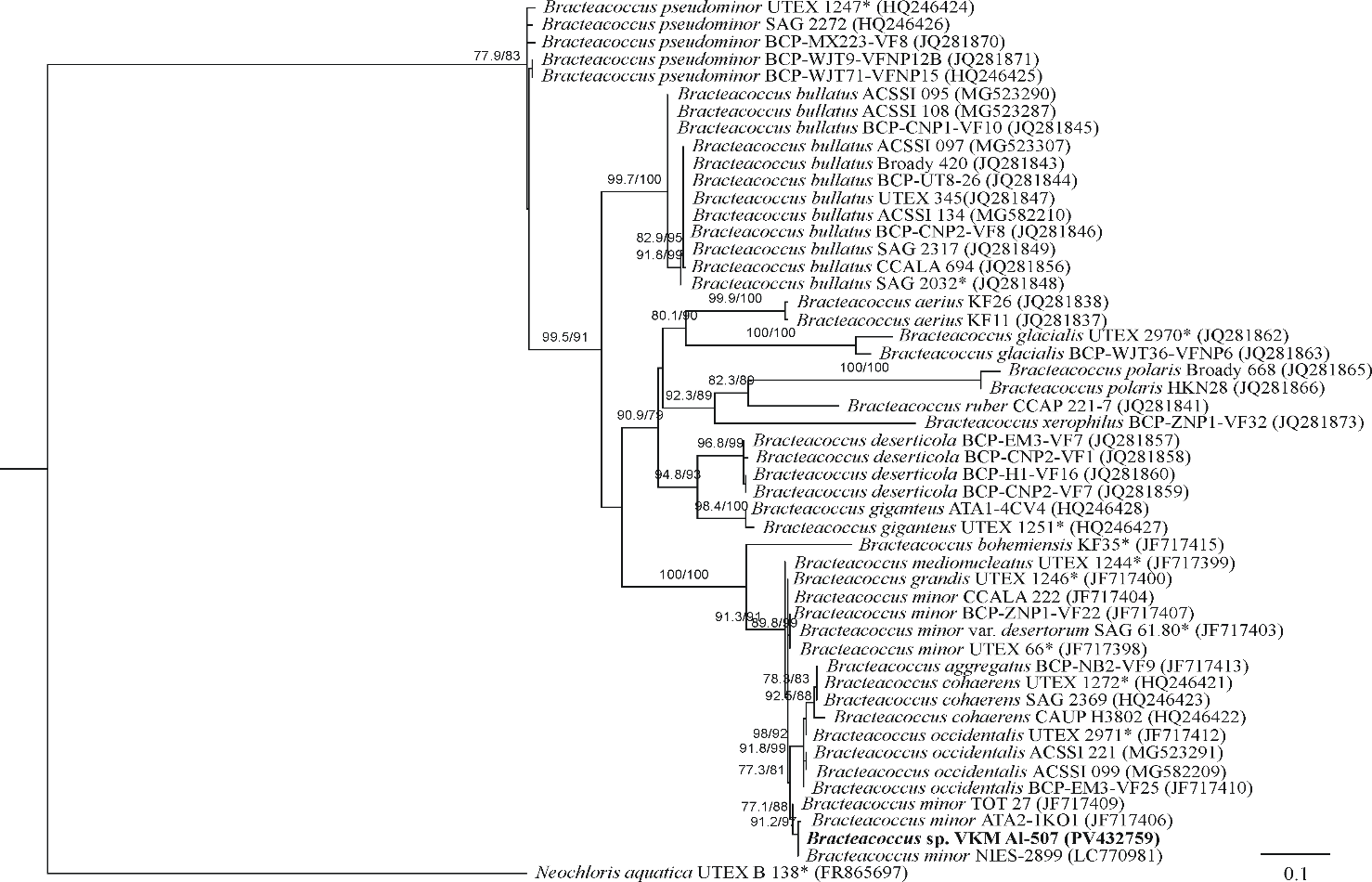
ITS2 rDNA sequence analysis demonstrated that strain VKM Al-507 shared an identical or nearly identical ITS2 sequence with the non-authentic strain Bracteacoccus minor NIES-2899. Together with other non-authentic strains (B. minor ATA2-1KO1 and B. minor TOT 27), these isolates formed a well-supported clade within the genus Bracteacoccus (Fig. 4). The genetic distances among these strains ranged from 0 to 0.7%, remaining below the accepted ITS2 intraspecific threshold of 2% [7], thereby confirming their conspecific relationship. In contrast, the authentic strain B. minor UTEX 66 occupied a distinct and phylogenetically separate lineage. The genetic distance between strain VKM Al-507 and B. minor UTEX 66 was 1.8%. The corresponding genetic distances between strain VKM Al-507 and authentic representatives of other Bracteacoccus species were 1.4% for B. grandis UTEX 1246, 1.8% for B. medionucleatus UTEX 1244, 2.5% for B. cohaerens UTEX 1272, and 3.2% for B. occidentalis UTEX 2971. Taken together, these findings indicate that strain VKM Al-507 belongs to the genus Bracteacoccus; however, a reliable species-level identification cannot presently be assigned because the corresponding phylogenetic lineage requires further taxonomic revision.
Figure 4. Rooted phylogenetic tree of green microalgae of the genus Bracteacoccus inferred by the Maximum Likelihood (ML) method based on internal transcribed spacer 2 (ITS2) sequences (401 nt). SH-aLRT/UB values are provided as statistical support at the tree nodes. SH-aLRT and UB values below 70% are not shown. Nucleotide substitution model: TIM2+F+G4. Designations: * indicates authentic strains; the studied strain is highlighted in bold
Strain Chlamydomonadales sp. VKM Al-508
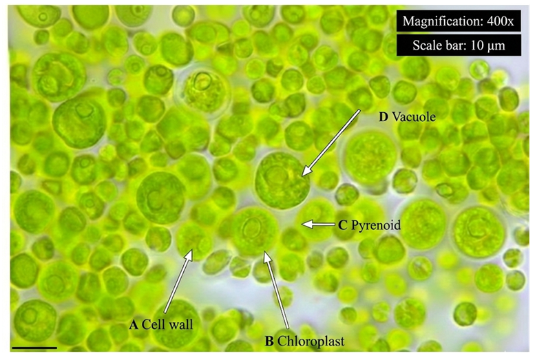
The strain exhibited morphological characteristics closely resembling those of Tetracystis sarcinalis (Fig. 5).
Figure 5. Morphology of Chlamydomonadales sp. strain VKM Al-508. A – cell wall, B – chloroplast C – pyrenoid, D – vacuoles. Magnification: 400x. Scale bar = 10 μm
Cells are spherical, measuring 5–14 μm in diameter, and contain a single parietal, cup-shaped chloroplast. In mature cells, the chloroplast becomes perforated by numerous openings and fissures. A single pyrenoid surrounded by a continuous starch sheath is located within the thickened region of the chloroplast. Each cell contains a single nucleus, while aging cells accumulate conspicuous lipid droplets. Asexual reproduction occurs through the formation of both autospores and zoospores. Zoospores are ellipsoidal, broadly ellipsoidal, or slightly obovate, measuring up to 9.6 μm in length and 6.7 μm in width. They possess a parietal chloroplast, a centrally positioned pyrenoid, and an anterior stigma, whereas a papilla is absent. Autospores are spherical and reach up to 5 μm in diameter.
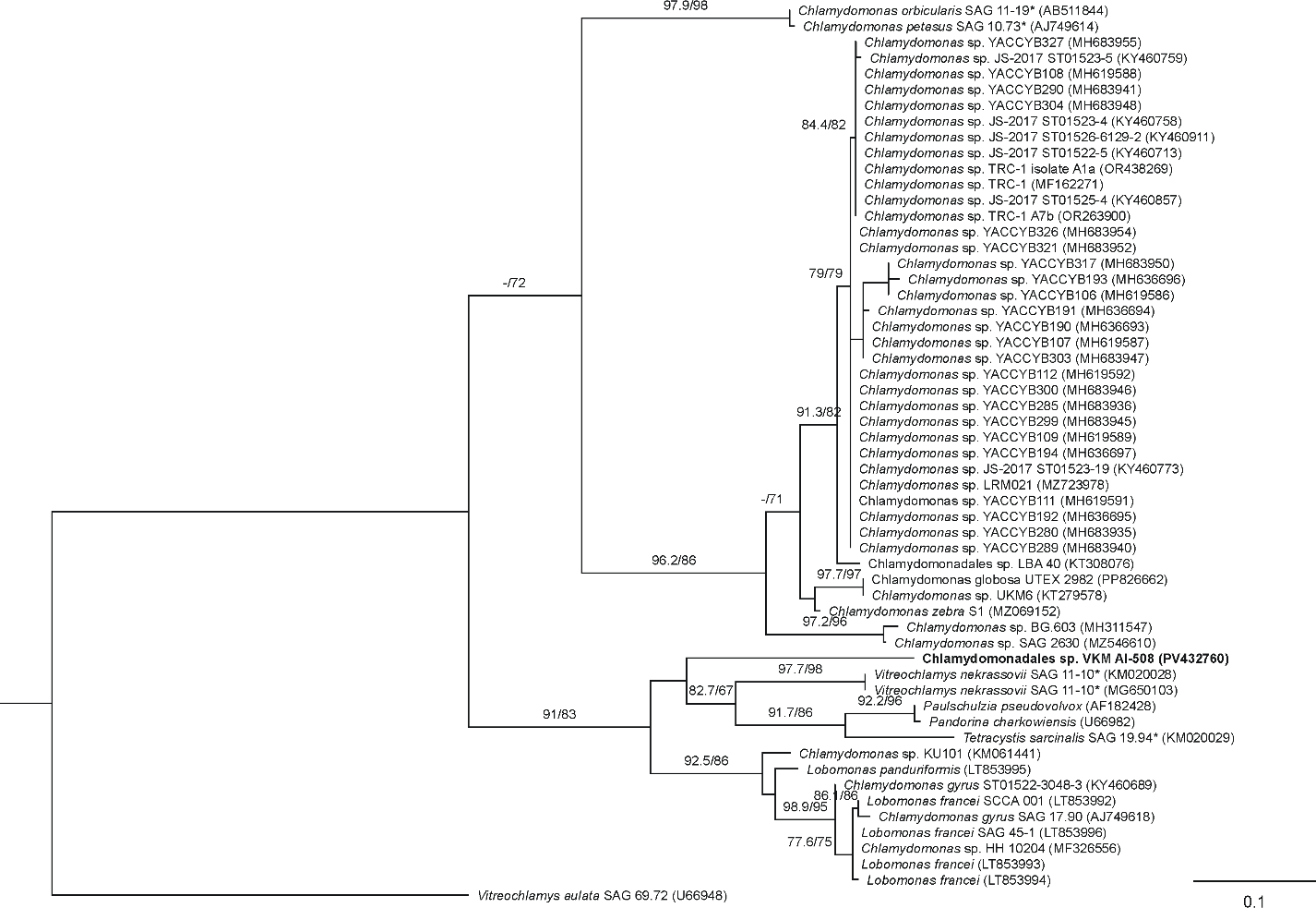
ITS2 rDNA sequence analysis placed strain VKM Al-508 as an independent phylogenetic lineage within a well-supported clade comprising Vitreochlamys nekrassovii, Lobomonas spp., and Tetracystis sarcinalis (Fig. 6). The genetic distance between strain VKM Al-508 and the authentic strain Tetracystis sarcinalis SAG 19.94 was 23.2%, indicating divergence at least at the generic level. These findings suggest that the strain may represent a distinct taxonomic lineage within the order Chlamydomonadales. However, additional phylogenetic analyses based on the 18S rRNA and rbcL gene sequences are required to resolve its precise taxonomic position. Therefore, pending further molecular evidence, the isolate was provisionally identified as Chlamydomonadales sp.
Figure 6. Rooted phylogenetic tree illustrating the phylogenetic position of strain VKM Al-508, inferred by the Maximum Likelihood (ML) method based on internal transcribed spacer 2 (ITS2) sequences (277 nt). SH-aLRT and UB values are provided at the nodes as statistical support. SH-aLRT and UB values below 70% are not shown. Nucleotide substitution model: TIM2e+G4. Designations: * indicates authentic strains; the studied strain is highlighted in bold
Strains Chlorella vulgaris VKM Al-509 and VKM Al-510
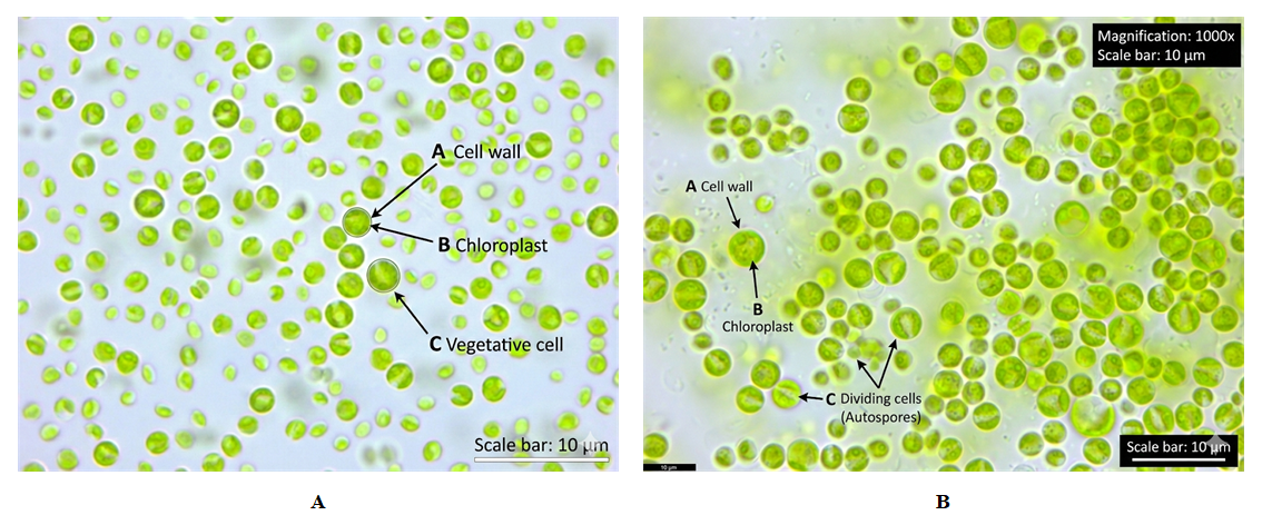
Strain VKM Al-509 consisted of solitary or loosely aggregated spherical cells measuring 3–7 μm in diameter (Fig. 7A). Each cell contained a single parietal, cup-shaped chloroplast with a centrally positioned pyrenoid surrounded by a starch sheath. Asexual reproduction occurred through the formation of 2–8 autospores.
Strain VKM Al-510 was characterized by solitary or loosely aggregated spherical cells measuring 3.2–8.4 μm, occasionally reaching 10.7 μm in diameter (Fig. 7B). The cells possessed a single parietal, cup-shaped chloroplast containing one pyrenoid enclosed by a starch sheath. Asexual reproduction occurred through the formation of 2, 4, or 8 autospores.
The observed morphological characteristics of both isolates were consistent with the published descriptions of Chlorella vulgaris (Chlorellales, Trebouxiophyceae), supporting their preliminary identification based on morphology.
Figure 7. Morphology of Chlorella vulgaris strains VKM Al-509 (A) and VKM Al-510 (B). A – cell wall, B – chloroplast, C – vegetative cell. Magnification: 1000x. Scale bar = 10 μm
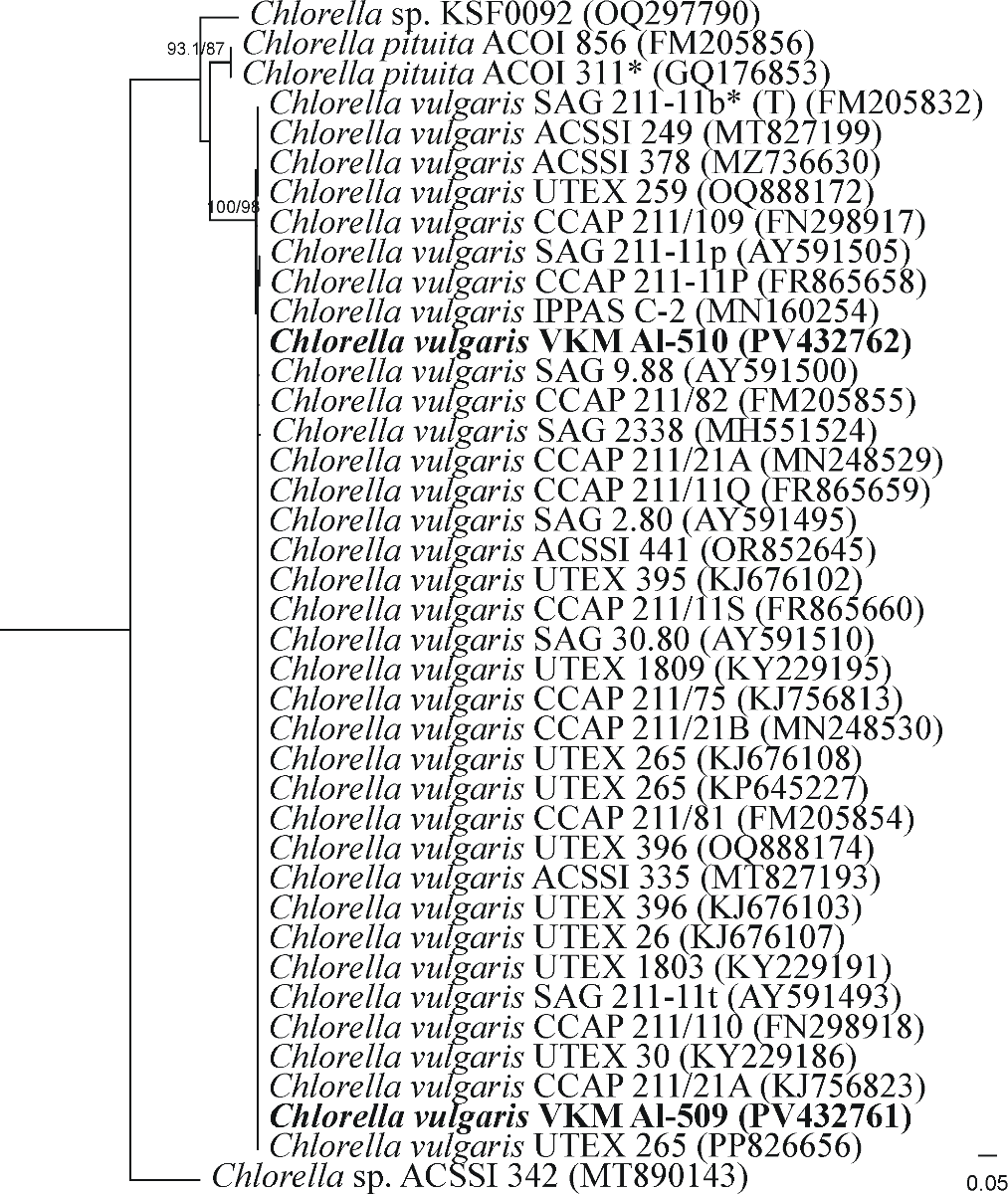
ITS2 rDNA sequence analysis placed strains VKM Al-509 and VKM Al-510 within the Chlorella vulgaris clade, which includes the authentic reference strain SAG 211-11b (Fig. 8). No sequence variation was detected between the two studied strains in the ITS2 region, indicating complete sequence identity. The genetic distance between both isolates and the authentic strain SAG 211-11b was 0.8%, which is well below the accepted ITS2 intraspecific threshold of 2% [7]. This level of genetic similarity confirms that both isolates belong to the species Chlorella vulgaris. Taken together, the morphological characteristics and ITS2 sequence data provide strong evidence supporting the identification of strains VKM Al-509 and VKM Al-510 as Chlorella vulgaris.
Figure 8. Rooted phylogenetic tree illustrating the phylogenetic position of strains VKM Al-509 and 510, inferred by the Maximum Likelihood (ML) method based on internal transcribed spacer 2 (ITS2) sequences (264 nt). SH-aLRT and UB values are provided at the nodes as statistical support. SH-aLRT and UB values below 70% are not shown. Nucleotide substitution model: HKY+F+G4. Designations: * indicates authentic strains; the studied strains are highlighted in bold
The present study represents the first polyphasic taxonomic investigation of cultivable soil microalgae isolated from rice field agroecosystems of the Surkhandarya region, Uzbekistan. By integrating detailed morphological observations with ITS2 rDNA sequence analysis, four microalgal strains representing three phylogenetically distinct taxa were successfully identified. The combined use of morphological and molecular approaches substantially improved taxonomic resolution, overcoming the limitations associated with morphology-based identification alone [6,7,15,20].
The molecular analyses demonstrated complete agreement between morphological and molecular identification for the two Chlorella vulgaris isolates, which clustered with the authentic reference strain SAG 211-11b and exhibited only 0.8% ITS2 sequence divergence. These findings are consistent with previous studies demonstrating that ITS2 is a reliable molecular marker for species delimitation within the genus Chlorella [6–8]. Likewise, strain VKM Al-507 was assigned to the genus Bracteacoccus based on both morphological characteristics and phylogenetic evidence. However, its uncertain relationship with authentic representatives of Bracteacoccus minor indicates that further taxonomic revision of this lineage is warranted, which agrees with previous phylogenetic studies of the genus [2–4].
In contrast, strain VKM Al-508 occupied an independent phylogenetic position within the order Chlamydomonadales. Similar taxonomic complexity has previously been reported for Lobomonas, Tetracystis, and other closely related green algae, emphasizing the importance of integrating multiple molecular markers for accurate species delimitation [11,12]. Consequently, additional analyses based on the 18S rRNA and rbcL genes will be necessary to determine the precise taxonomic position of this isolate.
The occurrence of phylogenetically diverse green microalgae in rice field soils demonstrates that agricultural ecosystems of southern Uzbekistan constitute valuable reservoirs of indigenous microalgal diversity. Comparable observations have been reported from agricultural soils in both Europe and Uzbekistan, where environmental conditions and agricultural management practices strongly influence soil algal community composition [17–19]. Furthermore, recent reviews have emphasized that terrestrial microalgae represent an important but still insufficiently explored component of agricultural ecosystems, highlighting the need for integrative taxonomic approaches combining morphology and molecular phylogeny [20–22].
Overall, the present study expands current knowledge of soil microalgal diversity associated with rice agroecosystems in Uzbekistan and confirms the value of polyphasic taxonomy for the identification of cultivable microalgae. The taxonomic information obtained provides a solid basis for future ecological, physiological, and biotechnological investigations of indigenous microalgal strains adapted to arid agricultural environments.

4. Conclusions

This study represents the first polyphasic taxonomic investigation of cultivable soil microalgae isolated from rice field agroecosystems in the Surkhandarya region of Uzbekistan. By integrating detailed morphological characterization with ITS2 rDNA sequence analysis, four microalgal strains representing three taxonomic groups within the phylum Chlorophyta were successfully identified.
Among the investigated isolates, two strains (VKM Al-509 and VKM Al-510) were confidently identified as Chlorella vulgaris based on both morphological characteristics and molecular phylogenetic evidence. Strain VKM Al-507 was assigned to the genus Bracteacoccus, whereas strain VKM Al-508 was provisionally identified as Chlamydomonadales sp., pending further molecular analyses using additional genetic markers.
The results demonstrate the importance of integrating classical morphology with molecular phylogenetic approaches for the accurate identification of soil microalgae. Furthermore, the study expands current knowledge of microalgal diversity associated with rice field agroecosystems in southern Uzbekistan and provides a valuable taxonomic foundation for future ecological, phylogenetic, and biotechnological investigations of indigenous microalgal resources.
Future studies should focus on the ecological characterization and physiological evaluation of the isolated strains, particularly their tolerance to salinity and their potential applications in sustainable agriculture and soil restoration under arid environmental conditions.
Funding: This research was supported by the Ministry of Higher Education, Science and Innovation of the Republic of Uzbekistan under Project No. AL-9424115044.
Institutional Review Board Statement: Not applicable.
Informed Consent Statement: Not applicable.
Data Availability Statement: The data are available upon reasonable request from the corresponding author.
Conflicts of Interest: The authors declare no conflict of interest.
Author ORCIDs
Yulduzkhon Tukhtaboeva https://orcid.org/0009-0008-2209-3582
Abdullayeva Gulnoza https://orcid.org/0009-0008-6953-6504

References

[1]  Ergashev, A. E. (1974). Opredelitel' sinyezelenykh vodorosley Sredney Azii. Determinant of blue-green algae of Central Asia. Tashkent: Fan. (In Russian).
[2]  Fuciková, K., Rada, J. C., & Lewis, L. A. (2011). The tangled taxonomic history of Dictyococcus, Bracteacoccus and Pseudomuriella (Chlorophyceae, Chlorophyta) and their distinction based on a phylogenetic perspective. Phycologia, 50(4), 422–429.
[3]  Fucikova, K., Flechtner, V. R., & Lewis, L. A. (2012). Revision of the genus Bracteacoccus Tereg (Chlorophyceae, Chlorophyta) based on a phylogenetic approach. Nova Hedwigia, 96(1-2), 15–59.
[4]  Fučíková, K., & Lewis, L. A. (2012). Intersection of Chlorella, Muriella and Bracteacoccus: Resurrecting the genus Chromochloris Kol et Chodat (Chlorophyceae, Chlorophyta). Fottea, 12(1), 83–93.
[5]  Guiry, M. D., & Guiry, G. M. (2025). AlgaeBase. World-wide electronic publication, University of Galway. https://www.algaebase.org. Accessed 28 February 2025.
[6]  Hepperle, D., & Krienitz, L. (2001). Systematics of Chlorella-like algae: combining morphology and 18S rDNA sequences. Journal of Phycology, 37, 34.
[7]  Hoshina, R. (2014). DNA analyses of a private collection of microbial green algae contribute to a better understanding of microbial diversity. BMC Research Notes, 7, 592. https://doi.org/10.1186/1756-0500-7-592.
[8]  Huss, V. A. R., Frank, C., Hartmann, E. C., et al. (1999). Chlorella vulgaris: a unified concept for the most famous of all microalgae. Archiv für Protistenkunde, 149(4), 473-480.
[9]  Johnson, J. L., Fawley, M. W., & Fawley, K. P. (2007). The diversity of Scenedesmus and Desmodesmus (Chlorophyceae) in Itasa State Park, Minnesota, USA. Phycologia, 46, 214–229. doi: 10.2216/05-69.1.
[10]  Muzaffarov, A. M. (1965). Flora vodorosley vodoyemov Sredney Azii [Flora of algae of water bodies of Central Asia]. Tashkent: Nauka. (In Russian).
[11]  Novis, P. M., & Visnovsky, G. (2012). Novel alpine algae from New Zealand: Chlorophyta. Phytotaxa, 39, 1–30.
[12]  Sausen, N., Malavasi, V., & Melkonian, M. (2018). Molecular phylogeny, systematics, and revision of the type species of Lobomonas, L. francei (Volvocales, Chlorophyta) and closely related taxa. Journal of Phycology, 54(2), 198–214. doi: 10.1111/jpy.12615.
[13]  Shoyakubov, R. Sh. (1979). Khlorella v Uzbekistane [Chlorella in Uzbekistan]. Tashkent: Fan. (In Russian).
[14]  Starr, R. C. (1955). A comparative study of Chlorococcum meneghini and other spherical, zoospore-producing genera of the Chlorococcales. Indiana University Publications Science, 20, 1–111.
[15]  Temraleeva, A. D., Mincheva, E. V., Bukin, Yu. S., & Andreeva, A. M. (2014). Sovremennyye metody vydeleniya, kul'tivirovaniya i identifikatsii zelenykh vodorosley (Chlorophyta) [Modern methods for isolation, cultivation and identification of green algae (Chlorophyta)]. Kostroma: Kostromskoy pechatnyy dom. 215 p. (In Russian).
[16]  Tereg, E. (1923). Einige neue Grünalgen. Beihefte zum Botanischen Centralblatt, 39, 179–195.
[17]  Zancan, S., Trevisan, R., & Paoletti, M. G. (2006). Soil algae composition under different agro-ecosystems (rice-paddy, orchard and vineyard) from NE Italy. Agriculture, Ecosystems & Environment, 112(1), 1–12.
[18]  Tukhtaboeva, Y. A., Krivina, E. S., Red'kina, V. V., & Temraleeva, A. D. (2024). Microalgae from eroded soils in the Northern Fergana Valley, Uzbekistan. Microbiology, 93(4), 421–433. https://doi.org/10.31857/S0026365624040024.
[19]  Tukhtaboeva, Y. A., Redkina, V. V., Bukin, Y. S., et al. (2025). Diversity of cyanobacteria and algae in eroded soils of the Fergana Valley (Uzbekistan) according to light microscopy data and high-throughput sequencing. Arid Ecosystems, 15, 232–245. https://doi.org/10.1134/S2079096125700143.
[20]  Joseph, J., & Ray, J. G. (2024). A critical review of soil algae as a crucial soil biological component of high ecological and economic significance. Journal of Phycology, 60(2), 229–253. https://doi.org/10.1111/jpy.13444.
[21]  Alvarez, A. L., Weyers, S. L., Goemann, H. M., Peyton, B. M., & Gardner, R. D. (2021). Microalgae, soil and plants: A critical review of microalgae as renewable resources for agriculture. Algal Research, 54, 102200. https://doi.org/10.1016/j.algal.2021.102200.
[22]  Ramakrishnan, B., Maddela, N. R., Venkateswarlu, K., & Megharaj, M. (2023). Potential of microalgae and cyanobacteria to improve soil health and agricultural productivity: A critical view. Environmental Science: Advances, 2, 586–611.