-
Paper Information
- Paper Submission
-
Journal Information
- About This Journal
- Editorial Board
- Current Issue
- Archive
- Author Guidelines
- Contact Us
International Journal of Virology and Molecular Biology
p-ISSN: 2163-2219 e-ISSN: 2163-2227
2026; 15(3): 63-66
doi:10.5923/j.ijvmb.20261503.01
Received: Apr. 28, 2026; Accepted: May 26, 2026; Published: May 28, 2026

Morphometric Parameters of Renal Nephrons in Rats During Postnatal Ontogenesis
 Abstract
 Reference
 Full-Text PDF
 Full-text HTML
Ilyasov A. S.1, Mamarajabova B. A.2
1Doctor of Biological Sciences, Professor, Navoi Innovation University, Navoi, Uzbekistan
2Independent Researcher, Berdakh Karakalpak University, Nukus, Uzbekistan
Copyright © 2026 The Author(s). Published by Scientific & Academic Publishing.
This work is licensed under the Creative Commons Attribution International License (CC BY).
http://creativecommons.org/licenses/by/4.0/

The kidneys are paired vital organs that play a central role in maintaining physiological homeostasis in the human body. Their fundamental structural and functional unit is the nephron, with approximately one million nephrons present in each kidney. Background: Nephrons are responsible for key renal processes, including blood filtration, urine formation, and regulation of water and electrolyte balance. Structurally, nephrons are distributed within the cortical and medullary regions of the kidney, where they collectively ensure the efficient execution of renal functions essential for systemic homeostasis. Morphometric parameters of renal nephrons in rats during postnatal ontogenesis were investigated. Method: The experiment was conducted on outbred albino rats. A total of 48 animals were included, categorized into the following age groups: newborns; 3-month-old rats representing the early reproductive (at reproductive maturity) period; 6-month-old rats corresponding to the phase of active reproduction; and 12-month-old rats representing the mature reproductive stage or the period of reproductive decline. Based on this age stratification, the dynamics of changes in the morphometric parameters of renal structural components during postnatal development were analyzed. Results: It was established that the most pronounced increase in morphometric parameters of the rat kidneys during postnatal development occurs by the age of 3 months compared to 6-month-old animals: the glomerular height, the thickness of the Bowman’s capsule, and the thickness of the distal tubular wall increased by up to 40.1%. From the neonatal period to 12 months of age, the growth rate of nephron morphometric parameters ranged from a 1.5-fold increase in the thickness of the collecting duct walls to a 4.1-fold increase in the internal diameter of distal tubules. Conclusion: In rats, the period of postnatal ontogenesis is characterized by significant changes in the structure of the nephron, with the most pronounced changes observed at 3 months of postnatal development, reflecting a critical stage in renal development. In subsequent periods, growth continues, but very slowly. This indicates a gradual stabilization of the structural and functional parameters of the nephron during the developmental process.
Keywords: Rat, Postnatal ontogenesis, Morphometry, Glomeruli, Tubules, Nephron
Cite this paper: Ilyasov A. S., Mamarajabova B. A., Morphometric Parameters of Renal Nephrons in Rats During Postnatal Ontogenesis, International Journal of Virology and Molecular Biology, Vol. 15 No. 3, 2026, pp. 63-66. doi: 10.5923/j.ijvmb.20261503.01.
1. Introduction
- Each nephron represents a functional unit capable of independently performing specialized transport processes. However, certain renal functions can only be achieved through the coordinated activity of the entire nephron population; a notable example is the concentration of urine. Three types of nephrons are distinguished, differing in their structural organization, topographical localization, and specific features of blood supply. Each nephron consists of the glomerular capsule (Bowman’s capsule), the glomerulus, and an interconnected system of renal tubules. [1]. Nephrons differ in their anatomical topography and structural organization. Accordingly, they are classified into three types: superficial (cortical) nephrons, which are located entirely within the renal cortex and constitute approximately 1% of the total nephron population; intracortical nephrons, accounting for about 79%; and juxtamedullary nephrons, situated at the corticomedullary junction, whose loops extend deeply into the medulla and comprise approximately 20% of all nephrons [2]. Within the nephron tubular system, cellular specializations including microvilli, cilia, and intercellular junctions are formed, which collectively contribute to the structural and functional cytoskeletal organization of renal tubular epithelial cells [3]. The proximal tubule descends toward the medullary region. Its apical membrane is characterized by a well-developed brush border composed of densely packed microvilli coated with a glycocalyx [4]. In the distal segment, the brush border is absent. Cells in this region are involved in the synthesis of hormonally active substances, including renin and angiotensin, which participate in the regulation of arterial blood pressure. This segment is lined by simple cuboidal epithelium and lies in close proximity to the afferent arteriole [5]. Under the influence of renin, the renin–angiotensin system is activated, exhibiting potent vasoconstrictive effects. In addition, renin stimulates the zona glomerulosa of the adrenal cortex, leading to the secretion of aldosterone. Aldosterone enhances the reabsorption of sodium ions in the distal convoluted tubule [6].The collecting ducts are lined by cuboidal epithelium, which transitions to a columnar epithelium in the distal segments. In this region, epithelial cells exhibit high water permeability due to a well-developed labyrinth of intracellular membranes. Two principal types of epithelial cells are distinguished in the collecting ducts: principal cells and intercalated cells. Intercalated cells are the predominant cell type and are primarily responsible for the secretion of hydrogen ions, a process mediated by the enzyme carbonic anhydrase. Principal cells of this segment are represented by cuboidal epithelium with numerous apical microvilli on their surface [7].
2. Materials and Methods
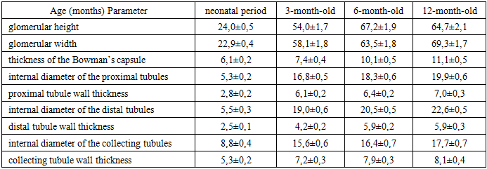
- The experiment was conducted on outbred albino rats. The laboratory animals underwent a mandatory veterinary examination to assess the presence of any existing pathological conditions, as well as to evaluate their body condition and age.A total of 48 rats were included in the study, distributed across different developmental stages: newborn animals (n = 11); 3-month-old rats representing the early reproductive period with established reproductive capability (n = 12); 6-month-old rats corresponding to the phase of active reproduction (n = 13); and 12-month-old rats representing the reproductive mature stage or period of reproductive decline (n = 12).Based on this age stratification, the study was designed to assess the dynamics of changes in the morphometric parameters of renal structural components during postnatal development in rats.Euthanasia of the rats was performed at neonatal, 3-, 6-, and 12-month age points under ether anesthesia. During the sacrifice and dissection of laboratory animals, all principles of biosafety and ethical standards for the use of experimental animals were strictly observed. Following laparotomy, the kidneys were excised, and the animals were weighed; both absolute and relative kidney masses were subsequently determined.Renal fixation was carried out in 10% neutral formalin. The samples were then processed through a graded series of ethanol concentrations and embedded in paraffin. Paraffin-embedded kidney sections (5–8 μm thick) were stained with hematoxylin and eosin as well as Van Gieson staining.Morphometric parameters were measured, including glomerular height and width; the thickness of the Bowman’s capsule; the internal diameter and wall thickness of proximal tubules; the internal diameter and wall thickness of distal tubules; and the internal diameter and wall thickness of collecting tubules. Measurements were performed using an eyepiece micrometer DN-107T / NLCD-307B model (Novel, China).
3. Results and Discussion
- The renal nephron is composed of several sequentially connected segments located within both the cortical and medullary regions of the kidney. Renal glomeruli consist of a vascular tuft enclosed between the afferent and efferent arterioles and its surrounding capsule. The initial portion of each nephron is the Malpighian corpuscle, which is formed by a network of interwoven capillary loops creating a vascular tuft of glomerular capillaries, surrounded by visceral and parietal layers of the Bowman’s capsule.The Malpighian glomerulus is formed by an arteriole that branches into multiple capillary loops. The glomerular capillaries subsequently converge to form the efferent arteriole.In addition to the glomerular capsule, the nephron forms the renal tubular system. The segments constituting this tubular system include the proximal tubule, the descending and ascending limbs of the nephron loop, the thin segment, the distal tubule, and the connecting tubule. Urine passes through this tubular network into the collecting ducts, the convergence of which forms the papillary ducts. The latter open on the surface of the renal papilla.In newborn rat pups, the height of renal glomeruli ranges from 21.0 to 27.0 μm, while their width varies from 21.0 to 26.0 μm. The thickness of the Bowman’s capsule ranges from 5.0 to 7.0 μm. The internal diameter of the proximal tubules of the kidney is 4.0–7.0 μm, with a tubular wall thickness of 2.0–4.0 μm. The internal diameter of the distal tubules is also 4.0–7.0 μm, while their wall thickness measures 2.0–3.0 μm.In the medullary region of the kidney, the internal diameter of the collecting tubules varies from 7.0 to 12.0 μm, and their wall thickness ranges from 4.0 to 7.0 μm.In sexually mature rats at 3 months of age, the height of renal glomeruli ranges from 42.0 to 61.0 μm, while their width varies from 48.0 to 70.0 μm. The thickness of the Bowman’s capsule ranges from 6.0 to 11.0 μm. The internal diameter of the proximal tubules is 14.0–20.0 μm, with a tubular wall thickness of 5.0–7.0 μm. At this developmental stage, the internal diameter of the distal tubules is 16.0–23.0 μm, while their wall thickness ranges from 3.0–6.0 μm.Examination of the medullary region of the kidney demonstrated that the internal diameter of the collecting tubules is 12.0–19.0 μm, with wall thickness varying from 5.0 to 9.0 μm. The structural organization of renal glomeruli in 3-month-old rats is presented in Figure 1.
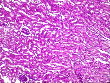 | Figure 1. Structure of renal glomeruli in kidneys of 3-month-old rats: Renal corpuscle Proximal tubule Collecting tubules. Staining: hematoxylin and eosin. Magnification: ocular 10×, objective 20× |
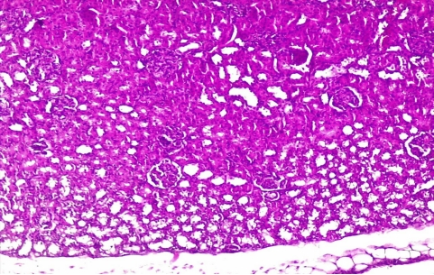 | Figure 2. Microscopic structure of the kidneys in 6-month-old rats: 1. Renal corpuscle 2. Proximal tubule 3. Collecting tubules. Staining: hematoxylin and eosin. Magnification: ocular 10×, objective 20× |
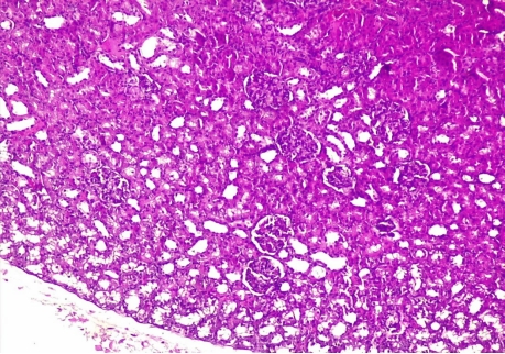 | Figure 3. Microscopic structure of the kidneys in 12-month-old rats: 1. Renal corpuscle 2. Proximal tubule 3. Collecting tubules. Staining: hematoxylin and eosin. Magnification: ocular 10×, objective 20× |
|