International Journal of Virology and Molecular Biology

p-ISSN: 2163-2219    e-ISSN: 2163-2227

2026;  15(1): 15-20

doi:10.5923/j.ijvmb.20261501.04

Received: Feb. 15, 2026; Accepted: Feb. 27, 2026; Published: Mar. 6, 2026


Inhibition of Pancreatic Lipase and α-amylase by Gingko Biloba Endophytes

Tashkhan Gulyamova1, Dilaram Ruziyeva1, Behruz Tojiyev1, Gulchekhra Rasulova1, Liliya Abdulmyanova1, Maftuna Yoldosheva2, Kseniya Kondrasheva1

1Institute of Microbiology, Academy of Sciences Republic of Uzbekistan, Tashkent, Uzbekistan

2University of Business and Science, Non-state Higher Education Institution, Tashkent, Uzbekistan

Copyright © 2026 The Author(s). Published by Scientific & Academic Publishing.

This work is licensed under the Creative Commons Attribution International License (CC BY).
http://creativecommons.org/licenses/by/4.0/

Abstract

Pancreatic lipase and pancreatic α-amylase inhibitors are effective drugs for obesity and diabetes. Endophytes are a promising but little-studied source of inhibitors of therapeutically essential enzymes. The activity of α-amylase and lipase was determined spectrophotometrically. Studies of extracts of endophytic isolates GBL4, GBL5 from leaves, and GBS7 from the stem of Ginkgo biloba have shown their high inhibitory activity against pancreatic lipase and α-amylase. IC50 values of GBL4 and GBL5 isolates were 2.6 and 5.6 mcg/ml, respectively, while the IC50 of Xenical as a positive control was 20.0 mсg/ml, and the IC50 of GBS7 extract was close to Xenical (26.5 mcg/ml). IC50 values of amylase inhibition by GBL4 and GBL5 were 5.2 and 5.95 mcg/ml, and 1,5 mcg/ml by GBS7 extract, which is two times lower than acarbose (3.7 mcg/ml) as a positive control. Considering that high enzyme inhibition values with low IC50 values indicate the effectiveness of the inhibitory effect of the substance, it can be assumed that the endophytic isolates Gingko biloba GBL4, GBL5 from the leaves, and GBL5 from the item of the plant have significant potential as sources of effective inhibitors of two pancreatic enzymes - lipase and amylase.

Keywords: Obesity, Pancreatic lipase, Pancreatic α-amylase, Inhibitors, Endophytic fungi, Ginkgo biloba, Ethyl acetate extracts

Cite this paper: Tashkhan Gulyamova, Dilaram Ruziyeva, Behruz Tojiyev, Gulchekhra Rasulova, Liliya Abdulmyanova, Maftuna Yoldosheva, Kseniya Kondrasheva, Inhibition of Pancreatic Lipase and α-amylase by Gingko Biloba Endophytes, International Journal of Virology and Molecular Biology, Vol. 15 No. 1, 2026, pp. 15-20. doi: 10.5923/j.ijvmb.20261501.04.

1. Introduction

Obesity and diabetes mellitus (DM) are essential medical and social problems that have reached the scale of a global epidemic [1,2]. According to the World Health Organization (WHO), the prevalence of obesity has increased significantly in recent decades, and this condition now affects millions of people around the world. According to the forecast of the International Diabetes Federation, the number of diabetics may increase to 552 million by 2030 [3], while WHO reports that more than 1 billion adults in the world are overweight, 300 million of them are obese [1]. Meanwhile, numerous studies indicate a close relationship between these two diseases. Obesity is one of the main risk factors for the development of metabolic disorders, including DM2 [4].
It is known that in the treatment strategy for both diabetes and obesity, the most effective method is the use of inhibitors of key enzymes of lipid and carbohydrate metabolism. Thus, pancreatic lipase (PL) plays a key role in the hydrolysis of triglycerides in the intestine, and its inhibition leads to a decrease in fat absorption, which makes PL inhibitors effective drugs to combat obesity [5,6]. Pancreatic α-amylase (PA) is an important enzyme involved in the hydrolysis of starch to maltose and glucose, and its inhibitors prevent postprandial hyperglycemia, which is a significant factor in the treatment of diabetes [7,8]. Currently, several enzyme inhibitors are available on the market and successfully used in therapeutic practice [9–11]. Such practical medicine in treating diabetes and obesity includes pancreatic alpha-amylase and lipase inhibitors, such as acarbose and orlistat [12]. However, long-term use of these inhibitors is accompanied by severe side effects, including hepatotoxicity, gallstones, kidney stones, acute pancreatitis, abdominal discomfort, bloating, flatulence, and diarrhea [13], which necessitates the development of new safe and effective drugs for natural nature.
Recent studies have convincingly proved that endophytes are a prosperous and valuable source of natural products with various chemical structures and biological activity, making them essential for discovering and developing new medicines [14,15]. In particular, it has been shown that endophytes of many medicinal plants produce compounds that can serve as inhibitors of several therapeutically essential enzymes, including pancreatic amylase and lipase [16–18].
Ginkgo biloba is one of the most famous and popular medicinal plants. Due to the wide range of bioactive properties of the compounds contained in G. biloba, many different therapeutic phytopreparations have been developed and are commercially widely available [19]. However, given that the plant grows for a long time, it is interesting to study the endophytes associated with it. Thus, many endophytic fungi have been isolated from the leaves, fruits, bark, stem, and roots of G. biloba seedlings, classified in various genera and producing such phytocompounds as flavonoids, terpenoids, and other compounds [20,21]. It has been shown that the secondary metabolites of G. biloba endophytes exhibit various bioactive properties and are of interest for developing commercial prodrugs.
Most importantly, from a therapeutic point of view, G. biloba has no side effects even after prolonged use. According to the available data on the endophytes inhabiting G. biloba, more than 30 different genera of microorganisms have been identified, among which three recently described species [22]. Over 60 secondary metabolites have been reported to have a wide range of activities, including antimicrobial, antioxidant, antitumor, and biocontrol. However, despite the variety of metabolites and their bioactive properties, there needs to be more information in the literature about G. biloba endophytes inhibiting amylase or lipase.
The objective of our study was isolation of endophytic fungi associated with Ginkgo biloba and studying the inhibitory activity of their secondary metabolites on pancreatic lipase (PL) and pancreatic α-amylase (PA) to assess the prospects of using G. biloba endophytes as a basis for the development of new prodrugs against obesity and diabetes.

2. Material and Methods

Objects of research
The object of the study was endophytic fungi isolated from the leaves and stem of G. biloba, growing in the Tashkent region.
Isolation of fungal endophytes
The isolation of endophytic fungi was carried out according to Hazalin et al. [23] from the roots, stems, leaves, and inflorescences of collected plants. After pretreatment each segment of the plant was aseptically crushed and placed on Petri dishes with agarized Chapek-Dox medium containing chlortetracycline at a concentration of 50 mg/ml and streptomycin sulfate at a concentration of 250 mg/ml to suppress the growth of bacterial microflora. The cups were incubated for 7–14 days at a temperature of 28°C. Individual colonies of endophytes grown after incubation were selected using a thin needle, transferred to tubes with agar and incubated at 28°C for seven days. A Chapek-Dox medium with an antibiotic was used as a control.
Endophyte fungal cultures are stored by periodic re-sowing on Chapek-Dox agar. All isolates are stored in the refrigerator at +4°C.
Cultivation of endophytes
Endophytes were grown submerged in Chapek-Dox medium on an orbital shaker at 120 rpm for seven days at 28°C. After the cultivation, the biomass was separated from the fermentation liquid by centrifugation at 6000 rpm for 15 minutes.
Extraction of secondary metabolites
About 5 g of biomass was homogenized and extracted twice with 25 ml ethyl acetate for 24 hours on an orbital shaker at room temperature. Then, the mixture was filtered through a paper filter (Watman paper No. 1), and Na₂SO₄ was added at a rate of 40 μg/ml to remove the water layer. The mixture was evaporated dry on a rotary evaporator, and 1 ml of dimethyl sulfoxide was added. The resulting extract was used as a stock and stored at +4°C.
Determination of lipase inhibitory activity
About 50 mg of bovine pancreatic lipase (“Sigma”, 100 U/ml) was suspended in 10 ml of tris-NaCl buffer, pH 7.4. The solution was shaken for 15 minutes, followed by centrifugation at 4000 rpm for 10 min. The supernatant was collected and used as an enzyme solution. Initial solutions of extracts and Xenical were prepared in DMSO with concentrations of 10 mg/ml. The final reaction mixture contained 875 µl of buffer, 100 µl of the enzyme, and 20 µl of extract, pre-incubated for 5 minutes at 37°C, followed by the addition of 10 µl of the substrate (4-nitrophenyl palmitate, 10 mM in acetonitrile). The optical density of the final mixture was measured after 5 min (SPEKOL–1300) at 405 nm [24]. The percentage of inhibition was calculated using the formula:
% inhibition of PL = [(Ae − At) / Ae] × 100,
where Ae is the optical density of the enzyme control (without an inhibitor), At is the difference between the optical density of the test sample with and without a substrate.
Determination of α-amylase inhibitory activity
The activity of α-amylase was determined spectrophotometrically at 630 nm as described by Picot et al. [25].
Determination of IC₅₀ value
The semi-maximal concentration of active extracts, with an inhibitory activity of more than 70%, was carried out within a range from 0.1 to 0.5 mg/ml. The IC₅₀ value was calculated from the regression curve using the least squares method of a semi-logarithmic graph relative to the percentage inhibition curves using GraphPad Prism version 4.0 [26].
All measurements were carried out in triple.

3. Results and Discussion

Ginkgo Biloba is a scarce plant in Uzbekistan. There are at most five such trees in the Tashkent region. In our work, we used the leaves and stems of one of the plants growing on the territory of the Kibray sanatorium, Tashkent. As a result, only ten isolates of endophytic fungi were isolated, which, according to morphophysiological characteristics, were previously classified as Aspergillus, Penicillium and Fusarium (Fig. 1).
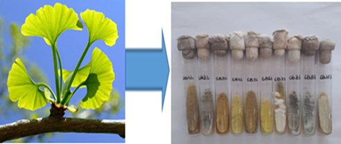
Figure 1. Isolates of Ginkgo biloba endophytic fungi on Chapek-Dox agar
The isolates were grown submerged on a Chapek-Dox medium, and the secondary metabolites from the biomass were extracted with ethyl acetate. The inhibitory activity of the extracts against PL and PA was evaluated in vitro spectrometrically using 4-nitrophenyl palmitate and starch as substrates. The results revealed that almost all extracts produce metabolites that, at varying degrees, inhibit the activity of both pancreatic enzymes.
The inhibitory activity of the extracts to PL was particularly significant. In general, the inhibitory activity of extracts of all isolates varied in a relatively narrow range from 53% to 87%, and inhibitory activity above 60% was observed in 7 out of 10 isolates. The most significant inhibitory activity, 82%, and 87% was shown by extracts of GL4 and GL 5 isolates, respectively, isolated from Gingko biloba leaves. Extracts of isolates GB 7, GB S 9, and GBS10 isolated from the stem of the plant showed inhibition at 74%, 76.3%, and 79.2%, respectively. The inhibitory activity of xenical as a standard under experimental conditions was 91.5% (Fig. 2a).
At the same time, PL-inhibitory activity above 50% was observed in 5 out of 10 isolates (fig. 2b). Of these, the GBS7 isolate from the plant stem showed the highest inhibitory activity, suppressing amylase activity by more than 88%, which is noticeably higher than for acarbose as a positive control (82.5%).
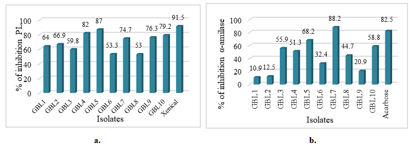
Figure 2. PL-inhibiting activity (a) and PA - inhibiting activity (b) of Gingko biloba endophytes
Thus, a comparative analysis with commercial lipase orlistat and α-amylase inhibitor acarbose, whose inhibitory activity was 91.5% and 82.5%, respectively, demonstrated for the first time that extracts of Ginkgo biloba endophytic fungi produce secondary metabolites with significant inhibitory activity against both pancreatic enzymes. Interestingly, relatively high inhibitory activity for amylase and lipase is observed in almost identical isolates.
The PL-inhibitory properties of secondary metabolites to pancreatic amylase of endophytes isolated from various plants have been reported in several studies [27]. Thus, the endophyte Aspergillus awamori was isolated from Acacia nilotica, producing compounds with inhibitory activity to α–amylase (81%), having low IC₅₀ values, and being resistant to extreme pH and temperature values [28]. Fungal isolates inhibiting α-amylase and α-glucosidase in vitro and having an IC₅₀ lower than acarbose have been isolated from antidiabetic plants Momordica charantia and Trigonella foenum-graceum [29]. While screening 46 endophytic fungal isolates isolated from 11 plants growing in Uzbekistan, A. terreus AF104S, A. egypticus HT166S, and Penicillium sp. CC200 were isolated, suppressing α-amylase activity by more than 80% under extracorporeal conditions [30]. There are also reports of endophytes suppressing pancreatic amylase [31].
Thus, as a result of screening 70 endophytic fungi from Aegle marmelos, Gupta et al. showed a high pancreatic lipase inhibitory potential of isolate 57TBBALM with an IC₅₀ value comparable to that of orlistat used as a positive control [32,33]. Screening of 39 endophytic fungi revealed a high pancreatic lipase inhibitory potential in two strains isolated from Citrus limon and Aegle marmelos, with 75% and 83% activity, respectively [34]. During differential extraction of inhibitory metabolites in various solvents, the hexane extract of the isolate from C. limon showed 87% inhibitory activity with an IC₅₀ of 15.46 μg/ml [35].
To assess the prospects of using the isolated isolates as potential enzyme inhibitors, the IC₅₀ values of five isolates of GBL4, GBL5, GBS7, GBS9, and GBS10 endophytes, which showed high inhibitory activity to pancreatic lipase and pancreatic α-amylase, were determined.
As can be seen from the data in Figure 3a, two of the five isolates have low IC₅₀ values of anti-influenza activity. Thus, the IC₅₀ of GBL4 and GBL5 isolates were 2.6 and 5.6 micrograms/ml, respectively, while the IC₅₀ of Xenical as positive control was 20.0 micrograms/ml, and the IC₅₀ of GBS7 extract was close to Xenical (26.5 micrograms/ml). The IC₅₀ of GBS9 and GBS10 isolates were much higher at 120.6 and 188.2 micrograms/ml, respectively (Fig. 3a).
It is interesting to note that isolates GBL4, GBL5, and GBS7, with low values of lipase inhibition, also exhibit low values of IC₅₀ inhibition of amylase activity. So, for GBL4 and GBL5 isolates, they are 5.2 and 5.95 micrograms/ml, and the IC₅₀ of GBS7 isolate is 1.5 micrograms/ml, which is two times lower than acarbose (3.7 mg/ml) as a positive control (Fig. 3b).
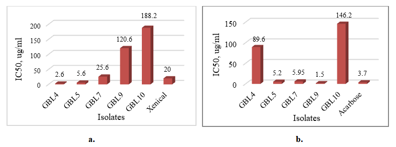
Figure 3. IC50 lipase inhibition (a) and IC50 α-amilase inhibition (b) by Gingko biloba endophytes
Considering that high enzyme inhibition values with low IC₅₀ values indicate the effectiveness of the inhibitory effect of the substance, it can be assumed that the endophytic isolates Ginkgo biloba GBL4, GBL5 from the leaves, and GBL5 from the stem of the plant have significant potential as sources of effective inhibitors of two pancreatic enzymes—lipase and amylase.
It is known that natural sources of amylase and lipase inhibitors contain substances belonging to various classes, including polyphenols, flavonoids, saponins, terpenoids, alkaloids, and other active substances. Several reviews provide data on the active ingredients and inhibitory activity of inhibitors of various predominantly plant sources. More information about endophytic sources of inhibitory compounds needs to be provided. Thus, when studying the nature of the metabolites of Penicillium brevicaule alba from Celosia cristata and Aspergillus egypticus isolated from Helianthus tuberosus, using HPLC and mass spectroscopy, it was found that the RA-inhibitory activity of the strains is associated with triterpene saponins and flavonoids, respectively. Mass spectral studies of metabolites of Aspergillus fischeri from the root of Viola odorata, suppressing pancreatic lipase by more than 80%, revealed the presence of three components of a polyphenolic nature—cinnamic acid (3-phenylpropenic acid), larycycuzinol sesquilignan, and hydroxyfloretin xyloxyl glycoside.
Preliminary data on the nature of secondary metabolites mediating inhibitory activity was obtained by qualitative analysis of the phytochemical composition of active extracts. It was found that extracts of GBL5 and GBS7 differ little in the spectrum of compounds, including alkaloids, flavonoids, terpenoids, tannins, and cardiac glycosides; unlike them, no cardiac glycosides were found in the composition of GBS7 (Table 1). Saponins, phenols, and anthraquinones were not detected in any of the extracts. According to preliminary data, GBL4, GBL5, and GBS7 isolates differ little in the spectrum of compounds, suggesting the need to study the active compounds responsible for inhibitory activity to lipase and amylase in all three isolates.
Table 1. Phytochemical composition of Gingko biloba endophyte extracts
     

4. Conclusions

Thus, it is shown for the first time that Ginkgo biloba endophytes represent a promising object for biotechnological and pharmaceutical research to create new effective inhibitors of pancreatic lipase and α-amylase. The high inhibitory activity of extracts of these endophytes indicates their potential effectiveness as natural inhibitors of these enzymes, which may contribute to developing new therapeutic agents for controlling lipid and carbohydrate metabolism.

ACKNOWLEDGEMENTS

The study was carried out within the framework of basic funding.

References

[1]  Cole, T. J., & Lobstein, T. (2022). Exploring an algorithm to harmonize International Obesity Task Force and World Health Organization child overweight and obesity prevalence rates. Pediatric Obesity, 17(7). https://doi.org/10.1111/ijpo.12905.
[2]  International Diabetes Federation. (2019). IDF diabetes atlas.
[3]  Huang, Y., Karuranga, S., Malanda, B., & Williams, D. R. R. (2018). Call for data contribution to the IDF Diabetes Atlas 9th edition 2019. Diabetes Research and Clinical Practice, 140, 351–352. https://doi.org/10.1016/j.diabres.2018.05.033.
[4]  Kahn, S. E., Hull, R. L., & Utzschneider, K. M. (2006). Mechanisms linking obesity to insulin resistance and type 2 diabetes. Nature, 444, 840–846. https://doi.org/10.1038/nature05482.
[5]  Birari, R. B., & Bhutani, K. K. (2007). Pancreatic lipase inhibitors from natural sources: Unexplored potential. Drug Discovery Today, 12, 879–889. https://doi.org/10.1016/j.drudis.2007.07.024.
[6]  Li, W. L., Zheng, H. C., Bukuru, J., & De Kimpe, N. (2004). Natural medicines used in the traditional Chinese medical system for therapy of diabetes mellitus. Journal of Ethnopharmacology, 92, 1–21. https://doi.org/10.1016/j.jep.2003.12.031.
[7]  Tundis, R., Loizzo, M. R., & Menichini, F. (2010). Natural products as α-amylase and α-glucosidase inhibitors and their hypoglycaemic potential. Medicinal Chemistry, 10, 315–331. https://doi.org/10.2174/138955710791331007.
[8]  Rabasa-Lhoret, R., & Chiasson, J. L. (2004). α-Glucosidase inhibitors. In International textbook of diabetes mellitus.
[9]  Malpathak, N., & Baikar, S. (2010). Secondary metabolites as DNA topoisomerase inhibitors: A new era towards designing of anticancer drugs. Pharmacognosy Reviews, 4, 12. https://doi.org/10.4103/0973-7847.65320.
[10]  Gupta, R. A., & Kumar, P. O. (2021). Development and characterization of pioglitazone nanoparticles for the effective treatment of diabetes mellitus. Journal of Pharmaceutical Research International, 33, 172–182. https://doi.org/10.9734/jpri/2021/v33i31A31678.
[11]  Aloko, S., & Bello, M. O. (2021). The role of alkaloids in the management of diabetes mellitus. In Structure and health effects of natural products on diabetes mellitus (pp. 267–278). Springer. https://doi.org/10.1007/978-981-15-8791-7_15.
[12]  Mukherjee, S. (2007). The role of oxidative stress in diabetes vascular disorders. Current Opinion in Lipidology, 18, 696–698. https://doi.org/10.1097/MOL.0b013e3282f1ed5f.
[13]  Padwal, R. S., & Majumdar, S. R. (2007). Drug treatments for obesity: Orlistat, sibutramine, and rimonabant. The Lancet, 369, 71–77. https://doi.org/10.1016/S0140-6736(07)60033-6.
[14]  Strobel, G., & Daisy, B. (2003). Bioprospecting for microbial endophytes and their natural products. Microbiology and Molecular Biology Reviews, 67, 491–502. https://doi.org/10.1128/MMBR.67.4.491-502.2003.
[15]  Schueffler, A., & Anke, T. (2014). Fungal natural products in research and development. Natural Product Reports, 31, 1425–1448. https://doi.org/10.1039/C4NP00060A.
[16]  Khan, A. L., Shahzad, R., Al-Harrasi, A., & Lee, I. J. (2017). Endophytic microbes: A resource for producing extracellular enzymes. In Endophytes: Crop productivity and protection (pp. 95–110). Springer. https://doi.org/10.1007/978-3-319-66544-3_5.
[17]  Kusari, S., Hertweck, C., & Spiteller, M. (2012). Chemical ecology of endophytic fungi: Origins of secondary metabolites. Chemistry & Biology, 19, 792–798. https://doi.org/10.1016/j.chembiol.2012.06.004.
[18]  Zhao, J., Shan, T., Mou, Y., & Zhou, L. (2011). Plant-derived bioactive compounds produced by endophytic fungi. Medicinal Chemistry, 11, 159–168. https://doi.org/10.2174/138955711794519492.
[19]  Mahadevan, S., & Park, Y. (2007). Multifaceted therapeutic benefits of Ginkgo biloba L. Journal of Food Science, 73. https://doi.org/10.1111/j.1750-3841.2007.00597.x.
[20]  Zhang, H. W., Song, Y. C., & Tan, R. X. (2006). Biology and chemistry of endophytes. Natural Product Reports, 23, 753. https://doi.org/10.1039/B609472B.
[21]  Qiu, M., Xie, R., Shi, Y., Zhang, H., & Chen, H. (2010). Isolation and identification of two flavonoid-producing endophytic fungi from Ginkgo biloba L. Annals of Microbiology, 60, 143–150. https://doi.org/10.1007/s13213-010-0016-5.
[22]  Liu, X., Dong, M., Chen, X., Jiang, M., Lv, X., & Yan, G. (2007). Antioxidant activity and phenolics of an endophytic Xylaria sp. from Ginkgo biloba. Food Chemistry, 105, 548–554. https://doi.org/10.1016/j.foodchem.2007.04.008.
[23]  Hazalin, N. A., Ramasamy, K., Lim, S. S. M., Wahab, I. A., Cole, A. L., & Abdul Majeed, A. B. (2009). Cytotoxic and antibacterial activities of endophytic fungi isolated from plants at the National Park, Pahang, Malaysia. BMC Complementary and Alternative Medicine, 9. https://doi.org/10.1186/1472-6882-9-46.
[24]  Bustanji, Y., Al-Masri, I. M., Mohammad, M., Hudaib, M., Tawaha, K., Tarazi, H., & AlKhatib, H. S. (2010). Pancreatic lipase inhibition activity of trilactone terpenes of Ginkgo biloba. Journal of Enzyme Inhibition and Medicinal Chemistry, 26, 453–459. https://doi.org/10.3109/14756366.2010.525509.
[25]  Picot, C. M. N., Subratty, A. H., & Mahomoodally, M. F. (2014). Inhibitory potential of five traditionally used antidiabetic medicinal plants. Advances in Pharmacological Sciences, 1–7. https://doi.org/10.1155/2014/739834.
[26]  Sebaugh, J. L. (2011). Guidelines for accurate EC50/IC50 estimation. Pharmaceutical Statistics, 10, 128–134. https://doi.org/10.1002/pst.426.
[27]  Ali, H., Houghton, P. J., & Soumyanath, A. (2006). α-Amylase inhibitory activity of some Malaysian plants used to treat diabetes. Journal of Ethnopharmacology, 107, 449–455. https://doi.org/10.1016/j.jep.2006.04.004.
[28]  Verma, V. C., Gond, S. K., Kumar, A., Mishra, A., Kharwar, R. N., & Gange, A. C. (2008). Endophytic actinomycetes from Azadirachta indica. Microbial Ecology, 57, 749–756. https://doi.org/10.1007/s00248-008-9450-3.
[29]  Ferreira, E. M. S., Corrêia, T. M., da Silva, J. F. M., & Pimenta, R. S. (2021). Endophytic fungi associated with medicinal plants of Amazonian forest. In Neotropical endophytic fungi (pp. 177–197). https://doi.org/10.1007/978-3-030-53506-3_9.
[30]  Ruzieva, D., Gulyamova, T., Nasmetova, S., Mukhammedov, I., & Rasulova, G. (2022). Identification of bioactive compounds inhibiting pancreatic α-amylase. Turkish Journal of Pharmaceutical Sciences, 19, 630–635. https://doi.org/10.4274/tjps.galenos.2021.05873.
[31]  Panda, S., Padhi, L., & Mohanty, G. (2011). Antibacterial activities and phytochemical analysis of Cassia fistula. Journal of Advanced Pharmaceutical Technology & Research, 2, 62. https://doi.org/10.4103/2231-4040.79814.
[32]  Gupta, R., Sharma, A. K., Sharma, M. C., Dobhal, M. P., & Gupta, R. S. (2012). Evaluation of antidiabetic and antioxidant potential of lupeol. Natural Product Research, 26, 1125–1129. https://doi.org/10.1080/14786419.2011.560845.
[33]  Gupta, M., Saxena, S., & Goyal, D. (2014). Lipase inhibitory activity of endophytic fungal species of Aegle marmelos. Symbiosis, 64, 149–157. https://doi.org/10.1007/s13199-015-0311-9.
[34]  Sarkar, U. J., Dioundi, D., & Gupta, M. (2017). Endophytic Pestalotiopsis species as pancreatic lipase inhibitors. Asian Journal of Pharmaceutical and Clinical Research, 10, 82–83. https://doi.org/10.22159/ajpcr.2017.v10i10.21148.
[35]  Mishra, Y., Sharma, L., Dhiman, M., & Sharma, M. M. (2021). Endophytic fungal diversity and biopotential applications. In Fungi bio-prospects in sustainable agriculture (pp. 227–283). Academic Press. https://doi.org/10.1016/B978-0-12-821394-0.00010-X.