-
Paper Information
- Next Paper
- Paper Submission
-
Journal Information
- About This Journal
- Editorial Board
- Current Issue
- Archive
- Author Guidelines
- Contact Us
International Journal of Virology and Molecular Biology
p-ISSN: 2163-2219 e-ISSN: 2163-2227
2025; 14(2): 13-17
doi:10.5923/j.ijvmb.20251402.01
Received: Apr. 11, 2025; Accepted: May 16, 2025; Published: May 30, 2025

Pathogenic Effects of the Genus Meloidogyne Goeldi, 1892 (Nematoda) on the Root Morphology and Tissue of Medicinal Dandelion (Taraxacum Officinale Wigg.)
 Abstract
 Reference
 Full-Text PDF
 Full-text HTML
Eshova X. S.1, Abdushukurova K. A.1, Mirzaliyeva G. R.2, Duschanova G. M.3, Abdinazarov S. Kh.4, Samadov I. N.4
1National University of Uzbekistan, Tashkent, Uzbekistan
2Tashkent State Pedagogical University, Tashkent, Uzbekistan
3National Center of Archeology of the Academy of Sciences of the Republic of Uzbekistan
4Tashkent Botanical Garden under the Institute of Botany of the Academy of Sciences of the Republic of Uzbekistan
Correspondence to: Abdushukurova K. A., National University of Uzbekistan, Tashkent, Uzbekistan.
| Email: |  |
Copyright © 2025 The Author(s). Published by Scientific & Academic Publishing.
This work is licensed under the Creative Commons Attribution International License (CC BY).
http://creativecommons.org/licenses/by/4.0/

This article presents the anatomical structure of the roots of medicinal dandelion (Taraxacum officinale Wigg.) affected and unaffected by nematodes of the genus Meloidogyne Göeldi, 1887. In the unaffected dandelion roots, biologically active compounds were found to be located in the cortical parenchyma, phloem fibers, and laticiferous vessels. In contrast, in the roots affected by root-knot nematodes, giant cell formations known as root galls were observed in the cortical parenchyma and secondary xylem of the woody tissue. It was determined that in the cortical parenchyma of both primary and lateral roots, 6 to 8 oothecae developed in the giant cells, containing numerous oval-shaped mature eggs. Under the pathogenic influence of the nematode, the damage centers in the roots of the medicinal dandelion enlarged, affecting the protective and mechanical tissues as well as the vascular system. Additionally, changes such as epidermal deformation, tearing, and hypertrophic growths were observed.
Keywords: Meloidogyne Göeldi, 1887, Medicinal dandelion (Taraxacum officinale Wigg.), Root tissue, Galls, Infestation, Giant cells, Cortical parenchyma, Woody tissue, Protective and mechanical tissues
Cite this paper: Eshova X. S., Abdushukurova K. A., Mirzaliyeva G. R., Duschanova G. M., Abdinazarov S. Kh., Samadov I. N., Pathogenic Effects of the Genus Meloidogyne Goeldi, 1892 (Nematoda) on the Root Morphology and Tissue of Medicinal Dandelion (Taraxacum Officinale Wigg.), International Journal of Virology and Molecular Biology, Vol. 14 No. 2, 2025, pp. 13-17. doi: 10.5923/j.ijvmb.20251402.01.
1. Introduction
- Nowadays, protecting promising medicinal, edible, and aromatic plants from pests and parasites is of significant importance worldwide. In the flora of Uzbekistan, 4,375 species of wild medicinal plants have been identified, of which nearly 1,200 possess medicinal properties and are used in medical practice [1]. Various pests and parasites from different systematic groups are found on medicinal plants, which negatively affect their medicinal properties. Among these parasites are phytonematodes belonging to the phylum Nematoda. Within this group, nematodes of the genus Meloidogyne Göeldi, 1887—commonly known as root-knot nematodes—parasitize the roots and cause serious damage to plants [11]. Therefore, studying the effects of Meloidogyne spp. nematodes on medicinal plants is of both scientific and practical significance.Currently, significant attention is being paid to cultivating and propagating promising medicinal, edible, and aromatic plants for industrial production worldwide. In some foreign countries, the medicinal dandelion plant is being cultivated, and its raw material is being exported. Promising medicinal plant species include Taraxacum officinale Wigg. and Taraxacum kok-saghyz Rodin [13]. These plants are considered valuable sources of medicinal and food-grade raw materials.Medicinal dandelion (Taraxacum officinale Wigg.) possesses healing properties. Its milky latex contains compounds such as taraxacin and taraxacerin, taraxerol, taraxasterol, andosterol, sterin, choline, saponin, ascorbic acid, carotene, apigenin, lutein, nicotinic acid, nicotinamide, 2–3% rubber substances, resins, inulin, fatty acids, essential oils, proteins, tannins, oleanolic acid, linolenic acid, palmitic acid, malic acid, mineral salts, alcohols, flavonoids, asparagine, iron salts, potassium and phosphorus salts [4,14].Nematodes of the genus Meloidogyne Göeldi, 1887 are among the most dangerous pathogens parasitizing both cultivated and wild plants. It is known that nematodes of this genus can infect more than 4,000 species of vegetables, melons, cereals, legumes, industrial crops, berries, and ornamental plants [18]. While the damage caused by Meloidogyne spp. to many agricultural crops has been studied, their pathogenic effect on medicinal plants—particularly on their root tissues—has not been investigated.For this reason, the pathogenic effects of Meloidogyne spp. on the root morphology and tissues of the medicinal plant dandelion (Taraxacum officinale Wigg.) were studied. The histological structure of healthy and infected dandelion roots was analyzed and compared.
2. Materials and Methods
- The materials for this study were collected from areas where medicinal dandelion (Taraxacum officinale) grows, specifically along the Bogishamol highway and within the Botanical Garden in Yunusabad district, Tashkent city. Samples of medicinal dandelion roots and rhizosphere soil were collected using the method proposed by E.S. Kir'yanova and E.L. Krall (1969) [6] from designated coordinates: 41.344720, 69.310595; 41.344518, 69.312264; 41.344146, 69.314490; 41.344998, 69.310330. Root-knot nematodes were extracted from plant roots and soil using a modified Baermann funnel technique [8].Sample fixation was carried out according to the method of A.A. Paramonov (1962) [10]. Permanent slides of root-knot nematodes were prepared by making sections through the anal–vulvar region and identifying the species using the Seinhorst technique. As controls, non-infected dandelion plants from the same sampling areas were used.To study the anatomical structure of Taraxacum officinale roots, the samples were fixed in 70% ethanol. Anatomical-histological slides were prepared by hand sectioning, stained with methylene blue, and mounted using a glycerin mixture to create temporary preparations [2]. The anatomical and histological structure of the roots was examined in cross-sections. The organization of major tissues and cells was described based on the methods of K. Esau (1969) [17] and N.S. Kiseleva (1971) [7]. Microphotographs were taken using a Canon A123 digital camera mounted on a computer-adapted microscope, specifically the Motic B1-220A-3 model.Samples were taken from both healthy and nematode-infected dandelion roots. Changes in tissue and cellular structure and function were studied using histological preparations of the root tissues.
3. Results and Discussion
- As a result of the study, it was determined that the roots of medicinal dandelion (Taraxacum officinale) naturally growing along the highway were infected with nematodes of the genus Meloidogyne. These nematodes cause a disease in plants known as Meloidogynosis. External symptoms of Meloidogynosis in plants include pale or yellowish-white leaves, wilting, reduced leaf size, leaf drop, small fruits, stunted growth, dwarfism, and in severe cases, complete plant wilting. The root system becomes deformed or decayed, and characteristic galls (swellings) form on the roots [8].During field observations, attention was paid to disease symptoms and external appearance of plants in the infected areas. Aboveground parts of nematode-infected dandelions were noticeably smaller in size and had paler leaves compared to healthy plants. Nematode infection was confirmed through examination of the root systems. Roots were excavated, cleaned from soil, and examined in detail. Morphological damage and the presence of root galls were recorded.Currently, 97 species of Meloidogyne are known worldwide [5]. In Uzbekistan, five species have been identified: Meloidogyne javanica, M. arenaria, M. incognita, M. acrita, and M. hapla [8,12,15,16]. In this study, three species—M. incognita, M. arenaria, and M. hapla—were found in associative form in infected dandelion roots. The global distribution of root-knot nematodes is as follows: M. incognita – 52%, M. javanica – 31%, M. arenaria – 8%, M. hapla – 7%, and other species – 2% [3]. Therefore, the nematode species identified in our research are among the most widespread. Some species of the genus Meloidogyne—M. incognita, M. javanica, M. arenaria, and M. hapla—are considered major plant parasites [9]. Accordingly, the M. incognita, M. arenaria, and M. hapla species found in this study are also classified as primary plant parasites.Nematodes of the genus Meloidogyne are sedentary endoparasites [10,19]. Their life cycle consists of four juvenile (larval) stages. The first stage (J1) occurs within the egg. The second-stage juvenile (J2), which is capable of infecting plant roots, hatches from the egg. Most invasive juveniles exit the infected root and move into the soil, while others remain in the root system. In the soil, J2 larvae become active and penetrate the plant root cortex. They settle near the vascular tissues with their head and begin feeding, then develop into third-stage (J3) and subsequently fourth-stage (J4) juveniles. As they mature, the larvae thicken and transform into spherical or pear-shaped adult females, reaching up to 1 mm in length [11].Once the invasive juveniles penetrate the root tissue and establish themselves, they begin secreting enzymatic fluids from their digestive glands. The saliva stimulates increased cell division, dissolving cell walls, and results in the formation of giant cells—5 to 10 times larger than normal plant cells [8]. As a result of parasitic activity, not only morphological but also histological changes occur in the plant. Based on our scientific analysis, it was observed that root-knot nematodes significantly alter the structure and function of root tissues and cells.In transverse sections, the anatomical structure of the healthy medicinal dandelion (Taraxacum officinale) root is round-shaped and classified as a non-stele (non-radial) type, indicating the absence of a radial structural organization. Based on its anatomical features, the root is composed of three main zones: the periderm, the cortical parenchyma, and the central cylinder. The outermost layer of the root is covered by the periderm, which consists of the phellogen (cork cambium), phellem (cork), and phelloderm. The periderm is relatively thin and composed of dark brown cells that are tightly packed.The cortical parenchyma is well-developed compared to the central cylinder and occupies approximately three-quarters of the root’s diameter. It is composed of both large and small round to oval-shaped parenchymal cells. Within the cortical parenchyma, layers of phloem fibers and laticiferous canals (latex ducts) are arranged concentrically, in which dark brown biologically active substances were observed (Figure 1).
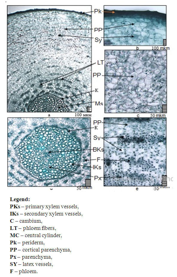 | Figure 1. Anatomical structure of the uninfected dandelion (Taraxacum officinale) root: a – general view of the root structure; b, c – cortical parenchyma of the root; d – xylem fibers (primary and secondary xylem); e – phloem fibers and latex vessels |
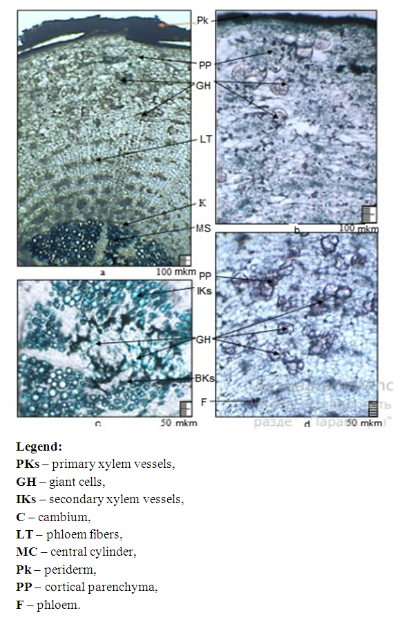 | Figure 2. Anatomical structure of the primary root of dandelion (Taraxacum officinale) infected with root-knot nematodes: a – general view of the root structure; b, c – formation of giant cells in the cortical parenchyma of the root; d – giant cells between primary and secondary xylem vessels |
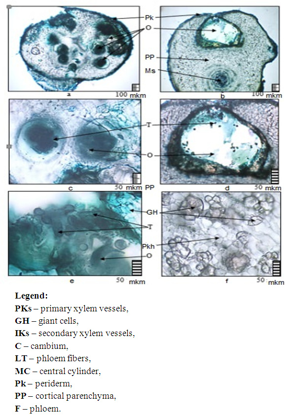 | Figure 3. Anatomical structure of the lateral root of dandelion (Taraxacum officinale) infected with root-knot nematodes: a, b – general view of the root structure; c, d, e – formation of giant cells and oothecae in the cortical parenchyma of the root |
4. Conclusions
- As a result of the study, it was determined that the roots of Taraxacum officinale (medicinal dandelion) naturally growing along the roadside were infected by nematodes of the genus Meloidogyne, specifically Meloidogyne incognita, M. arenaria, and M. hapla, which were found to occur in association. For the first time, the anatomical structure of both infected and uninfected Taraxacum officinale roots was studied, and species-specific structural diagnostic features were identified. A comparative anatomical analysis revealed that in uninfected roots, biologically active substances were located in the cortical parenchyma, phloem fibers, and laticiferous canals. In contrast, in roots infected by root-knot nematodes, tumor-like structures with giant cells, referred to as "root galls," were observed in the cortical parenchyma and the secondary xylem of the woody tissues.In both main and lateral roots, 6 to 8 oothecae (egg sacs) were formed within the giant cells of the cortical parenchyma, each containing numerous oval-shaped eggs. These newly identified anatomical features including the presence of outgrowths in the cortical parenchyma and giant cells in the central cylindercan be utilized for the diagnosis, treatment, and prevention of Taraxacum officinale root infections caused by root-knot nematodes.