International Journal of Virology and Molecular Biology

p-ISSN: 2163-2219    e-ISSN: 2163-2227

2024;  13(6): 81-86

doi:10.5923/j.ijvmb.20241306.01

Received: Sep. 28, 2024; Accepted: Oct. 22, 2024; Published: Oct. 30, 2024


Morphological and Molecular Identification of New Findings of Green Algae (Scenedesmaceae, Chlorophyta) from River Naryn, Uzbekistan

N. B. Ikramov

PhD student, Namangan State University, Namangan, Uzbekistan

Correspondence to: N. B. Ikramov, PhD student, Namangan State University, Namangan, Uzbekistan.

Copyright © 2024 The Author(s). Published by Scientific & Academic Publishing.

This work is licensed under the Creative Commons Attribution International License (CC BY).
http://creativecommons.org/licenses/by/4.0/

Abstract

For the first time, the presence of green algal species Coelastrella tenuitheca, Neodesmus danubialis and Tetradesmus obliquus (Scenedesmaceae, Chlorophyta) was identified in the phytoplankton of the Naryn River (Uzbekistan) based on morphological and molecular analysis. Some strains showed discrepancies with their primary morphological diagnoses. This study confirms that ITS2 can be successfully used as a DNA barcode for future monitoring of freshwater ecosystems.

Keywords: Morphological identification, New findings, Green algae, Molecular identification, Scenedesmaceae, Primary morphological diagnoses, Chlorophyta, Naryn River, ITS2, Phytoplankton, DNA barcode, Freshwater ecosystems

Cite this paper: N. B. Ikramov, Morphological and Molecular Identification of New Findings of Green Algae (Scenedesmaceae, Chlorophyta) from River Naryn, Uzbekistan, International Journal of Virology and Molecular Biology, Vol. 13 No. 6, 2024, pp. 81-86. doi: 10.5923/j.ijvmb.20241306.01.

1. Introduction

Coccoid green algae, which reproduce solely through the production of autospores, are among the most complex groups in taxonomy. Based on morphological analysis, these algae were initially classified as belonging to the order Chlorococcales [1]. However, subsequent molecular genetic studies, primarily based on the 18S rRNA gene, have shown that green algae that produce autospores form many independent and unrelated lineages. Most of these lineages belong to the class Trebouxiophyceae or order Sphaeropleales within the Chlorophyceae [2-4].
One of the most common types of green algae found in freshwater phytoplankton is Scenedesmaceae, also known as Coelastraceae. These algae consist of flat or curved colonies with oval or spindle-shaped cells. Occasionally, they can form three–dimensional clusters or syncoenobia. Since Oltmann (1904), other authors have significantly revised the scope of this family [1,5]. Currently, the Scenedesmaceae family includes 41 genera: Acutodesmus, Asterarcys, Chodatodesmus, Closteriococcus, Coelastrella, Coelastropsis, Coelastrum, Comasiella, Crucigeniopsis, Danubia, Desmodesmus, Dimorphococcus, Enallax, Flechtneria, Gilbertsmithia, Gloeoactinium, Hariotina, Hofmania, Hylodesmus, Komarekia, Lauterborniella, Neodesmus, Pectinodesmus, Pediludiella, Pseudodidymocystis, Scenedesmus, Sceneoocystis, Schmidledesmus, Schroederiella, Scotiellopsis, Soropediastrum, Staurogenia, Steinedesmus, Tetradesmus, Tetrallantos, Tetranephris, Tetrastrum, Verrucodesmus, Westella, Westellopsis, Yadavaea [6]. Thus, this family is the richest in the order Sphaeropleales, with 367 taxa that have been transferred to new genera due to reclassification of previously known genera such as Scenedesmus, Cohniella, and Crucigenia. Additionally, it includes several genera that have been described de novo, such as Hylodesmus [7], Flechtneria [8], Pediludiella [9].
Representatives of the Scenedesmaceae family can be found in various aquatic and terrestrial habitats, including water, soil, stones, sand, lichens, and biocrusts [10-11]. Their isolates have repeatedly demonstrated a high biotechnological potential [12-19] and the possibility of use in monitoring and assessing the quality of freshwater ecosystems [20] and for their bioremediation [21-22].
Molecular identification has become an essential and universal tool for algal research [23]. The ITS2 sequence has been widely used as a DNA barcode to confirm the taxonomic identity of freshwater green microalgal strains [24-26]. The aim of this research was to identify and characterize three strains of green algae isolated from phytoplankton samples collected from the Naryn River through a combination of morphological and molecular genetic analyses.

2. Materials and Methods

Study area and water sampling. Naryn is a river that flows through the territory of the republics of Kyrgyzstan and Uzbekistan in Central Asia. It is formed by the confluence of two other rivers, the Big Naryn and Small Naryn, originating from glaciers in the Central Tien Shan mountains. Naryn has a length of 807 km and a basin area of 59,900 km2, and it plays a significant role in the region's economy, providing water for irrigation and generating hydroelectric power. Water sampling was conducted in 2022 using the Apstein plankton net at two locations (Figure 1):
1. Observation point 1 (Nosh) is located on the border with Kyrgyzstan, near the village of Yangiyer (41.160003, 72.145856). The height above sea level is 505 m. Water transparency in the slower parts of the current is 80–100 cm, and in some shallow places, it reaches the bottom. In winter, the water temperature is 0–1.8°C; in summer, the average temperature is 22–23°C.
2. Observation point 3 (C01) is located near the bridge–dam in Uchkurgan (41.113258, 72.067386) at an altitude of 490 m above sea level. The water in this area has a transparency of up to 80–90 cm and a temperature of 1–2°C in winter and 22–23°C on average in summer.
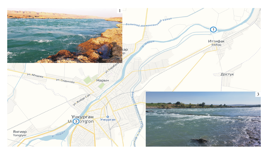
Figure 1. River Naryn with observation points
Algal isolation, medium, and culture conditions. The algae were isolated by inoculating phytoplankton onto solid and liquid BG-11 media. Monocultures were then obtained by streak–inoculating and isolating individual colonies using a Pasteur pipette [27]. The strains were then cultivated in a climate chamber under standard conditions (23–25°C, 60–75 μmol quanta m–2 s–1, 12–hour illumination) on BG–11 agar media containing nitrogen (pH=7.0, 1% agar). All strains studied were deposited in the All–Russian Collection of Microorganisms, Russia.
Microscope observations. Cell morphology and life cycles were investigated using a Leica DM750 light microscope (Germany). The results were documented using a Leica Flexacam C3 color digital camera (Germany), and the observation was carried out for 1 to 12 weeks. Important diacritic characteristics used to identify microalgal strains included the type of thallus organization, cell shape and size, chloroplast number and type, presence of pyrenoids, presence and thickness of mucilaginous envelopes, and reproduction type. Morphometric measurements were performed using the Leica Application Suite X software package, and for size comparison, 100 cells from each strain were measured. The algal system accepted in the Algae Base international electronic database [6] was the basis of this work.
DNA extraction, amplification, and sequencing. DNA extraction from the strains was carried out using a commercial kit “FitoSorb” (Syntol, Russia), following the manufacturer’s instructions. To amplify the internal transcribed spacer ITS2, primers from Johnson et al. [28] were used: ITS–AF (5’–CGTTTCCGTAGGTGAACTGC–3’) and ITS–BR (5’–CATATGCTTAGTTCAGCGGG–3’). The PCR reaction was performed at 95°C for 3 min, followed by 35 cycles of 95°C for 30 s, 57.6°C for 30 s, 72°C for 1 min, and 72.0°C for 10 min. Detection of target products was performed electrophoretically on a 1% agarose gel, and amplicons were purified using the Cleanup Standard Kit (Evrogen, Russia). Evrogen, Russia, performed sequencing of nucleotides.
Phylogenetic analysis. To determine the studied strains' phylogenetic relationship, we searched for nucleotide sequence homology in the ITS2 region using the BLASTn algorithm from GenBank [29]. We selected sequences based on several criteria, including maximum relatedness (≥95% similarity), quality of reads (no degenerate or unknown nucleotides), read length (at least 210 base pairs), and membership in the type species and authentic collection strains. A representative of the Scenedesmaceae family, Desmodesmus armatus (Chlorophyta), was chosen as an outgroup in this study. Multiple alignments were performed using the ClustalW algorithm in BioEdit [30], and the jModelTest program was used to select a nucleotide substitution model. The minimum Akaike Information Criterion (AIC) value was used to determine the best model. Phylogenetic reconstruction was performed using maximum likelihood in PhyML [31], and statistical support for tree topology was assessed through bootstrap analysis with 1,000 replicates. Phylogenetic trees were visualized using FigTree v1.3.1. Genetic differences between the nucleotide sequences of ITS2 were characterized using genetic distances. These distances were calculated as the percentage of mismatched nucleotides in pairwise alignments of the sequences using the MEGA 11.0 program [32]. We also consulted articles by Hegewald and Hanagata [33], Wang et al. [34], Suarez–Montes et al. [35], as well as Mai et al. [36], when constructing phylogenetic trees.
All sequences obtained in the work were deposited to NCBI GenBank (https://www.ncbi.nlm.nih.gov/) under accession numbers PP937762–PP937764.

3. Results and Discussions

Three strains of Scenedesmus–like green microalgae were isolated, one from observation point 1 (Nosh) and two from observation point 3 (C01). The morphological descriptions of these strains are provided below.
Strain VKM Al–486 from observation point 1 (Figure 2 a–c): The cells are mostly solitary (coenobia were not observed in culture), fusiform, 7.4–11.3 x 4.2–8.1 μm, or very rarely almost spherical–up to 13 μm in diameter. The cell membrane appears smooth under light microscopy (LM), with two (rarely one) polar thickenings. There is a parietal chloroplast with one pyrenoid. Reserve products are numerous grains of starch and drops of oil. They reproduce with 2–4–8 autospores. Remains of the mother cell wall remain in the culture for some time. Sexual reproduction has not been observed. Morphologically, the strain was identified as Tetradesmus obliquus.
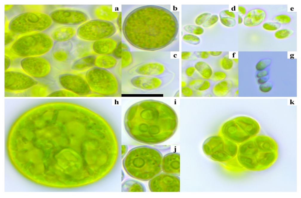
Figure 2. LM micrographs of the studied strains. a–c–T. obliquus; d–g–N. danubialis; h–k–C. tenuitheca. g–сells are stained with a 0.1% methylene blue solution to reveal the mucus structure. Scale 10 μm
Strain VKM Al–485 from observation point 3 (Figure 2, d–g): Cells are single or in bicellular coenobia. The cells are drop–shaped, one end is rounded and the other is pointed, measuring 4.3–6.9 x 2.7–3.6 μm in size. The cell wall is covered with a layer of mucus, and there is a single parietal chloroplast and a pyrenoid. Asexual reproduction occurs by two autospores or rarely by four. The unique characteristics of the division of the parent cell determine the arrangement of cells within the coenobium, with opposite ends facing each other. Sexual reproduction has not been observed. Based on the listed characteristics, the strain has been identified as Neodesmus danubialis. Our strain differs from the original species description in its shorter cell length. Hindak [37] provided dimensions of 7–9x2.5–3 μm. In addition, he noted that autospores always form in pairs, while we have encountered autosporangia containing four autospores. Furthermore, in our strain culture, we have not observed syncenobia forming long chains of cells as described by Hindak for natural samples. However, the author stated that when cultured even in liquid media, syncenobia are not long and contain no more than six cells.
Strain VKM Al–484 from observation point 3 (Figure 2, h–k): Cells are single, broadly oval or spherical, with a 6–11 (up to 28) μm diameter. The cell membrane is thin and smooth. The chloroplast is parietal in young cells, whereas in mature cells, it is lobed or segmented with one pyrenoid consisting of two segments. There are cells with several pyrenoids in culture. Mature cell contents are vacuolated, and spare products are drops of oil. Asexual reproduction occurs by 2–6 autospores, and the remains of the mother cell wall persist in culture for a while. Sexual reproduction has not been observed. Based on morphology, the strain was identified as Coelastrella tenuitheca. It was distinguished from the original description of the species by mature cells that are a much larger (up to 28 μm) than indicated in the diagnosis–up to 18 μm [38].
All the described species are typical small phytoplankton that can be found in freshwater environments and almost everywhere [39]. An exception is the recently described C. tenuitheca, for which not yet enough data have been collected.
Nevertheless, most green algae do not have good discriminative characters for the ease of taxonomy [9]. The family Scenedesmaceae is not an exception to this rule. Difficulties in identifying Scenedesmaceae strains are due to their extreme phenotypic plasticity and relatively simple morphology, which leads to cryptic species [40–45]. No wonder Krienitz and Bock [46] described the genus Scenedesmus as “a nightmare for field ecologists who wish to determine a taxon in a fixed sample under the inverted microscope”. And this is true for the entire Scenedesmaceae family. We were able to identify the species using only molecular analysis accurately.
According to the results of the ITS2 analysis, all the studied strains were members of the Scenedesmaceae family (Figure 3):
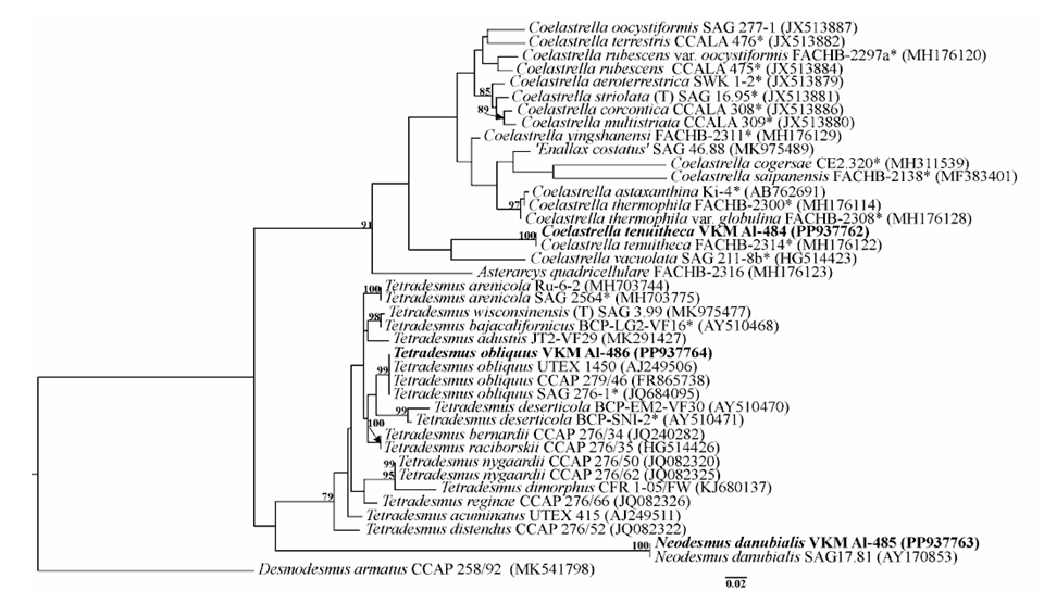
Figure 3. A rooted phylogenetic tree of the studied strains was constructed using the maximum likelihood method (ML), based on ITS2 sequences (275 nt). Bootstrap values for ML are provided as statistical support for tree nodes, with values <70% not being shown. The model of nucleotide substitutions is GTR+I+G. The designations are: *–authentic strains; T–type species
Thus, the strain VKM Al–486 was closely related to the authentic strain of T. obliquus SAG 276–1 (BP–99%), and there were no genetic differences between them. Strain VKM Al–485 was clustered with N. danubialis SAG 17.81 (BP–100%). There were also no genetic differences between them. Strain VKM Al–484 was included in the genus Coelastrella and clustered with authentic strain C. tenuitheca FACHB–2314 with maximum statistical support. The ITS2 genetic distance was 0%. However, we would like to report on the difficulties encountered during the molecular analysis of strains within this family. Firstly, there is a lack of sequence data for type species and authentic strains, as with N. danubialis. Secondly, several molecular data have been obtained from genetic databases, including species that have not been verified morphologically. Thirdly, the complexity arises from incorrectly identified sequences.

4. Conclusions

Thus, we confirmed for the first time the presence of the species C. tenuitheca, N. danubialis and T. obliquus in the phytoplankton of the Naryn River based on morphological and molecular analyses. The ITS2 as a DNA barcode can be successfully used in the future to monitor the state of freshwater ecosystems. The molecular identification of algae has greatly enhanced our understanding of their taxonomy, phylogeny, and ecology. Accurate taxonomic identification is essential in biology and related fields such as evolution, biogeography, biotechnology and conservation.

ACKNOWLEDGEMENTS

The author expresses gratitude to the All–Russian Collection of Microorganisms (VKM) staff for their assistance with conducting research and providing consultations.

References

[1]  Komarek, J. and Fott, B., 1983, Familie Scenedesmaceae. In Das Phytoplankton des Sußwassers 7–Chlorococcales, Edited by G. Huber–Pestalozzi, Stuttgart, Schweizerbart.
[2]  Hanagata, N., 1998, Phylogeny of the subfamily Scotiellocystoideae (Chlorophyceae, Chlorophyta) and related taxa inferred from 18S ribosomal RNA gene sequence data, J Phycol, 34, 1049–1054.
[3]  Krienitz, L., Hegewald, E., Hepperle, D., Wolf, M., 2003, The systematics of coccoid green algae: 18S rRNA gene sequence data versus morphology, Biologia (Bratisl), 58, 437–446.
[4]  Wolf, M., Hegewald, E., Hepperle, D., Krienitz, L., 2003, Phylogenetic position of the Golenkiniaceae (Chlorophyta) as inferred from 18S rDNA sequence data, Biologia (Bratisl), 58, 433–436.
[5]  Hegewald, E. and Hanagata, N., 2000, Phylogenetic studies on Scenedesmaceae (Chlorophyta), Arch Hydrobiol, 136, 29–49.
[6]  Guiry, M.D. and Guiry, G.M. AlgaeBase. World–Wide Electronic Publication. National University of Ireland, Galway. Available online: https://www.algaebase.org (accessed on 17 June 2024).
[7]  Elias, M., Nemcova, Y., Skaloud, P., Neustupa, J., Kaufnerova, V., Sejnohova, L., 2009. Hylodesmus singaporensis gen. et sp. nov., a novel autosporic subaerial green alga (Scenedesmaceae, Chlorophyta) from Singapore, International Journal of Systematic and Evolutionary Microbiology, 60, 1224–1235.
[8]  Sciuto, K., Lewis, L.A., Verleyen, E., Moro, I., LaRocca, N., 2015, Chodatodesmus australis sp. nov. (Scenedesmaceae, Chlorophyta) from Antarctica, with the emended description of the genus Chodatodesmus, and circumscription of Flechtneria rotunda gen. et sp. nov., Journal of Phycology, 51(6), 1172–1188.
[9]  Hoshina, R., Hayakawa, M.M., Kobayashi, M., Higuchi, R., Suzaki, T., 2020, Pediludiella daitoensis gen. et sp. nov. (Scenedesmaceae, Chlorophyceae), a large coccoid green alga isolated from a Loxodes ciliate, Scientific Reports, 10, 628.
[10]  Lewis, L. A. and Flechtner, V. R., 2004, Cryptic species of Scenedesmus (Chlorophyta) from desert soil communities of western North America, J. Phycol., 40, 1127–1137.
[11]  Tsarenko, P. M., and John, D. M., 2011, “Phylum Chlorophyta. Order Sphaeropleales sensu lato,” in The freshwater algal flora of the British Isles. An identification guide to freshwater and terrestrial algae, 2nd ed., Ed. B. A. Whitton, New York, USA, Cambridge University Press.
[12]  Abe, K., Hattori, H., Hirano, M., 2007, Accumulation and antioxidant activity of secondary carotenoids in the aerial microalga Coelastrella striolata var. multistriata, Food Chem., 100, 656–661.
[13]  Hu, C–W, Chuang, L–T, Yu, P–C, Chen, C–NN, 2013, Pigment production by a new thermotolerant microalga Coelastrella sp. F50, Food Chem. 138, 2071–2078.
[14]  Kawasaki, S., Mizuguchi, K., Sato, M., Kono, T., Shimizu, H., 2013, A novel astaxanthin–binding photooxidative stress–inducible aqueous carotenoprotein from a eukaryotic microalga isolated from asphalt in midsummer, Plant Cell Physiol., 54 (7), 1027–1040.
[15]  Ho, S.–H., Ye, X., Hasunuma, T., Chang, J.–S., Kondo, A., 2014, Perspectives on engineering strategies for improving biofuel production from microalgae–A critical review, Biotechnol. Adv., 32, 1448–1459.
[16]  Thao, T.Y., Linh, D.T.N., Si, V.C., Carter, T.W., Hill, R.T., 2017, Isolation and selection of microalgal strains from natural water sources in Vietnam with potential for edible oil production, Mar Drugs, 15, 194.
[17]  Bracharz, F., Helmdach, D., Aschenbrenner, I., Funck, N., Wibberg, D., Winkler, A., Bohnen, F., Kalinowski, J., Mehlmer, N., Brück, T.B., 2018, Harvest of the Oleaginous Microalgae Scenedesmus obtusiusculus by Flocculation From Culture Based on Natural Water Sources, Front Bioeng Biotechnol., 6, 200.
[18]  Goecke, F., Noda, J., Paliocha, M., Gislerød H.R., 2020, Revision of Coelastrella (Scenedesmaceae, Chlorophyta) and first register of this green coccoid microalga for continental Norway, World J Microbiol Biotechnol, 36, 149.
[19]  Calhoun, S., Bell, T.A.S., Dahlin, L.R., Kunde, Y., LaButti, K., Louie, K.B., Kuftin, A., Treen, D., Dilworth, D., Mihaltcheva, S., Daum, C., Bowen, B.P., Northen, T.R., Guarnieri, M.T., Starkenburg, S.R., Grigoriev, I.V., 2021, A multi–omic characterization of temperature stress in a halotolerant Scenedesmus strain for algal biotechnology, Commun. Biol., 4, 333
[20]  Omar, W.M.W., 2010, Perspectives on the use of algae as biological indicators for monitoring and protecting aquatic environments, with special reference to Malaysian freshwater ecosystems, Trop. Life Sci. Res., 21(2), 51–67.
[21]  Kumar, K.S., Dahms, H.U., Won, E.J., Lee, J.S., Shin, K.H, 2015, Microalgae–A promising tool for heavy metal remediation, Ecotoxicol. Environ. Saf., 113, 329–352.
[22]  Luo, L., He, H., Yang, C., Wen, S., Zeng, G., Wu, M., Zhou, Z., Lou, W., 2016, Nutrient removal and lipid production by Coelastrella sp. in anaerobically and aerobically treated swine wastewater, Bioresour Technol., 216, 135–141.
[23]  Manoylov, K.M., 2014, Taxonomic identification of algae (morphological and molecular): species concepts, methodologies, and their implications for ecological bioassessment, Journal of Phycology, 50: 409–424.
[24]  Timmins, M, Thomas–Hal,l S.R., Darling, A., Zhang, E., Hankamer, B., Marx, U.C., Schenk, P.M., 2009, Phylogenetic and molecular analysis of hydrogen–producing green algae, Journal of Experimental Botany, 60, 1691–1702.
[25]  Hegewald, E, Bock, C, Krienitz, L., 2013, A phylogenetic study on Scenedesmaceae with the description of a new species of Pectinodesmus and the new genera Verrucodesmus and Chodatodesmus (Chlorophyta, Chlorophyceae), Fottea, 13(2), 149–164.
[26]  Hadi, S.I.I.A., Santana, H., Brunale, P.P.M., Gomes, T.G., Oliveira, M.D., Matthiensen, A., Oliveira, M.E.C., Silva, F.C.P., Brasil, B.S.A.F., 2016, DNA Barcoding Green Microalgae Isolated from Neotropical Inland Waters, PLoS ONE 11, e0149284.
[27]  Temraleeva, A.D., Mincheva, E.V., Bukin, Yu.S., Andreeva, A.M., 2014, Sovremennye metody vydeleniya, kultivirovaniya i identifikatsii zelenykh vodoroslei (Chlorophyta) (Modern Methods for Isolation, Cultivation, and Identification of Green Algae (Chlorophyta)), Kostroma, Kostrom. Pechat. Dom.
[28]  Johnson, J.L., Fawley, M.W., Fawley, K.P., 2007, The diversity of Scenedesmus and Desmodesmus (Chlorophyceae) in Itasa State Park, Minnesota, USA, Phycologia, 46, 214–229.
[29]  Sayers, E.W., Bolton, E.E., Brister, J.R., et al. 2022. Database resources of the national center for biotechnology information. Nucleic Acids Res. 50(D1): D20–D26.
[30]  Hall, T.A., 1999, BioEdit: a user–friendly biological sequence alignment editor and analysis program for Windows 95/98/NT, Nucleic Acids Symp Ser, 41, 95–98.
[31]  Guindon, S. and Gascuel, O., 2003, A simple, fast, and accurate algorithm to estimate large phylogenies by maximum likelihood, Syst Biol, 52(5), 696–704.
[32]  Tamura, K., Stecher, G., Kumar, S., 2021, MEGA11: Molecular Evolutionary Genetics Analysis version 11, Mol. Biol. Evol., 38, 3022–3027.
[33]  Hegewald, E. and Hanagata, N., 2002, Validation of the new combinations of Coelastrella and Neodesmus and the description of the new subfamily Desmodesmoideae of the Scenedesmaceae (Chlorophyta), Algological Studies/Archiv für Hydrobiologie, 105, 7–9.
[34]  Wang, Q., Song, H., Liu, X., Liu, B., Hu, Z., Liu, G., 2019, Morphology and molecular phylogeny of coccoid green alga Coelastrella sensu lato (Scenendesmaceae, Sphaeropeales), including the description of three new species and two new varieties, Journal of Phycology, 55(6):1290–1305.
[35]  Suarez–Montes, D., Borrell, Y.J., Gonzalez, J.M., Rico, J.M., 2022, Isolation and identification of microalgal strains with potential as carotenoids producers from a municipal solid waste landfill, Science of the Total Environment, 802, 149755.
[36]  Mai, X.C., Shen, C. –R.,Liu, C.–L., Trinh, D.M., Nguyen, M.L., 2023, DNA signaturing database construction for Tetradesmus species identification and phylogenetic relationships of Scenedesmus–like green microalgae (Scenedesmaceae, Chlorophyta), Journal of Phycology, 59, 775–784.
[37]  Hindak, F., 1976, Neodesmus, eine neue Gattung der Familie Scenedesmaceae (Chlorococcales, Chlorophyceae), Preslia, 48, 207–215.
[38]  Wang, Q., Song, H., Liu, X., Liu, B., Hu, Z., Liu, G., 2019, Morphology and molecular phylogeny of coccoid green algae Coelastrella sensu lato (Scenedesmaceae, Sphaeropeales), including the description of three new species and two new varieties, Journal of phycology, 55(6), 1290–1305.
[39]  GBIF, 2024, Available online: https://www.gbif.org/ (accessed on 20 June 2024). Trainor, F.R., Egan, P.F., 1990, Phenotypic plasticity in Scenedesmus (Chlorophyta) with special reference to S. armatus unicells, Phycologia, 29, 461–469.
[40]  Trainor, F. R., 1991, Scenedesmus plasticity: facts and hypotheses, J. Phycol., 27, 555–556.
[41]  Trainor, F. R., 1998, Biological Aspects of Scenedesmus (Chlorophyceae)–Phenotypic Plasticity, Nova Hedw. Beiheft 117, J. Cramer, Berlin, 367 pp.
[42]  An, S.S., Friedl, T., Hegewald, E., 1999, Phylogenetic relationships of Scenedesmus and Scenedesmus–like coccoid green algae as inferred from ITS–2 rDNA sequence comparisons, Plant Biol., 1, 418–428.
[43]  Lewis, L. A. and Flechtner, V. R., 2004, Cryptic species of Scenedesmus (Chlorophyta) from desert soil communities of western North America, J. Phycol., 40, 1127–1137.
[44]  Ismagulova, T., Chekanov, K., Gorelova, O., Baulina, O., Semenova, L., Selyakh, I, Chivkunova, O., Lobakova, E., Karpova, O., Solovchenko, A., 2018, A new subarctic strain of Tetradesmus obliquus–Part I: Identification and fatty acid profiling, J. Appl. Phycol., 30, 2737–2750.
[45]  Krienitz, L. and Bock, C., 2012, Present state of the systematics of planktonic coccoid green algae of inland waters, Hydrobiologia, 698, 295–326.