-
Paper Information
- Next Paper
- Paper Submission
-
Journal Information
- About This Journal
- Editorial Board
- Current Issue
- Archive
- Author Guidelines
- Contact Us
International Journal of Virology and Molecular Biology
p-ISSN: 2163-2219 e-ISSN: 2163-2227
2022; 11(3): 33-36
doi:10.5923/j.ijvmb.20221103.02
Received: Jul. 9, 2022; Accepted: Jul. 22, 2022; Published: Aug. 15, 2022

Determining the Speed of Neurodegenerative Processes in Patients with Chronic Alcohol Consumption
 Abstract
 Reference
 Full-Text PDF
 Full-text HTML
Inoyatova Feruza1, Abdullaeva Mashkhura1, Muminova Guyokhon2
1Tashkent Medical Academy, Tashkent, Uzbekistan
2Andijan State Medical Institute, Andijan, Uzbekistan
Correspondence to: Inoyatova Feruza, Tashkent Medical Academy, Tashkent, Uzbekistan.
| Email: |  |
Copyright © 2022 The Author(s). Published by Scientific & Academic Publishing.
This work is licensed under the Creative Commons Attribution International License (CC BY).
http://creativecommons.org/licenses/by/4.0/

Relevance: In recent years, among the pathological changes of the nervous system, neurodegenerative disorders caused by diseases of various organs have been taking the leading place. Detection of neurotropic autoantibodies helps in early diagnosis of neurodegenerative processes and thus helps to explain chronic alcoholism and changes in brain activity and mental disorders. Purpose: early diagnosis of neurodegenerative processes by determining the indicators of autoantibodies against neurospecific proteins and neurotransmitter receptors in the blood serum of alcohol-dependent patients. Material and methods: Blood serum was obtained from 30 chronic alcoholic patients to achieve the aim of the study. Neurospecific proteins such as GFAP, S-100, VGCC, NF-200 and MBP and Glu-R, DA-R, GABA-R, m-OR, The amount of autoantibodies against Ser-R, Chol-R and β-end receptors was determined. Results: When examining chronic alcoholic patients, the majority of them had GFAP, S-100, VGCC, NF-200, MBP neurospecific proteins and Glu-R, DA-R, GABA-R, m-OR, Ser-R, Chol-R , it was found that the amount of autoantibodies against the receptors of neurotransmitters such as b-end deviates from the normal values. Summary: According to the results of the study, the manifestation of autoimmune reactions of studied neurotropic autoantibodies depends on the effect of chronic alcohol intoxication. Changes in the amount of autoantibodies against neurospecific proteins and neurotransmitter receptors indicate pathogenetic changes in the function of the immune system and can be used as a predictor of brain damage in alcohol intoxication.
Keywords: Alcoholism, Neurotropic autoantibodies, GFAP, S-100, VGCC, NF-200, MBP, Glu-R, DA-R, GABA-R, m-OR, Ser-R, Chol-R, β-end
Cite this paper: Inoyatova Feruza, Abdullaeva Mashkhura, Muminova Guyokhon, Determining the Speed of Neurodegenerative Processes in Patients with Chronic Alcohol Consumption, International Journal of Virology and Molecular Biology, Vol. 11 No. 3, 2022, pp. 33-36. doi: 10.5923/j.ijvmb.20221103.02.
Article Outline
1. Introduction
- Currently, millions of patients in the world suffer from chronic neurodegenerative diseases, which, regardless of treatment, end in death or disability. At the basis of many neurological and psychological diseases are disorders of the structure of the nervous system and neurodegenerative processes. Also, neurodegenerative processes can occur as a result of various pathologies. Currently, neurological disorders caused by liver, cardiovascular, kidney diseases, diabetes, alcoholism, hypothyroidism and other diseases are becoming widespread [1,2,3,4]. In particular, it is noted that neurodegenerative processes occur as a result of the direct effect of free radicals and acetaldehyde on the brain in alcoholism [5,6]. At the same time, changes in the immune status in cases of alcohol dependence, as in other diseases accompanied by the destruction of nerve tissue, have been found in studies [7,8]. Also, the authors note that the activation of the immune system, which produces autoantibodies against brain antigens, occurs as a result of the "excitotoxicity" effect of glutamate in hypothyroidism. For this reason, one of the urgent tasks is to improve the methods of early diagnosis and treatment of disorders of the structure of the nervous system in various diseases that are widespread nowadays. According to data, autoimmune mechanisms play an important role in the pathogenesis of neurodegenerative diseases. Autoimmune processes directed against nerve tissue antigens occur in nervous and immune system dysfunction. In these processes, there is an increase or decrease in the amount of neurotropic autoantibodies. Deviations of neurotropic autoantibody indicators indicate early signs of disorders of the specific structure of nervous tissue [9]. For this reason, recently many researchers are studying the pathogenetic and diagnostic value of autoantibodies against neurospecific proteins and neurotransmitter receptors in order to diagnose neurodegeneration early.It is known that an immunoenzyme test based on the comprehensive determination of the level of autoantibodies in the blood serum against brain tissue proteins has been developed, with the help of which changes in immunoreactivity in the blood serum characteristic of various neuropsychiatric diseases are shown, and it is recommended to use it in determining the degree of the disease and in the comprehensive diagnosis of diseases of the central nervous system possible Changes in serum immunoreactivity to proteins of nervous tissue are characteristic of many patients with various diseases of the nervous system, and occur not only with an increase in immunoreactivity, but also with its decrease from the norm.
2. Purpose of the Research
- The purpose of this study is to diagnose neurodegeneration by determining neurotropic autoantibody parameters in patients suffering from chronic alcoholism.
3. Materials and Methods
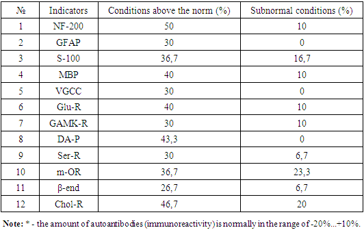
- To achieve the goal of the study, blood serum was collected from 30 middle-aged alcoholics (42±2.7). NF-200 (specific protein of axons), GFAP (specific glial fibrillary acidic protein of the brain, which forms the intermediate filaments of the astrocyte cytoskeleton system), MBP (the main protein of the myelin sheaths of axons) in the blood serum using the "ELI-N-Test" kit (Russia)), S100b (highly specific Ca2+-binding protein for the nervous system located mainly in the cytoplasm of astrocytes), VGCC (potential-dependent calcium channels) proteins and neurotransmitter receptors: glutamate receptor (Glu-R), dopamine receptor (DA-R), GAMK - The amount of neurotropic autoantibodies belonging to class G against receptor (GABA-R), opiate receptors (m-OR), serotonin receptor (Ser-R), acetylcholine receptor (Chol-R) and β-endorphin (β-end) was determined [10,11]. Pre-antigen components are incubated in the cells of the sorbed tablet to establish a balance between free and bound antibodies against the corresponding antigens, diluted with the control and the analyzed blood serum. When the contents of the cell are lost, the conjugate solution containing the relevant autoantibodies with peroxidase is dripped, and the conjugate molecules are sorbed proportionally to the amount of bound autoantibodies. An enzymatic reaction of peroxidase is carried out with hydrogen peroxide in the presence of a chromogen (tetramethylbenzidine) to determine the activity of peroxidase when the unbound conjugate is lost. The color intensity of the chromogen is proportional to the concentration of antibodies in the studied sample. After the peroxidase reaction is stopped with a stop-reagent, the optical density is determined and the immunoreactivity for the analyzed blood serum is calculated according to the formula.In this case, the amount of autoantibodies against neurospecific proteins and receptors of neurotransmitters (immunoreactivity) is normally in the range of -20%...+10%, and an increase or decrease from these indicators is considered anomalous.
4. Results and Discussion
- We studied the amount of autoantibodies against neurospecific proteins and neurotransmitter receptors in the blood serum of 30 middle-aged male and female alcoholic patients. Table shows that the amount of autoantibodies against NSO in alcoholic patients has changed compared to normal values. However, since the range of changes in the number of indicators is large, we tried to express the analysis in percentages, since most of the literature also gives percentages according to the deviation of indicators [10]. The highest deviation of autoantibodies was determined against NF-200 protein. In particular, it was found that 50% of patients were above the norm, 10% of the patients were below the norm, and the remaining 40% of the patients were within the norm. The amount of autoantibodies against MBP, S-100, GFAP and VGCC proteins is 40%, respectively, compared to normal values in blood serum; 36.7%; 30%; It was higher in 30% of patients. Autoantibodies against MBP, S-100 were found to be partially decreased in 10% and 16.7% of patients, while autoantibodies to GFAP and VGCC proteins were not abnormally decreased.We also found significant changes in the amount of autoantibodies against various neurotransmitter receptors in the blood serum of alcohol-dependent patients.In particular, the highest values were determined in relation to Khol-R, DA-R and Glu-R receptors, and the amount of autoantibodies was 46.7%, respectively; 43.3%; It was found that 40% of patients have an increase in blood serum. The reduction of autoantibodies was found in 23.3% and 20% of patients compared to m-OR and Xol-R. There were no cases of decreased autoantibodies against DA-R.Mechanisms of brain injury during ethanol intake include the processes of changes in autoantibody indicators against neurospecific proteins.
|