-
Paper Information
- Next Paper
- Paper Submission
-
Journal Information
- About This Journal
- Editorial Board
- Current Issue
- Archive
- Author Guidelines
- Contact Us
International Journal of Virology and Molecular Biology
p-ISSN: 2163-2219 e-ISSN: 2163-2227
2015; 4(1): 1-3
doi:10.5923/j.ijvmb.20150401.01
Caulerpa taxifolia (M.Vahl) C. Agardh against Tobacco Necrotic Virus (TNV)
 Abstract
 Reference
 Full-Text PDF
 Full-text HTML
Poonam Sethi
Guru Nanak College, Chennai, India
Correspondence to: Poonam Sethi, Guru Nanak College, Chennai, India.
| Email: |  |
Copyright © 2015 Scientific & Academic Publishing. All Rights Reserved.
The seaweed Caulerpataxifolia was tested for activity against Tobacco necrotic virus (TNV) a virus that causes leaf necrosis and affects the yield of the host plant,Cyamopsistetragonoloba Taub.(Linn.). Local lesion method was adopted and half leaf comparisons were made in cotyledonary leaves of Ctetragonoloba Taub.(Linn.). Extracts of the experimental algae prepared with water, methanol and toluene were tested for antiviral activity against the plant virus TNV. Various concentrations were tested and the toluene exhibited good activity at a concentration of 1000 µg/mL and reduced the number of lesion formed by TNV by nearly 64%. These results clearly depict the potential of the algae abundant in the shores of Kanyakumari, India during the month of April.
Keywords: Caulerpa, Chlorophyceae, Cyamopsis, Phytoviral, Tobacco necrotic virus
Cite this paper: Poonam Sethi, Caulerpa taxifolia (M.Vahl) C. Agardh against Tobacco Necrotic Virus (TNV), International Journal of Virology and Molecular Biology, Vol. 4 No. 1, 2015, pp. 1-3. doi: 10.5923/j.ijvmb.20150401.01.
Article Outline
1. Introduction
- Caulerpa taxifolia (M. Vahl) C. Agardh, (Chlorophyceae) a marine seaweed, native to the Indian Ocean, used as an ornamental in aquariums as it is attractive but it is also listed as a federal noxious weed or world’s worst invasive algal species and is of not much use due to its antagonistic effect. They are unusual because they consist of only one cell with many nuclei, making them among the biggest single cells in the world. The recent increase in compounds isolated from land plants, has open doors to the poorly exploited marine ecosystem which appears to be a good candidate of natural resource [1]. The aquatic ecosystem covers about 70 % of the earth’s surface and India has a vast coastline of 6100 km supporting a rich flora of marine plants such as seaweeds, mangroves and sea grasses [2]. Seaweeds offer us amazing health benefits. They are the most nutritious and rich in vitamins and minerals than any other food. They are also one of nature’s richest sources of vegetable protein and provide full spectrum of carotenes, chlorophyll, enzymes, amino acids and fibre in large quantities. The distinctive salty taste of marine plants is due to a balanced chelated combination of sodium, potassium, calcium, magnesium, phosphorus, iron and trace minerals. The levels of these minerals are ten to twenty times the total mineral content of land plants [1]. As a part of our ongoing research a natural source providing mankind with various secondary metabolites and having activity against plant viruses, was tested and Caulerpa proved to be a very promising seaweed. Earlier marine algae has been used in traditional and folk medicine in China for more than two thousand years and several species of marine macroalgae are listed in Chinese Materia Medica for their antifebrile, antiedema, diuretic and expectorant properties. Various compounds that include organic and fatty acids, terpenes, carbonyls, bromophenols, halogenated aliphatic and sulfur-containing heterocyclic compounds, isoprenyated and brominated hydroquinones and phlorotannins have been reported to be responsible for the antiviral activities of many marine algae. [3]. The results of the study suggest that the algae which are abundantly available in this vast Indian coastline not only have a considerable amounts of carbohydrates, amino acids, proteins, phenols and lipids for their use in food and pharmaceutical industry but also in controlling plant viral diseases. As suggested by [4] the efficacy of lipid extracts was tested hence toluene extracts were used.
2. Materials and Method
2.1. Sampling and Virus Isolation
- The experimental algae Caulerpa taxifolia (M. Vahl) C. Agardh, (Chlorophyceae) was collected in the month of April from the shores of Kanyakumari, India. Fresh material was shade dried and crushed in mixer to obtain a coarse powder. Local strain of the TNV (Tobacco Necrosis Virus) were obtained from Madurai Kamaraj University, Tamil Nadu. The procedures outlined by [5] were followed in the assay. Seeds of Cyamopsis tetragonoloba Taub.(Linn.) var. Navbahar were purchased from a government certified seed centre and surface sterilized with 0.1% acidified mercuric chloride for 3 to 5 minutes. The seeds were then sown in sterilized soil and grown in earthen pots.
2.2. Preparation of Various Extracts
- Ten grams of the dried alga was extracted with 100 mL of the solvents double distilled water, methanol and toluene and later kept in a shaker for 48 hours[5] The three extracts were then filtered through Whatman No.1 filter paper and dried in an oven at 40℃. The residues obtained were then weighed and stored at 4℃ and used for the antiviral assay.
2.3. Inoculum and Inoculation
- The inoculum was prepared by grinding 1.0 g of virus-infected cotyledonary leaves, the sap strained through cheese cloth, celite was added as an abrasive at the rate of 2 mg/ mL. Inoculation was carried out by rubbing the leaves from the base of the leaf towards the tip with the forefinger previously dipped in the inoculum.
2.4. Antiviral Assay
- The extract residues of the experimental algae were weighed and dissolved in 0.25% DMSO (Dimethyl sulphoxide) to obtain 1.0 mg/1.0 mL concentrations (1:1) from this solution, 1: 10 water dilutions were made and the inhibitory effect of these solutions were tested against tobacco necrosis virus (TNV) on the cluster bean plant, Cyamopsis tetragonoloba. Inoculations were performed on 9-day old cotyledonary leaves [6, 7, 9] all the tests were made in replicates of ten, and half-leaf comparisons were made. The experimental half-leaves were inoculated with known concentrations of the extract and virus suspension. The plants were monitored for 72 hours and the number of local lesions induced by TNV was estimated using the following formulainhibition = A – B / A x 100where, control-A, experimental-BInhibition is based on the reduction of the number of local lesions induced by TNV, in the absence (A) or the presence (B) of the algal extract. The readings were taken on the basis of dark green spots of 1 mm diameter which developed as a result of infection through inoculation were observed at every 24 hour interval. The intensity of the viral infection was scored after 72 hours based on the number of lesions formed. Results were analysed for statistical significance using SPSS (Statistical Package for Scientific Studies) programme.
3. Results
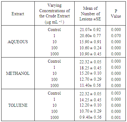
- Extracts of the experimental algae prepared with water, methanol and toluene were tested and toluene were best in comparison to water and methanol extracts against the plant virus. The effect of the extract residues on TNV are given in Table 1. At a concentration of 1000 µg/mL, the toluene extract residue of Caulerpa was able to reduce the lesion formation by nearly 64%.
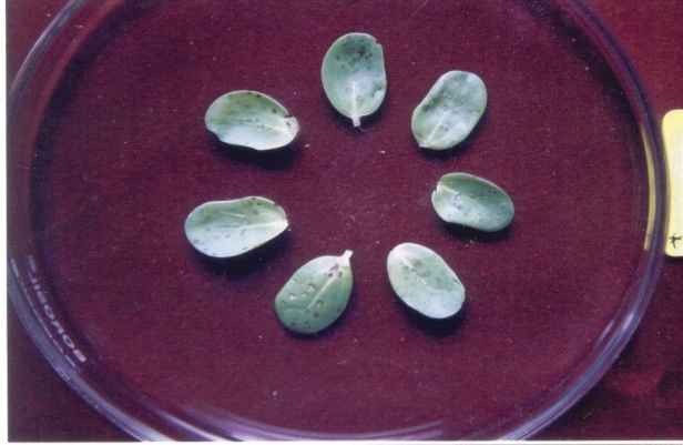 | Plate 1. Cotyledonary leaves of Cyamopsis tetragonoloba showing TNV induced lesion |
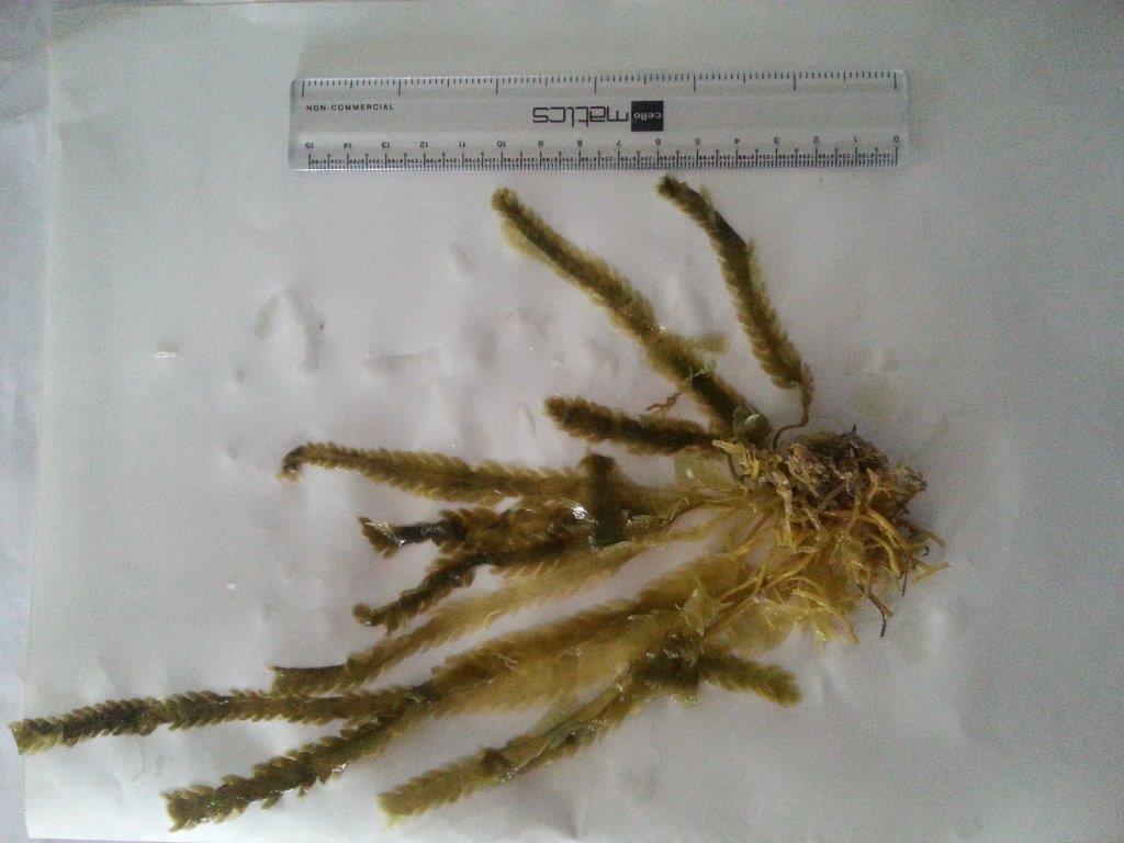 | Plate 2. Experimental plant |
|
4. Discussion
- Earlier it was reported that the methanolic extracts of brown algae when assayed against Potato virus X inhibited the infectivity by more than 80% [6], contradictory to this Caulerpa was active against TNV with toluene (2:1 v/v) than methanolic extract. Organic extracts obtained from the brown-alga Lessonia trabeculata inhibited bacterial growth and reduced both the number and size of the necrotic lesion in tomato leaves following infection with Botrytis cinerea whereas aqueous and ethanolic extracts from the red-alga Gracillaria chilensis prevent the growth of Phytophthora cinnamomi, showing a response which depends on doses and collecting-time. Similarly, aqueous and ethanolic extracts from the brown-alga Durvillaea antarctica were able to diminish the damage caused by tobacco mosaic virus (TMV) in tobacco leaves [8] but not much work has been done on Caulerpa taxifolia a chlorophycean or green alga against microbes hence this report with the world’s worst invasive species being highly active against necrotic virus was the first one. Thus it is a promising antiphytoviral agent. Recently marine sources are receiving much attention mainly because of functional ingredients such as polyunsaturated acids, carotene and their pigment carotenoids, sulphated polysaccharide and sterol. Among different compounds with functional properties, anti oxidants and antibacterials are mostly widely studied.