International Journal of Prevention and Treatment

p-ISSN: 2167-728X    e-ISSN: 2167-7298

2014;  3(2): 17-25

doi:10.5923/j.ijpt.20140302.01

Concerted Dysregulation of 5 Major Classes of Blood Leukocyte Genes in Diabetic ZDF Rats: A Working Translational Profile of Comorbid Rheumatoid Arthritis Progression

Richard M. Kream1, Kirk J. Mantione1, Federico M. Casares1, George B. Stefano1, 2

1Neuroscience Research Institute, State University of New York, College at Old Westbury, NY, U.S.A.

2Center for Molecular and Cognitive Neuroscience, 1st Faculty of Medicine, Charles University in Prague, Prague, Czech Republic

Correspondence to: Kirk J. Mantione, Neuroscience Research Institute, State University of New York, College at Old Westbury, NY, U.S.A..

Email:

Copyright © 2014 Scientific & Academic Publishing. All Rights Reserved.

Abstract

The key objective of the present study was to evaluate developmental alterations of leukocyte gene expression in the blood of diabetic ZDF rats in comparison to healthy ZL controls as a working index of potential comorbid mechanisms between Type II diabetes, rheumatoid arthritis (RA), and associated autoimmune disorders. Paired gene expression analyses were performed on whole blood samples from twelve-week old male homozygous (Fa/Fa) leptin receptor-deficient ZDF rats exhibiting clinically relevant Type II symptomatology in comparison to heterozygous (Fa/fa) ZL healthy lean controls. Gene expression values were derived from DNA microarray analyses and quantified as normalized fold changes utilizing a stable reference gene and confirmed by RT-PCR analyses of selected genes.At 12 weeks of age, ZDF rats presented a full-fledged Type II diabetic phenotype highlighted by hyperglycemia, hyperlipidemia, liver hypertrophy, increased water consumption and urine output. These translational/clinical data were complemented and mechanistically supported by the observed state-dependent differential changes in the expression of functionally clustered genes within 5 major classes of pro- and anti-inflammatory, pro- and anti-apoptotic, and glucocorticoid-responsive leukocyte genes. The overall pattern of change of leukocyte gene expression in the blood of diabetic ZDF rats is consistent with and reflects profound dysregulation of metabolic homestasis and immune competence leading to comorbid RA/autoimmune disease progression in human populations afflicted with Type II diabetes.

Keywords: Zucker Diabetic Fatty Rat, Type II Diabetes, Rheumatoid Arthritis, Pro-inflammation, Apoptosis, Glucocorticoid, Leukocyte Gene expression, DNA microarray, RT PCR

Cite this paper: Richard M. Kream, Kirk J. Mantione, Federico M. Casares, George B. Stefano, Concerted Dysregulation of 5 Major Classes of Blood Leukocyte Genes in Diabetic ZDF Rats: A Working Translational Profile of Comorbid Rheumatoid Arthritis Progression, International Journal of Prevention and Treatment, Vol. 3 No. 2, 2014, pp. 17-25. doi: 10.5923/j.ijpt.20140302.01.

1. Introduction

In metabolically compromised adult populations, the pathogenesis of rheumatoid arthritis (RA) is driven by markedly enhanced inflammatory processes converging on dysregulation of T cell autoantigen presentation and processing functionally linked to aberrant B cell immunoglobulin production [1]. For example, activated pro-inflammatory Th17 lymphocytes, beyond their protective role in the clearance of extracellular pathogens, have been functionally linked to the pathogenesis of RA [2, 3]. Furthermore, selective deletion of a RA susceptibility gene within highly polarized M1 murine macrophages has been observed to trigger a spontaneous erosive polyarthritis that simulates RA in humans [4]. Operationally, a pernicious cycle of pro-inflammatory chemical mediators and region-specific immunoglobulins targeting synovial joint epitopes engender chronic autoimmune processes linked to destruction of articular cartilage and fusion of joints (ankylosis).
Detailed epidemiological studies have demonstrated that both RA and osteoarthritis (OA) are strongly associated with cardiovascular diseases in the general population [5, 6], thereby establishing a clear functional connection between metabolic rundown encountered in Type II diabetics and the etiology of rheumatoid autoimmune diseases [7-9]. In the present study, dependent measure data sets were derived from paired DNA microarray gene expression analyses performed on whole blood samples from eleven-week old male homozygous (Fa/Fa) leptin receptor-deficient diabetic ZDF rats in comparison to heterozygous (Fa/fa) healthy lean control ZL rats. The diabetic ZDF rat has been well established in the biomedical literature as a high resolution translationalmodel for elucidation of underlying pathophysiological mechanisms critically linked to advanced therapeutic development for major human disorders including Type II diabetes [10, 11], cardiovascular disease [7], renal disease [12], and atherosclerosis [13, 14].
To underscore the translational validity of the ZDF diabetic rat model to present a cogent translational model of comorbid RA/rheumatoid autoimmune disease progression, the present study monitored state-dependent differential changes in the expression of functionally clustered genes within 5 major classes of pro- and ant-inflammatory, pro- and anti-apoptotic, and glucocorticoid-responsive leukocyte genes. Accordingly, we hypothesized that the overall pattern of change of leukocyte gene expression in the blood of diabetic ZDF rats would be reflective of profound dysregulation of metabolic homestasis and immune competence and provide a working translational profile of comorbid RA/autoimmune disease progression in human populations afflicted with Type II diabetes.

2. Material and Methods

2.1. Experimental Animals

Seven to eight-week old, male homozygous (Fa/Fa) leptin receptor-deficient ZDF rats and their heterozygous (Fa/fa) healthy lean control ZL rats were purchased from Charles Rivers Laboratories (Wilmington, MA) and maintained by PhysioGenix, Inc. (Waukesha, WI). Animal care and all technical procedures were performed PhysioGenix, Inc. staff in accordance with IUCAC approval and established protocols described in Guide for Care and Use of Laboratory Animals (Eighth Edition). Animals were housed at 2 per cage and maintained in the Innovive caging system (San Diego, CA) upon arrival at PhysioGenix, Inc and were allowed an acclimation period of 4 days prior to baseline blood collections. Cages were monitored daily to ensure the Innovive system maintained 80 air changes per hour and positive pressure. rat rooms were maintained at temperatures of 66-75 degrees Fahrenheit and relative humidity between 30% and 70%. The rooms were lit by artificial light for 12 hr per day (7:00 AM - 7:00 PM). Animals had free access to water and Purina 5008 rodent food (Waldschimdt’s, Madison, WI) for the duration of the study. At 8, 10, and 12 week time points, blood aliquots were collected for analyses of true (non-fasting) glucose and serum lipids (total cholesterol, triglycerides, fractionated HDL and LDL cholesterol).
The study lasted for 28 days and ZDF diabetic and ZL lean healthy control animals were euthanized by isoflurane overdose and thoracotomy at 11 weeks of age. For molecular biological analyses, blood was collected via descending vena cava into tubes containing RNAase inhibitor lysis buffer, immediately frozen at -20°C, and shipped on ice packs to the SUNY Neuroscience Research Institute. For analyses of serum lipids (total cholesterol, triglycerides, fractionated HDL and LDL cholesterol), blood was collected via descending vena cava and processedserum was frozen at -20°C and shipped to Comparative Clinical Pathology Services, LLC. For each study animal, at day 28 fasted blood glucose was quantified using a Bayer Contour glucometer and after sacrifice liver and abdominal fat were collected and weighed.

2.2. DNA Microarray Analyses

Total RNA extracted from rat blood samples was isolated using the PAX RNA kit (Qiagen, Valencia, CA). DNA microarray analyses were performed using a system provided by Agilent. Arrays included four arrays per chip (Agilent 4X44K chip). Total RNA was reverse transcribed (1000 ng) using T7 primers and labeled and transcribed using Cyanine-3 dye. Each array was hybridized with at least 1.65 μg of labeled cRNA at 65°C for 18 hours. Arrays were scanned using an Agilent array scanner. The microarray platform has been previously determined to reliably quantify a minimum 1.5 fold change in gene expression.
For all ZDF and ZL data sets, raw fluorescent signals were normalized to those of an empirically validated and stably expressed reference gene [15], i.e., Ppia (rat leukocyte eptidylprolyl isomerase A, also termed cyclophilin A), and paired gene expression fold changes were quantified using GeneSpring version 12.6.1 (Agilent, Santa Clara, CA). Statistical significance of paired ZDF/ZL group data was determined by calculating corrected (Benjamini-Hochberg) p-values of 2-way ANOVA.

2.3. Real-Time PCR Analyses

Representative Real-time PCR analyses of forkhead box O3 (Foxo3) and mitochondrial superoxide dismutase 2 (Sod2) gene expression and nitric oxide synthase, inducible (Nos2) in the blood of ZDF and ZL control rats were performed to validate the DNA microarray data sets. Ppia was used as a reference gene. The real-time PCR master mix included 25 μL 2x universal master mix, 2.5 μL 20x detector set (with the primer and probe), and 21.5 μL of water. PCR was performed in an Applied Biosystems 7500 sequence detection system. The thermocycler conditions included denaturation at 95°C for 15 seconds and annealing/extension at 60°C for 60 seconds. Forty cycles of PCR were preceded by 95°C for 10 minutes. Reactions were performed in triplicate. The relative quantities of genes were determined using the formula 2-∆∆Ct using the Applied Biosystems 7500 software.

3. Results and Discussion

3.1. Presentation of a Clinically Cogent Translational Model of Type II Diabetes in 12-week ZDF Rats

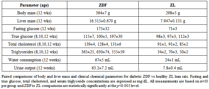
At 12 weeks, fasted blood glucose concentrations of ZDF rats were calculated to be 175±3 mg/dL, an approximate 50% increase in comparison to values calculated for non-diabetic ZL control rats (Table 1). The developmental expression of a clinically relevant diabetic phenotype was supported by comparative true (non-fasting) serum glucose concentrations monitored at 8, 10, and 12 weeks. Interestingly, the approximate 75% increase in true serum glucose concentration of ZDF vs ZL control rats at 12 weeks was complemented by approximate 200 and 700% increases in daily water consumption and urine output, respectively. The presentation of a full-fledged Type II diabetic phenotype was also supported by the statistically significant approximate doubling of serum triglyceride concentrations between 8 and 12 weeks in ZDF vs ZL control rats and is consistent with a development profile of insulin resistance [10]. Additionally, the ostensible doubling of liver mass in ZDF vs ZL control rats indicates diabetes-related abnormal intrahepatic lipid storage functionally linked to incipient steatotic liver disease [16, 17]. In sum, basic physiological and clinical chemical parameters presented here support the validity of the ZDF rat as a high resolution, clinically cogent, translational model of Type II diabetes in humans [7, 18].
Table 1. Presentation of a Cogent Translational Model of Type II Diabetes in ZDF Rats
     

3.2. Differential and Functionally Concerted Changes in the Gene Expression of Major Anti-Inflammatory and Pro-Inflammatory Signaling Complexes and Enzyme Activities in ZDF Rat Blood

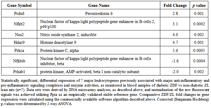
As depicted in Table 2, DNA microarray analyses yielded a statistically significant differential expression of 7 major leukocytegenes previously associated with major anti-inflammatory and pro-inflammatory signaling complexes and enzyme activities in ZDF, as compared to non-diabetic ZL rat blood. The dramatic enhancements of Prdx6, Nfkb2, Nos2, Hdac9, and Prkcapro-inflammatory gene expression, were calculated at 2.7, 3.1, 4.0, 4.7 and 5.6 fold, respectively, and contrasted with the observed reductions of Nfkbib and Prkab1 anti-inflammatory gene expression of -1.6 and -2.0 fold, respectively. The overall pattern of gene expression changes clearly indicated a causal association of the ZDF diabetic phenotype with an accelerated pathophysiological pro-inflammatory state in blood leukocyte populations leading to accelerated RA/rheumatic autoimmune processes.
Table 2. Differential Expression of Leukocyte Genes Previously Associated with Major Anti-inflammatory and Pro-inflammatory Signaling Complexes and Enzyme Activities in ZDF Rat Blood
     
The leukocyte NFKB complex functions as a key regulator/activator of genes involved in inflammation and immune function [19]. Accordingly, the observed differential decrease of the NFKIB inhibitor protein [20] and increase of the NFKB2 enhancer protein [21] promotes pro-inflammatory-mediated RA progression. The PRDX6 gene encodes a bifunctional protein with both glutathione peroxidase and calcium-independent phospholipase A2 activities. Aberrant PRDX6 phospholipase A2 activity linked to cyclooxygenase-2 mediated proinflammatory prostaglandin release has been functionally linked to the destruction of both articular bone and cartilage in RA and osteoarthritis [22-24]. Cardiovascular disease linked to metabolic and rheumatoid autoimmune disorders is characterized by characterized by uncoupling of physiologically beneficial constitutive NO production leading to marked enhancement of pro-inflammatory NOS2 gene expression and enhanced IL-1β secretion. Mechanistically, high glucose was observed to enhance NOS2 expression and NO generation via PKCA activation [25].
The enzyme expressed from the HDAC9 gene is an epigenetic regulator of multiple gene expression. HDAC inhibitors have been demonstrated to enhance FOXO3 gene expression, as well as the production of regulatory anti-inflammatory T cells. Accordingly, enhanced HDAC9 expression limits the anti-inflammatory suppressive functions of FOXO3-positive regulatory T cells in RA patients [26, 27]. In a diabetic rodent model, high glucose promotes iNOS expression linked to enhanced IL-1β secretion. Mechanistically, high glucose was observed to enhance iNOS expression and NO generation via PKCA activation [28, 29], thereby indicating that inhibition of PKCA gene expression will promote profound ameliorative effects on the development and progression of RA and associated rheumatoid autoimmune diseases.
Finally, AMP-activated protein kinase (AMPK) is a preeminent serine/threonine kinase that regulates energy homeostasis and metabolic stress in cellular and integrated organ systems. Functional down-regulation of AMPK-mediated cellular processes via decreased PRKAB1 gene expression will promote RA and associated rheumatoid autoimmune disease progression.

3.3. Differential and Functionally Concerted Changes in the Gene Expression of Major Anti-Inflammatory and Pro-Inflammatory Cytokines and Growth Factors in ZDF Rat Blood

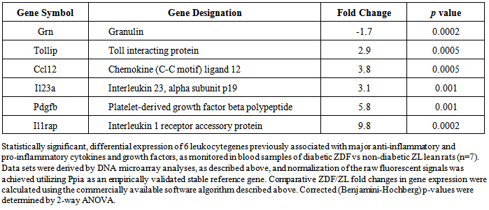
As depicted in Table 3, DNA microarray analyses yielded a statistically significant differential expression of 6 major leukocyte genes previously associated with major anti-inflammatory and pro-inflammatory cytokines and growth factors in ZDF, as compared to non-diabetic ZL rat blood. The dramatic enhancements of Tollip, Il23a, Ccl12, Pdgfb, and Il1rap pro-inflammatory gene expression, were calculated at 2.9, 3.1, 3.8, 5.8 and 9.8 fold, respectively, and contrasted with the observed reduction of Grn anti-inflammatory gene expression of -1.7 fold. In support of the data set presented in Table 2, the differential enhancement of pro-inflammatory cytokines and growth factors and reduction of granulin gene expression indicates that at 12 weeks of age diabetic ZDF rats present a cogent translational profile consistent with profound dysregulation of metabolic homestasis leading to comorbid RA disease progression.
Table 3. Differential Expression of Leukocyte Genes Previously Associated with Major Anti-inflammatory and Pro-inflammatory Cytokines and Growth Factors in ZDF Rat Blood
     
Progranulin expressed from the GRN gene inhibits LPS-mediated pro-inflammatory signaling in endothelial cells through attenuation of the NF-κB signaling pathway [30] and TNFα signalling [31, 32]. The TOLLIP gene encodes a ubiquitin-binding protein that interacts with several Toll-like receptor (TLR) signaling cascade components and the expressed protein promotes pro-inflammatory signaling and is involved in interleukin-1 receptor trafficking and in the turnover of IL1R-associated kinase. In LPS-primed macrophages, Tollip-dependent mechanisms promote profound pro-inflammatory effects linked to initiation and progression of RA and associated rheumatoid autoimmune diseases [33].
The Il23a gene encodes a subunit of the heterodimeric cytokine interleukin 23 (IL23). Interleukin-23 serum levels are markedly elevated in patients affected by severe peripheral arterial disease and the cytokine is involved in differentiation of pro-inflammatory Th17 cells in the presence of TGF-β and IL-6. Inflammatory macrophages express IL-23R and are activated by IL-23 to produce IL-1, TNF-α, and IL-23 itself, thereby identifying IL-23 as a central cytokine involved in the etiology and progression of RA and associated autoimmune diseases [34-36]. CCL12, also known as MCP-5, belongs to the monocyte chemotactic protein (MCP) subfamily of the CC chemokines. Overexpression of monocyte chemotactic proteins have been demonstrated to promote enhanced pro-inflammatory activities in human disease states including RA [37], and inhibition of CCL12 gene expression will functionally inhibit pro-inflammatory-mediated RA progression.
Several key studies have demonstrated that PDGFRB-mediated signaling pathways are critically involved in RA progression [38]. Accordingly, enhancement of PDGFB gene expression will functionally mediate cellular inflammatory insults in RA. The IL1RAP gene encodes the interleukin 1 receptor accessory protein. The protein is a necessary part of the interleukin 1 receptor complex, which initiates signaling events that result in the activation of interleukin-1 responsive pro-inflammatory genes [39]. Accordingly, augmentation of ILRAP gene expression will have profound effects on RA progression.

3.4. Differential and Functionally Concerted Changes in Glucocorticoid-Responsive Gene Expression in ZDF Rat Blood

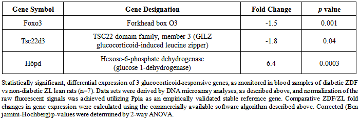
DNA microarray analyses yielded a statistically significant differential expression of 3 major glucocorticoid-responsive leukocytegenes previously associated with RA disease progression in ZDF, as compared to non-diabetic ZL rat blood (Table 4). The dramatic enhancement of H6pd gene expression calculated at 6.4 fold contrasted with the observed reductions Foxo3 and Tsc22d3 gene expression of anti-inflammatory gene expression of -1.5 and -1.8 fold, respectively.
Table 4. Differential Expression of Glucocorticoid-Responsive Genes in ZDF Rat Blood
     
The FOXO3 gene encodes a key transcription factor involved in the regulation of multiple cellular processes including protection against oxidative stress and conservation of energy metabolism. FOXO3 expression is functionally linked to glucocorticoid action via glucocorticoid receptor activation and dysregulation of FOXO3 signaling is associated with RA processes mediated by proinflammatory cytokines [40, 41]. The TSC22D3 encodes a leucine zipper transcriptional regulator, also designated GILZ, which is stimulated by glucocorticoid receptor activation and anti-inflammatory cytokines. Thus, TSC22D3 activation regulates the anti-inflammatory and immunosuppressive effects of glucocorticoids [42, 43]. In murine lymphocytes, glucocorticoids induce expression of glucocorticoid-induced leucine zipper (GILZ), which prevents the nuclear factor kappaB-mediated activation of pro-inflammatory gene transcription [44]. Accordingly, diminished TSC22D3, as currently determined, gene expression will exacerbate RA and its inflammatory progression. Hexose-6-phosphate dehydrogenase expressed from the H6PD gene supplies a crucial cofactor, reduced nicotinamide adenine dinucleotide phosphate (NADPH), which allows the hydroxysteroid reductase expressed by the HSD11B1 gene to maintain cortisone reductase activity. Mutations in the H6PD gene are functionally associated with cortisone reductase deficiency [45, 46]. High H6PD gene expression may function to support metabolic disease-related dysregulation of normative glucocorticoid signaling systems involved in the pathogenesis of RA.

3.5. Differential and Functionally Concerted Changes in the Gene Expression of Major Anti-Apoptotic and Pro-Apoptotic Regulatory Proteins and Enzymes in ZDF Rat Blood

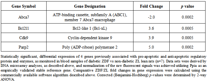
As depicted in Table 5, DNA microarray analyses yielded statistically significant differential changes in the expression of 4 major anti-apoptotic and pro-apoptotic regulatory proteins and enzymes in ZDF, as compared to non-diabetic ZL rat blood. The dramatic enhancements of Bcl21 (Bcl-xL), Cdk9, and Parp2 anti-apoptotic gene expression, were calculated at 3.6, 3.9, and 5.0 fold, respectively, and contrasted with the observed reduction of Abca7 pro-apoptotic gene expression of -2.0 fold.
Table 5. Differential Expression of Genes Previously Associated with Pro-apoptotic and Anti-apoptotic Regulatory Proteins and Enzymes in ZDF Rat Blood
     
Dysregulation of the critical cellular balance of pro-apoptotic and anti-apoptotic regulatory protein expression is predictive of unchecked deleterious cellular damage linked to aberrant antigen presentation and is functionally linked to theonset RA processes. The ATP-binding cassette protein encoded by the ABCA7 gene when expressed by anti-inflammatory M2 macrophages has been empirically demonstrated to facilitate the clearance of pro-inflammatory apoptotic cells [47, 48]. In light of these data, state-dependent reduction of ABCA7 gene expression will promote significant pro-rheumatoid arthritis effects via prolongation of the lifespan of polarized macrophages. The BCL-2 family of proteins plays a critical role in controlling immune responses by regulating the expansion and contraction of activated lymphocyte clones by apoptosis. It encodes two alternatively spliced transcript variants BCL-xs and BCL-xl, which express apoptotic activator and apoptotic inhibitor proteins, respectively. BCL-xs promotes apoptosis of pro-inflammatory myeloid cells involved in the etiology and progression of RA [49]. Conversely, inhibition of anti-apoptotic BCL-xl expression results in potent inhibition of lymphocyte proliferation as measured by in vitro mitogenic or ex vivo Ag-specific stimulation [50].
Accordingly, the observed increases in Bcl-xl expression will promote development and progression of RA via markedly enhanced proliferation of activated T cells.
CDK9 activity regulates neutrophil lifespan and inhibition of its activity increased apoptosis and caused a rapid decline in the level of the anti-apoptotic protein MCL-1, indicating that pharmacological inhibition of CDK9 might be a valuable anti-inflammatory strategy [51-53]. Because inappropriate inhibition of neutrophil apoptosis contributes to chronic inflammatory diseases such as RA, development of CDK9 inhibitors represents a powerful therapeutic approach for treatment of RA and associated rheumatoid autoimmune diseases. The approximate 4-fold enhancement of Cdk9 gene expression in ZDF vs ZL rats is indicative of a marked prolongation of a deleterious pro-inflammatory cellular environment. The protein expressed from the PARP2 gene is involved in DNA repair and transcriptional regulation and is now recognized as a key regulator of cell survival and cell death as well as a master component of a number of transcription factors involved in pro-inflammatory processes [54]. The observed 5-fold enhancement of Parp2 gene expression in ZDF vs ZL rats is indicative of a marked synergy underlying the prolonged lifespans of activated classes of leukocytes within a pro-inflammatory cellular environment.

3.6. Differential and Functionally Concerted Enhancement in the Gene Expression of 4 Major Pro-Inflammatory Receptor Proteins in ZDF Rat Blood

DNA microarray analyses yielded statistically significant enhancement in the expression of 4 major pro-inflammatory receptor proteins in ZDF, as compared to non-diabetic ZL rat blood. The dramatic enhancements of Ager, Nod2, Erbb3, and Casr, pro-inflammatory receptor gene expression were calculated at 2.8, 2.9, 4.0 and 8.6 fold, respectively (Table 6).
Table 6. Differential Enhancement in the Expression of Genes Previously Associated with Pro-inflammatory Receptor Proteins in ZDF Rat Blood
     
The RAGE (receptor for advanced glycosylation end-products) protein expressed by the AGER gene and is found on multiple cell types implicated in the progression of atherosclerosis and diabetes-associated atherosclerosis as well as RA [55, 56]. Importantly, RAGE is activated by released the soluble pro-inflammatory protein encoded by the HMGB1 gene, thereby amplifying proinflammatory signaling in the development and progression of RA. The receptor protein expressed from the NOD2 gene is a prominent member of the NLR (NOD-LRR) family of proteins that contain nucleotide-binding NACHT domains and receptor-like LRR domains. The protein is primarily expressed in the peripheral blood leukocytes and mediates signal transduction mechanisms that include NF-KB and p38 MAPK14. Hereditary polymorphisms in the genes encoding NOD1 and NOD2 have been associated with an increasing number of chronic inflammatory diseases including RA and inhibition of NOD2 gene expression represents a highly promising treatment for amelioration of multiple symptoms of rheumatoid autoimmune diseases [57-59].
The gene ERBB3 encodes a member of the epidermal growth factor receptor (EGFR) family of receptor tyrosine kinases that contains a neuregulin binding domain but not an active kinase domain. EGFR activation plays a central role in the pathogenesis of RA [60, 61]. The concentration of CASR protein encoded by the CASR gene is markedly elevated on the surface of monocytes of patients with Type II diabetes mellitus co-afflicted with peripheral artery disease [62] and CASR receptor protein expression in circulating monocytes is markedly increased in RA patients with severe coronary artery calcification [63]. Accordingly, the observed enhancements of Egrf and Casr gene expression in ZDF vs ZL is again indicative of a marked synergy underlying the prolonged lifespans of activated classes of leukocytes within a pro-inflammatory cellular environment functionally linked to RA progression.

3.7. Representative Real-time PCR analyses of Forkhead box O3 (Foxo3) and Inducible Nitric Oxide Synthase (Nos2) Gene Expression in ZDF Rat Blood

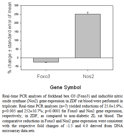
As depicted in Fig.1, representative real-time PCR analyses confirmed the validity of the fold changes in gene expression derived from DNA microarray data sets. Real-time PCR analyses yielded a differential reduction of 25.0±1.9% for Foxo3 and enhancement of 252±10.7% for Nos2 gene expression in ZDF, as compared to non-diabetic ZL controls. The comparative reductions in Foxo3 and Sod2 gene expression were consistent with the respective fold changes of -1.5 and +4.0 derived from DNA microarray data sets (Tables 2 and 4).
Figure 1. Representative Real-time PCR analyses of forkhead box O3 (Foxo3) and nitric oxide synthase, inducible (Nos2) gene expression in ZDF rat blood

4. Conclusions

The present report has monitored profound state-dependent differential changes in the expression of functionally clustered genes within 5 major classes of pro- and ant-inflammatory, pro- and anti-apoptotic, and glucocorticoid-responsive leukocyte genes in the blood of diabetic ZDF rats. The overall pattern of change of leukocyte gene expression in the blood of diabetic ZDF rats is consistent with and reflects profound dysregulation of metabolic homestasis and immune competence leading to comorbid RA/autoimmune disease progression in human populations afflicted with Type II diabetes. Importantly, we have provided an initial working translational profile of functionally clustered genes by which to evaluate potentially novel therapeutic interventions for treatment of comorbid RA progression in human populations afflicted with Type II diabetes.

References

[1]  Wilson, C.L., Hine, D.W., Pradipta, A., Pearson, J.P., van, E.W., Robinson, J.H., and Knight, A.M., 2012, Presentation of the candidate rheumatoid arthritis autoantigen aggrecan by antigen-specific B cells induces enhanced CD4(+) T helper type 1 subset differentiation, Immunology, 135(4), 344-354.
[2]  Cosmi, L., Liotta, F., Maggi, E., Romagnani, S., and Annunziato, F., 2014, Th17 and Non-Classic Th1 Cells in Chronic Inflammatory Disorders: Two Sides of the Same Coin, Int. Arch. Allergy Immunol., 164(3), 171-177.
[3]  O'Connell, R.M., Kahn, D., Gibson, W.S., Round, J.L., Scholz, R.L., Chaudhuri, A.A., Kahn, M.E., Rao, D.S., and Baltimore, D., 2010, MicroRNA-155 promotes autoimmune inflammation by enhancing inflammatory T cell development, Immunity., 33(4), 607-619.
[4]  VandeWalle, L., Van Opdenbosch, N., Jacques, P., Fossoul, A., Verheugen, E., Vogel, P., Beyaert, R., Elewaut, D., Kanneganti, T.D., van, L.G., and Lamkanfi, M., 2014, Negative regulation of the NLRP3 inflammasome by A20 protects against arthritis, Nature, 512(7512), 69-73.
[5]  Hollan, I., Meroni, P.L., Ahearn, J.M., Cohen Tervaert, J.W., Curran, S., Goodyear, C.S., Hestad, K.A., Kahaleh, B., Riggio, M., Shields, K., and Wasko, M.C., 2013, Cardiovascular disease in autoimmune rheumatic diseases, Autoimmun. Rev., 12(10), 1004-1015.
[6]  Ong, K.L., Wu, B.J., Cheung, B.M., Barter, P.J., and Rye, K.A., 2013, Arthritis: its prevalence, risk factors, and association with cardiovascular diseases in the United States, 1999 to 2008,Ann.Epidemiol., 23(2), 80-86.
[7]  Carley, A.N. and Severson, D.L., 2005, Fatty acid metabolism is enhanced in type 2 diabetic hearts, Biochim. Biophys. Acta, 1734(2), 112-126.
[8]  Donath, M.Y., 2014, Targeting inflammation in the treatment of type 2 diabetes: time to start, Nat. Rev. Drug Discov., 13(6), 465-476.
[9]  Lu, M.C., Yan, S.T., Yin, W.Y., Koo, M., and Lai, N.S., 2014, Risk of rheumatoid arthritis in patients with type 2 diabetes: a nationwide population-based case-control study, PLoS. One., 9(7), e101528.
[10]  Kakimoto, T., Kimata, H., Iwasaki, S., Fukunari, A., and Utsumi, H., 2013, Automated recognition and quantification of pancreatic islets in Zucker diabetic fatty rats treated with exendin-4, J. Endocrinol., 216(1), 13-20.
[11]  Wang, F., Guo, X., Shen, X., Kream, R.M., Mantione, K.J., and Stefano, G.B., 2014, Vascular Dysfunction Associated with Type II Diabetes and Alzheimer's Disease: A Potential Etiological Linkage, Med SciMonit Basic Res, 20, 118-129.
[12]  Zanchi, C., Locatelli, M., Benigni, A., Corna, D., Tomasoni, S., Rottoli, D., Gaspari, F., Remuzzi, G., and Zoja, C., 2013, Renal expression of FGF23 in progressive renal disease of diabetes and the effect of ACE inhibitor, PLoS. One., 8(8), e70775.
[13]  Mierzecki, A., Kloda, K., Bukowska, H., Chelstowski, K., Makarewicz-Wujec, M., and Kozlowska-Wojciechowska, M., 2013, Association between low-dose folic acid supplementation and blood lipids concentrations in male and female subjects with atherosclerosis risk factors, Med SciMonit, 19, 733-739.
[14]  Stohr, R. and Federici, M., 2013, Insulin resistance and atherosclerosis: convergence between metabolic pathways and inflammatory nodes, Biochem.J., 454(1), 1-11.
[15]  Spalenza, V., Girolami, F., Bevilacqua, C., Riondato, F., Rasero, R., Nebbia, C., Sacchi, P., and Martin, P., 2011, Identification of internal control genes for quantitative expression analysis by real-time PCR in bovine peripheral lymphocytes, Vet. J., 189(3), 278-283.
[16]  Gholam, P.M., Flancbaum, L., Machan, J.T., Charney, D.A., and Kotler, D.P., 2007, Nonalcoholic fatty liver disease in severely obese subjects, Am. J. Gastroenterol., 102(2), 399-408.
[17]  Tiniakos, D.G., Vos, M.B., and Brunt, E.M., 2010, Nonalcoholic fatty liver disease: pathology and pathogenesis, Annu. Rev. Pathol., 5, 145-171.
[18]  Cho, Y.E., Basu, A., Dai, A., Heldak, M., and Makino, A., 2013, Coronary endothelial dysfunction and mitochondrial reactive oxygen species in type 2 diabetic mice, Am. J. Physiol Cell Physiol, 305(10), C1033-C1040.
[19]  Welters, I.D., Fimiani, C., Bilfinger, T.V., and Stefano, G.B., 1999, NF-kB, nitric oxide and opiate signaling, Medical Hypotheses, 54(2), 263-268.
[20]  Majdalawieh, A. and Ro, H.S., 2010, Regulation of Ikappa Balpha function and NF-kappaB signaling: AEBP1 is a novel proinflammatory mediator in macrophages, Mediators. Inflamm., 2010, 823821.
[21]  Shen, J., Yang, M., Ju, D., Jiang, H., Zheng, J.P., Xu, Z., and Li, L., 2010, Disruption of SM22 promotes inflammation after artery injury via nuclear factor kappaB activation, Circ.Res, 106(8), 1351-1362.
[22]  Ikeda, D., Ageta, H., Tsuchida, K., and Yamada, H., 2013, iTRAQ-based proteomics reveals novel biomarkers of osteoarthritis, Biomarkers, 18(7), 565-572.
[23]  Yoon, H.Y., Lee, E.G., Lee, H., Cho, I.J., Choi, Y.J., Sung, M.S., Yoo, H.G., and Yoo, W.H., 2013, Kaempferol inhibits IL-1beta-induced proliferation of rheumatoid arthritis synovial fibroblasts and the production of COX-2, PGE2 and MMPs, Int. J. Mol. Med, 32(4), 971-977.
[24]  Yun, H.M., Park, K.R., Lee, H.P., Lee, D.H., Jo, M., Shin, D.H., Yoon, D.Y., Han, S.B., and Hong, J.T., 2014, PRDX6 promotes lung tumor progression via its GPx and iPLA2 activities, Free Radic. Biol. Med, 69, 367-376.
[25]  Luiking, Y.C., Ten Have, G.A., Wolfe, R.R., and Deutz, N.E., 2012, Arginine de novo and nitric oxide production in disease states, Am. J. Physiol Endocrinol. Metab, 303(10), E1177-E1189.
[26]  Beier, U.H., Wang, L., Han, R., Akimova, T., Liu, Y., and Hancock, W.W., 2012, Histone deacetylases 6 and 9 and sirtuin-1 control Foxp3+ regulatory T cell function through shared and isoform-specific mechanisms, Sci Signal., 5(229), ra45.
[27]  Tao, R., de Zoeten, E.F., Ozkaynak, E., Chen, C., Wang, L., Porrett, P.M., Li, B., Turka, L.A., Olson, E.N., Greene, M.I., Wells, A.D., and Hancock, W.W., 2007, Deacetylase inhibition promotes the generation and function of regulatory T cells, Nat. Med, 13(11), 1299-1307.
[28]  Hua, K.F., Wang, S.H., Dong, W.C., Lin, C.Y., Ho, C.L., and Wu, T.H., 2012, High glucose increases nitric oxide generation in lipopolysaccharide-activated macrophages by enhancing activity of protein kinase C-alpha/delta and NF-kappaB, Inflamm. Res, 61(10), 1107-1116.
[29]  Jancinova, V., Perecko, T., Nosal, R., Kostalova, D., Bauerova, K., and Drabikova, K., 2009, Decreased activity of neutrophils in the presence of diferuloylmethane (curcumin) involves protein kinase C inhibition, Eur. J. Pharmacol., 612(1-3), 161-166.
[30]  Liu, C.J., 2011, Progranulin: a promising therapeutic target for rheumatoid arthritis, FEBS Lett., 585(23), 3675-3680.
[31]  Tang, W., Lu, Y., Tian, Q.Y., Zhang, Y., Guo, F.J., Liu, G.Y., Syed, N.M., Lai, Y., Lin, E.A., Kong, L., Su, J., Yin, F., Ding, A.H., Zanin-Zhorov, A., Dustin, M.L., Tao, J., Craft, J., Yin, Z., Feng, J.Q., Abramson, S.B., Yu, X.P., and Liu, C.J., 2011, The growth factor progranulin binds to TNF receptors and is therapeutic against inflammatory arthritis in mice, Science, 332(6028), 478-484.
[32]  Thurner, L., Preuss, K.D., Fadle, N., Regitz, E., Klemm, P., Zaks, M., Kemele, M., Hasenfus, A., Csernok, E., Gross, W.L., Pasquali, J.L., Martin, T., Bohle, R.M., and Pfreundschuh, M., 2013, Progranulin antibodies in autoimmune diseases, J. Autoimmun., 42, 29-38.
[33]  Deng, H., Maitra, U., Morris, M., and Li, L., 2013, Molecular mechanism responsible for the priming of macrophage activation, J. Biol. Chem., 288(6), 3897-3906.
[34]  Chang, L.H., Huang, H.S., Wu, P.T., Jou, I.M., Pan, M.H., Chang, W.C., Wang, D.D., and Wang, J.M., 2012, Role of macrophage CCAAT/enhancer binding protein delta in the pathogenesis of rheumatoid arthritis in collagen-induced arthritic mice, PLoS. One., 7(9), e45378.
[35]  Duvallet, E., Semerano, L., Assier, E., Falgarone, G., and Boissier, M.C., 2011, Interleukin-23: a key cytokine in inflammatory diseases, Ann. Med, 43(7), 503-511.
[36]  Jawien, J., Toton-Zuranska, J., Kus, K., Pawlowska, M., Olszanecki, R., and Korbut, R., 2012, The effect of AVE 0991, nebivolol and doxycycline on inflammatory mediators in an apoE-knockout mouse model of atherosclerosis, Med SciMonit, 18(10), BR389-BR393.
[37]  Jenkins, E., Brenner, M., Laragione, T., and Gulko, P.S., 2012, Synovial expression of Th17-related and cancer-associated genes is regulated by the arthritis severity locus Cia10, Genes Immun., 13(3), 221-231.
[38]  Rosengren, S., Corr, M., and Boyle, D.L., 2010, Platelet-derived growth factor and transforming growth factor beta synergistically potentiate inflammatory mediator synthesis by fibroblast-like synoviocytes, Arthritis Res Ther., 12(2), R65.
[39]  Radons, J., Gabler, S., Wesche, H., Korherr, C., Hofmeister, R., and Falk, W., 2002, Identification of essential regions in the cytoplasmic tail of interleukin-1 receptor accessory protein critical for interleukin-1 signaling, J. Biol. Chem., 277(19), 16456-16463.
[40]  Lee, J.C., Espeli, M., Anderson, C.A., Linterman, M.A., Pocock, J.M., Williams, N.J., Roberts, R., Viatte, S., Fu, B., Peshu, N., Hien, T.T., Phu, N.H., Wesley, E., Edwards, C., Ahmad, T., Mansfield, J.C., Gearry, R., Dunstan, S., Williams, T.N., Barton, A., Vinuesa, C.G., Parkes, M., Lyons, P.A., and Smith, K.G., 2013, Human SNP links differential outcomes in inflammatory and infectious disease to a FOXO3-regulated pathway, Cell, 155(1), 57-69.
[41]  Lutzner, N., Kalbacher, H., Krones-Herzig, A., and Rosl, F., 2012, FOXO3 is a glucocorticoid receptor target and regulates LKB1 and its own expression based on cellular AMP levels via a positive autoregulatory loop, PLoS.One., 7(7), e42166.
[42]  Berrebi, D., Bruscoli, S., Cohen, N., Foussat, A., Migliorati, G., Bouchet-Delbos, L., Maillot, M.C., Portier, A., Couderc, J., Galanaud, P., Peuchmaur, M., Riccardi, C., and Emilie, D., 2003, Synthesis of glucocorticoid-induced leucine zipper (GILZ) by macrophages: an anti-inflammatory and immunosuppressive mechanism shared by glucocorticoids and IL-10, Blood, 101(2), 729-738.
[43]  Cheng, Q., Morand, E., and Yang, Y.H., 2014, Development of novel treatment strategies for inflammatory diseases-similarities and divergence between glucocorticoids and GILZ, Front Pharmacol., 5, 169.
[44]  Cheng, Q., Fan, H., Ngo, D., Beaulieu, E., Leung, P., Lo, C.Y., Burgess, R., van der Zwan, Y.G., White, S.J., Khachigian, L.M., Hickey, M.J., and Morand, E.F., 2013, GILZ overexpression inhibits endothelial cell adhesive function through regulation of NF-kappaB and MAPK activity, J. Immunol., 191(1), 424-433.
[45]  Chapman, K.E., Coutinho, A.E., Zhang, Z., Kipari, T., Savill, J.S., and Seckl, J.R., 2013, Changing glucocorticoid action: 11beta-hydroxysteroid dehydrogenase type 1 in acute and chronic inflammation, J. Steroid Biochem. Mol. Biol., 137, 82-92.
[46]  Moon, S.S., Lee, Y.S., Kim, J.G., Kim, S.W., Jeong, J.Y., Jeon, E.J., Seo, H.A., Kwak, S.H., Park, K.S., and Lee, I.K., 2011, Relationship of 11beta-hydroxysteroid dehydrogenase type 1 and hexose-6-phosphate dehydrogenase gene polymorphisms with metabolic syndrome and type 2 diabetes, Endocr.J., 58(11), 949-959.
[47]  Jehle, A.W., Gardai, S.J., Li, S., Linsel-Nitschke, P., Morimoto, K., Janssen, W.J., Vandivier, R.W., Wang, N., Greenberg, S., Dale, B.M., Qin, C., Henson, P.M., and Tall, A.R., 2006, ATP-binding cassette transporter A7 enhances phagocytosis of apoptotic cells and associated ERK signaling in macrophages, J. Cell Biol., 174(4), 547-556.
[48]  Wu, B.J., Ong, K.L., Shrestha, S., Chen, K., Tabet, F., Barter, P.J., and Rye, K.A., 2014, Inhibition of arthritis in the Lewis rat by apolipoprotein A-I and reconstituted high-density lipoproteins, Arterioscler. Thromb. Vasc. Biol., 34(3), 543-551.
[49]  Sawatzky, D.A., Willoughby, D.A., Colville-Nash, P.R., and Rossi, A.G., 2006, The involvement of the apoptosis-modulating proteins ERK 1/2, Bcl-xL and Bax in the resolution of acute inflammation in vivo, Am.J.Pathol., 168(1), 33-41.
[50]  Bardwell, P.D., Gu, J., McCarthy, D., Wallace, C., Bryant, S., Goess, C., Mathieu, S., Grinnell, C., Erickson, J., Rosenberg, S.H., Schwartz, A.J., Hugunin, M., Tarcsa, E., Elmore, S.W., McRae, B., Murtaza, A., Wang, L.C., and Ghayur, T., 2009, The Bcl-2 family antagonist ABT-737 significantly inhibits multiple animal models of autoimmunity, J.Immunol., 182(12), 7482-7489.
[51]  Rossi, A.G., Sawatzky, D.A., Walker, A., Ward, C., Sheldrake, T.A., Riley, N.A., Caldicott, A., Martinez-Losa, M., Walker, T.R., Duffin, R., Gray, M., Crescenzi, E., Martin, M.C., Brady, H.J., Savill, J.S., Dransfield, I., and Haslett, C., 2006, Cyclin-dependent kinase inhibitors enhance the resolution of inflammation by promoting inflammatory cell apoptosis, Nat.Med, 12(9), 1056-1064.
[52]  Schmerwitz, U.K., Sass, G., Khandoga, A.G., Joore, J., Mayer, B.A., Berberich, N., Totzke, F., Krombach, F., Tiegs, G., Zahler, S., Vollmar, A.M., and Furst, R., 2011, Flavopiridol protects against inflammation by attenuating leukocyte-endothelial interaction via inhibition of cyclin-dependent kinase 9,Arterioscler.Thromb.Vasc.Biol., 31(2), 280-288.
[53]  Wang, K., Hampson, P., Hazeldine, J., Krystof, V., Strnad, M., Pechan, P., and J, M., 2012, Cyclin-dependent kinase 9 activity regulates neutrophil spontaneous apoptosis, PLoS. One., 7(1), e30128.
[54]  Peralta-Leal, A., Rodriguez-Vargas, J.M., Aguilar-Quesada, R., Rodriguez, M.I., Linares, J.L., de Almodovar, M.R., and Oliver, F.J., 2009, PARP inhibitors: new partners in the therapy of cancer and inflammatory diseases, Free Radic. Biol. Med, 47(1), 13-26.
[55]  Chen, Y., Sun, W., Gao, R., Su, Y., Umehara, H., Dong, L., and Gong, F., 2013, The role of high mobility group box chromosomal protein 1 in rheumatoid arthritis, Rheumatology.(Oxford), 52(10), 1739-1747.
[56]  Koulis, C., Kanellakis, P., Pickering, R.J., Tsorotes, D., Murphy, A.J., Gray, S.P., Thomas, M.C., Jandeleit-Dahm, K.A., Cooper, M.E., and Allen, T.J., 2014, Role of bone-marrow- and non-bone-marrow-derived receptor for advanced glycation end-products (RAGE) in a mouse model of diabetes-associated atherosclerosis, Clin.Sci (Lond), 127(7), 485-497.
[57]  Correa, R.G., Milutinovic, S., and Reed, J.C., 2012, Roles of NOD1 (NLRC1) and NOD2 (NLRC2) in innate immunity and inflammatory diseases, Biosci. Rep., 32(6), 597-608.
[58]  Ospelt, C., Brentano, F., Jungel, A., Rengel, Y., Kolling, C., Michel, B.A., Gay, R.E., and Gay, S., 2009, Expression, regulation, and signaling of the pattern-recognition receptor nucleotide-binding oligomerization domain 2 in rheumatoid arthritis synovial fibroblasts, Arthritis Rheum., 60(2), 355-363.
[59]  Yao, Q., 2013, Nucleotide-binding oligomerization domain containing 2: structure, function, and diseases, Semin. Arthritis Rheum., 43(1), 125-130.
[60]  Gompels, L.L., Malik, N.M., Madden, L., Jin, P., Feldmann, M., Shepard, H.M., and Paleolog, E.M., 2011, Human epidermal growth factor receptor bispecific ligand trap RB200: abrogation of collagen-induced arthritis in combination with tumour necrosis factor blockade, Arthritis Res Ther., 13(5), R161.
[61]  Swanson, C.D., Akama-Garren, E.H., Stein, E.A., Petralia, J.D., Ruiz, P.J., Edalati, A., Lindstrom, T.M., and Robinson, W.H., 2012, Inhibition of epidermal growth factor receptor tyrosine kinase ameliorates collagen-induced arthritis, J. Immunol., 188(7), 3513-3521.
[62]  Malecki, R., Fiodorenko-Dumas, Z., Jakobsche-Policht, U., Malodobra, M., and Adamiec, R., 2013, Altered monocyte calcium-sensing receptor expression in patients with type 2 diabetes mellitus and atherosclerosis, J. Physiol Pharmacol., 64(4), 521-527.
[63]  Paccou, J., Boudot, C., Renard, C., Liabeuf, S., Kamel, S., Fardellone, P., Massy, Z., Brazier, M., and Mentaverri, R., 2014, Total calcium-sensing receptor expression in circulating monocytes is increased in rheumatoid arthritis patients with severe coronary artery calcification, Arthritis Res Ther., 16(5), 412.