-
Paper Information
- Paper Submission
-
Journal Information
- About This Journal
- Editorial Board
- Current Issue
- Archive
- Author Guidelines
- Contact Us
International Journal of Psychology and Behavioral Sciences
p-ISSN: 2163-1948 e-ISSN: 2163-1956
2014; 4(2): 70-78
doi:10.5923/j.ijpbs.20140402.02
People’s Psycho-physiological Responses to Plantscape Colors Stimuli: A Pilot Study
 Abstract
 Reference
 Full-Text PDF
 Full-text HTML
Mohamed Elsadek1, 2, Eijiro Fujii1
1Graduate School of Horticulture, 648 Matsudo, Matsudo-shi Chiba 271-8510, Chiba University, Japan
2Department of Horticulture, Faculty of Agriculture, Suez Canal University, Egypt
Correspondence to: Mohamed Elsadek, Graduate School of Horticulture, 648 Matsudo, Matsudo-shi Chiba 271-8510, Chiba University, Japan.
| Email: |  |
Copyright © 2014 Scientific & Academic Publishing. All Rights Reserved.
Interior planting has become increasingly popular in the working environment during the last 30 years. To better understand how plant variegation may affect human health. The study addresses question of whether different kind of plant variegation have particular effects on psycho-physiological parameters, such as wellbeing, and the potential role of colors variegation in these effects. The present study was designed to determine the subjective preference to three color variations of plants in male and female subjects. The data of eye-tracking information and cerebral blood flow recordings within the right hemisphere of the brain were compared to attempt to correlate the influence of plant variegation on the function of different brain regions. Additionally, each participant was asked to clarify whether or not he preferred the particular plant. The results showed that different plantscape variations stimulate different psycho-physiological reactions. In addition, with regard to the plant color, the green-red plant was less appealing, while, the green plant was more favorable to both male and female subjects, where it was most effective for promoting positive responses such as, wellbeing, pleasure and relaxation evidenced by the sedation of cerebral blood flow at the right prefrontal cortex. Furthermore, cerebral blood flow increment was correlated with the degree of the participants’ attention to the visual stimuli based on the eye-tracking information. On the other hand, during visual stimulation with the favorite color, it is apparent from the results that cerebral blood flow was activated at the occipital lobe especially the visual area. On the contrary, during stimulation with the unfavorite color, the cerebral blood flow was sedated at the judgment and somatosensory areas. The findings indicate that the differential brain activation patterns associated with color preference are assumed to reflect the human emotional status.
Keywords: Cerebral blood flow, Near infrared spectroscopy, Eye movements, Psycho-physiological responses, Plant variegation, Color preference
Cite this paper: Mohamed Elsadek, Eijiro Fujii, People’s Psycho-physiological Responses to Plantscape Colors Stimuli: A Pilot Study, International Journal of Psychology and Behavioral Sciences, Vol. 4 No. 2, 2014, pp. 70-78. doi: 10.5923/j.ijpbs.20140402.02.
Article Outline
1. Introduction
- The positive effects of plants on human being can be traced back to the European healing garden in the middle ages [1]. Certain types of plants have been considered physically, mentally, and socially beneficial for people, yet, recently, there have been no studies to verify such claims. In the past thirty years, a number of scientific studies started documenting the relationships between people and plants, showed positive emotional, and physiological responses when people react with plants and nature [2]. Fjeld [3] reported that there was a positive correlation between plants in the workplace and the health of the worker. Furthermore, a decline in the frequency of several health problems such as, fatigue, headaches, symptoms of dry throat and dry hands was found when plants were placed in the office. Plant presence also seems to have a positive effect on cognitive functioning in terms of recovering concentration. Moreover, there is evidence that people become calmer and more relaxed when there are plants nearby [4]. A Sweden research showed that people who frequently visited urban green spaces reported less stress-related illness than the other groups of people, who did not [5]. Therefore, contact with plants has been found to be associated with health benefits, including improvements in physical, cognitive, psychological and social functioning [6]. Colors are one of the important components to humans in its perceptual and cognitive properties associated with subjective preference. Furthermore, color preference is influenced by various factors such as gender [7], personality, geographical region and culture [8]. Additionally, plants with various colors have been shown to have psycho-physiological benefits for human health, for example, green and purple plantscape were found to have more positive psychological impacts, as evidenced by lower ratings of irritability, reduction in anxiety, and improvement in mood, compared with red, yellow and white plantscapes [9]. Therefore, it has become urgent to understand how users perceive plants as a part of their visual environment. Whereas previous studies in this domain almost exclusively focused on self-report variables, the research presented herein emphasizes psychological and physiological indicators. Although, people receive information from the environment through five senses, it is estimated that for the sight, more than 70% occurs through visual perception [10]. Meanwhile, eye movement should be used as indicator of humans’ psychological status [11]. The eye movement system is closely related to cognitive functions such as perception, attention and memory. This is not surprising since eye movements provide the easiest and the most accurate way to extract information from the visual environment and the eye movement system largely determines what information is selected for further processing. Additionally, it is widely known that big areas of the brain are involved in the visual recognition of objects [12]. As the visual information perceived by the eyes is transferred to the brain, so, studying the impacts of colors on eye movements and brain activity is being actively pursued in order to determine color effects on human psycho-physiological responses. The functional neuroanatomy of emotion was widely examined using the blood oxygen level-dependent functional magnetic resonance imaging [13-15]. The use of near-infrared spectroscopy (NIRS) is being widely explored and used for different types of data collection and analysis. This non-invasive technique uses near-infrared light to evaluate the fluctuation of oxygenated hemoglobin and/or deoxygenated hemoglobin in tissues below the body surface. Here, we employed this technique for functional mapping of human brain activity. NIRS signal and eye tracking parameters can serve as indicators for which psycho-physiological reaction/correlated. There are growing evidences to support the notion that plants can play an important role in providing a higher quality living environment. The potential impact of indoor environment on human health nationally is considerable, for several reasons. People, on average, spend approximately 90 percent of their time indoors. Interior planting has become urgently needed in the indoor spaces. In recent years, there has been an increasing interest to study the psycho-physiological impacts of plant colors on human being.Although plant colors have been largely unexamined, as well as colors able to stimulate positive feelings, variegated plants might be able to stimulate desire responses such as promoting calm and relaxation. Therefore, the study aims to a more thorough explanation for human psycho-physiological changes in response to a range of different foliage colors, by testing eye movement and brain activity. By relating eye movement behavior to the ongoing brain activity it is possible to see how perceptual and cognitive processes unfold in time, being able to predict how brain activity eventually leads to behavior. Additionally, the effects of the colors on male and female participants will be compared. And further to discriminate the differential activation patterns of the brain in response to favorite and unfavorite colors. The three tested plants can be used to reduce the concentrations of pollutants in closed spaces. The findings presented here help provide a better understanding of the human visual cognitive responses to different foliage colors. Furthermore, it will be useful to generate practical applications for incorporating plants of particular color into built and natural environments.
2. Materials and Methods
2.1. Interior Plants
- In order to present different plant color variations, Spathiphyllum wallisii (green), Cordyline terminalis (green-red) and Aglaonema pictum (green-white), were selected as visual stimuli Fig 1. The images given to the subjects were live plants which commonly used for interior landscaping, improving indoor air quality and which are known for their potential to reduce rates of toxins such as formaldehyde, ammonia and carbon monoxide from tainted indoor air. All plants do not emit any odor.
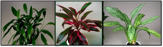 | Figure 1. Spathiphyllum wallisii (left), Cordyline terminalis (center) and Aglaonema pictum (right) |
2.2. Participants and Experimental Setting
- The participants were 28 right-handed Japanese students (14 males and 14 females) in their twenties (mean±SD 21.42±1.72 years) with normal or corrected-to-normal visual acuity and no history of neurological illness were recruited from the school of Horticulture, Chiba University in Japan. Volunteer students included both undergraduate and graduate, each volunteer was asked to sign a liability consent form, and was given a brief outline of the procedure of the experiment without identifying which plant colors would be included. This study was conducted in accordance with the ethics regulations of Chiba University. The participants were tested individually in a shield room at Chiba University, where the participants would be exposed to minimal external influences and go through the test under the same conditions. In the middle of a shielded room (59.4 m2) with a white walls and indoor lighting consisting of fluorescent light bulbs (700±4 lux), a chair was set up for the subject 150 cm away from the tested plants (Fig. 2). The psycho-physiological recording devices were placed behind the participant to decrease the disturbance of the machines. A 23°C temperature and 55% relative humidity were maintained throughout the experiment.
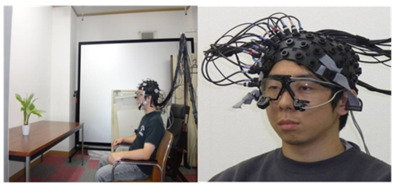 | Figure 2. Experimental setting and physiological measures, eye mark recorder and multi channel near infrared spectroscopy |
2.3. Psycho-physiological Measurements
- The study used a combination of biological measures and verbal responses. While each subject was exposed to the visual stimuli, eye-tracking information and cerebral activity were recorded, after taking the biological measurements, verbal evaluation was used to determine which plant the subject preferred and which one he did not. Eye movement, reflecting conscious and unconscious reactions to visual stimuli, was tracked to investigate cognitive characteristics of the plant color. Eye tracking and eye fixation were recorded using an eye mark recorder with the cornea and pupillary reflex method (EMR-9, NAC Image Technology Co., Ltd. Japan). Portable goggles were employed to simultaneously measure both eye movement and cerebral activity (Fig. 2). As the visual information perceived by the eyes transferred to the brain, the cerebral blood flow (CBF) was recorded while the participant was viewing the plant. Changes in cerebral activity resulting from the visual stimuli were measured by multichannel near-infrared spectroscopy (NIRS, OMM-2001, Shimadzu, Co., Ltd. Japan). NIRS directly monitors regional relative changes of hemoglobin concentration in the cerebral blood flow [16]. Measurement was limited to the right brain hemisphere as the seat of activities related to emotions and image creation [17-21] Fig. 3. A total of 47 measurement locations, referred to as channels (e.g., ch1, ch25, ch47), were located in the frontal, parietal, temporal, and occipital lobes. These brain locations correspond with feeling, judgment, premotor, motor, somatosensory, cognition, visual, auditory, and memory functions and indicate how the fragrance influenced subjects’ memory and emotions [19,22] (fig.3 A and B).
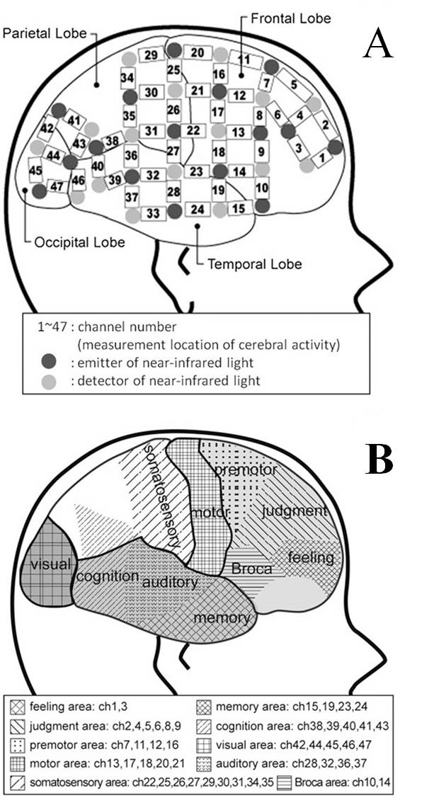 | Figure 3. Measurement of cerebral activity using multichannel near-infrared spectroscopy (NIRS). (A) The cerebral activity was detected in a total of 47 areas in cerebral cortex using electrodes (emitter and detector) of near-infrared light. The channel is the area between emitter and detector and indicates the location of cerebral activity. The 47 channels were located in the frontal, parietal, temporal, and occipital lobes. (B) According to the theory of localization of the brain function, 47 channels corresponded with feeling (ch1, 3), judgment (ch2, 4, 5, 6, 8, 9), premotor (ch7, 11, 12, 16), motor (ch13, 17, 18, 20, 21), somatosensory (ch22, 25, 26, 27, 29, 30, 31, 34, 35),memory (ch15, 19, 23, 24, 33), cognition (ch38, 39, 40, 41, 43), visual (ch42, 44, 45, 46, 47), auditory (ch28, 32, 36, 37), and speech (Broca, ch10, 14) functions |
2.4. Experimental Procedure
- Figure 4 describes the overall experimental protocol, which took place over a 30 min period. The participants were first informed about the procedure and how the instruments will be used, while helping them to adapt to the experimental environment. They were also instructed to turn off mobile phones and to focus on the tested object. An informed consent was signed, stating they could withdraw from the experiment anytime. During each trial, the near infrared spectroscopy (NIRS) electrodes were attached to the right-hemisphere brain regions of the participant as well as, eye movement detector was installed. The measuring conditions of cerebral activities were checked, and calibration for eye movement was carried out. The participant was then instructed to relax fully during a rest period 1 to 2 min with eyes closed to adjust his mood to the experimental environment, and while the participant rested with closed eyes, the first plant category was placed on a table covered with a black cloth at the participant’s eye level to ensure a straight line of the vision without having to move the head. After the NIRS monitor confirmed that cerebral activity was stable, the participant was asked to open his eyes. The brain activity and eye movement were recorded for 1 min. Then the participant was asked to close his eyes, the instruments stopped recording eye movement and brain activity. Then the second plant category takes place, the sequence being repeated with each color. The order in which the plants were presented was random. After completing the psycho-physiological responses, each participant was asked to indicate whether or not they prefer the particular plants.
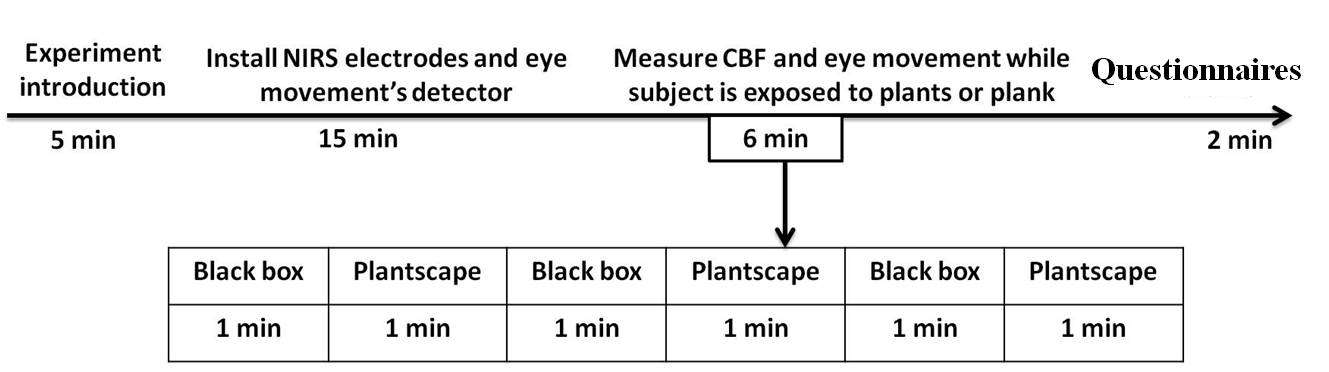 | Figure 4. Timeline (in minutes) of the physiological and psychological measurements during exposure to different plant color variations |
2.5. Statistical Analysis
- Eye fixation numbers (the visual points fixed for 0.2 sec or longer on the visual stimulus is based on the fact that more than 0.2 sec is required to consciously recognize the stimuli) [23] and eye fixation durations 1 min after exposure to the visual stimuli were analyzed by EMR-dFactory ver. 2.0. Steel-Dwass multiple comparison tests were used to statistically compare among the tested plants. Regarding CBF, the data were analyzed separately for each channel (ch). The cerebral changes during exposure to each plant category were analyzed by comparing the means of each 30-sec interval starting with the last 30 sec of the rest period before plants presentation. This was assumed to represent the most stable states of the brain and physiological activity during the rest period. A paired t-test (two-sided) was used to compare the physiological changes between rest and exposure periods. The cerebral activity analysis used fluctuations in oxygenated hemoglobin (oxy-Hb) as the index of cerebral changes, where increased oxy-Hb is associated with increased cerebral activity. The changes were separately computed in 47 measurement locations of the brain (Fig 4).
3. Results and Discussions
3.1. Effects of Plant Colors Stimuli on Eye Movements
- As can be seen from table 1 and figs 5 and 6, eye fixation durations and numbers were affected by plant colors. Regarding male participants, there was a significant difference between green and green-red plants (P=0.028), there appears to be a trend that green plant resulted in a longer duration of eye movement, compared with the green-red plant. On the other hand, regarding eye fixation numbers, it was found that there are significant differences between green-red and green plants (P=0.049), as well as between green-red and green-white plants (P=0.091). The results indicate that male participants carefully observed the details of green plant, because of long duration of eye movement. Moreover they appeared to be calm in the presence of the green plant, evidently by lower numbers of eye fixation. While, the green-red plant was exciting and stimulating for the participants eye movements evidently by higher fixation numbers.
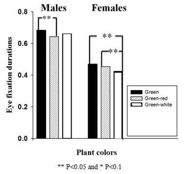 | Figure 5. Comparison of eye fixation durations among S. wallisii (left), C. terminalis (center) and A. pictum (right) for male and female participants |
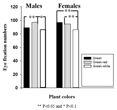 | Figure 6. Comparison of eye fixation numbers among S. wallisii (left), C. terminalis (center) and A. pictum (right) for male and female participants |
3.2. Color Preference
- As expected, people expressed a range of preferences among the presented plantscape colors. A variety of different comments were made by the participants. In some cases the reason given as to why someone preferred a plant was the same reason another person gave for disliking it, and vice versa. Overall, figure 7 clearly indicates that, so far as conscious impressions of participants were concerned, the most favorite color for both genders was the green color. In contrast, the green-red was less appealing for both male and female participants, while the green-white resulted in a rather muted and neutral reaction.
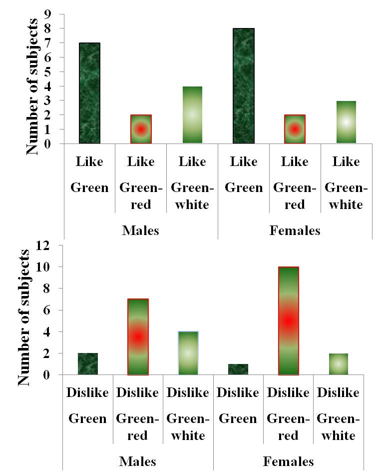 | Figure 7. Subjective responses to the double-sided questionnaire for color preference, which were evaluated on the basis of like and dislike colors in 28 participants (14 males and 14 females) |
3.3. Effect of Plant Colors Stimuli on Brain Activity
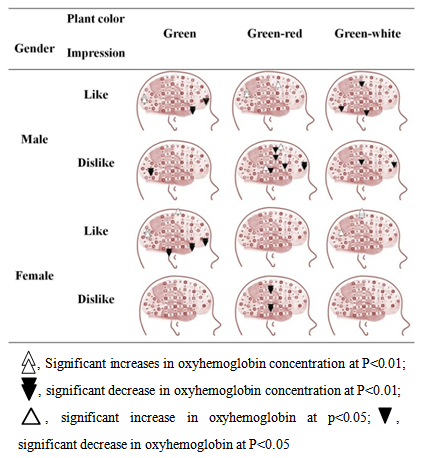
- Figure 8 demonstrates fluctuation in brain activity, and shows the channels where significant fluctuations in oxy-Hb were observed when the participants were viewing the tested plant.
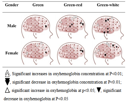 | Figure 8. Changes to cerebral activities by vision of each plant between male and female subjects |
3.3.1. Male Participants
- While male participants were viewing the green plant, the CBF was significantly sedated in the feeling area (ch3, P<0.01) which is especially important for stimulating the feeling of relaxation. On the other hand, during the stimulation with the green-red plants, the CBF was significantly sedated in the judgment (ch2 and ch4, P<0.01), which, controls concentration and attention, as well as, in the somatosensory area (ch27, P<0.05). In contrast, the CBF was notably increased in the motor area (ch21 P<0.05), which controls muscles, premotor area (ch11 and ch16, P<0.01), which is especially important in the conditional motor tasks and controls motor and movement skills, and in the cognition area (ch41, P<0.05) which controls object recognition. In contrast, the stimulation with the green-white plants showed sedation in brain areas including the motor (ch20, P<0.01), somatosensory (ch25, P<0. 01) and memory areas (ch24 and 33 P<0.01), while the CBF was activated in the cognition area (ch43, P<0.01) which involves specifically perception of color and vision motion.
3.3.2. Female Participants
- Female participants showed some different patterns of cerebral response while viewing the same plant colors compare to males. When female participants were viewing the green plant, the CBF was activated in the judgment (ch8 and 9 P <0.05), premotor (ch11, P <0.01), motor (ch21, P <0.01) and visual areas (ch46 P <0.01). On the other hand, the CBF was sedated in the feeling area (ch3 P <0.01). While, in the presence of the green-red plants, the CBF was sedated in the judgment area (ch2 and 4 P <0.01 and 0.05 respectively), but, it was activated in the motor area (ch17 P <0.05). When the participants were shown the green-white plants, CBF was activated in the motor (ch20 and 21, P<0.05 and 0.01 respectively), somatosensory (ch25 P <0. 01), visual (ch42 P <0.05) and cognition areas (ch43 P <0. 01). While, it was sedated in the judgment area (ch5 and 6 P<0.05). For both genders, nevertheless, green-red plants were effective in promoting a sense of strength, as evidenced by the cerebral activation in the motor area which controls muscles. These results are in accord with those of Kuller [26], who found that red color put the brain into a more excited state. In contrast, it was clear that CBF was significantly decreased in the judgment area when the green-red plant was viewed. These results suggest that a decrease in the concentration and creativity were associated with the lower activity in this part of the brain, which, controls attention and interest. In words, the participants were less concentrated in the presence of green-red plant. In addition, it was clearly observed that the brain activity was sedated in the somatosensory area, the results indicating that the green-red plant could result in diminishing the sensation of the body; it means that participants did not pay attention for the vision of this plant color [27].In general, the results showed that people prefer green to green-red and green-white plants. Furthermore, people exhibit different responses to plantscape colors. The outcome is of practical applicability for selecting subjective favorite colors in conjunction with the interior and living environments. Plant colors can be used to help people release stress and improve emotional status. Green plant could be incorporated into parks or hospitals in order to create relaxing environment. While, green- red plants, which stimulated the motor area in the brain which controls muscles, can be used in office environment to improve employees’ productivity and in children area.Relationship between color preference and brain activities Figs 9, 10 and 11 demonstrate brain activities in the presence of favorite and unfavorite color. As mentioned above, each participant was asked to identify which plant color he like and which one he dislike, here we try to quantify the relationship between brain activity and color preference. In words, the brain activation pattern association with favorite and unfavorite color. The participants were divided into two groups, the first group was the participants who preferred the plant color and the second group was who did not prefer it, and then data were analyzed and compared to find out the significant differences.
 | Figure 9. Cerebral blood flow activation resulting from the two-sample t-test of favorite and unfavorite colors for male and female subjects |
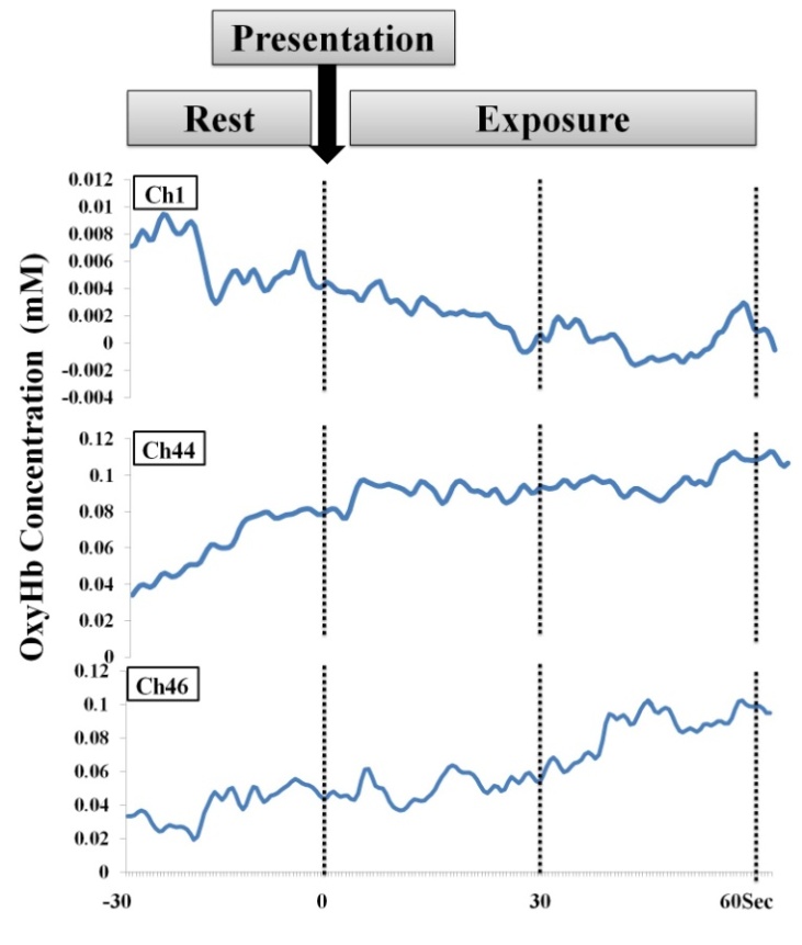 | Figure 10. Time-series changes in oxygenated hemoglobin (oxyHb) concentration during exposure to favorable stimuli |
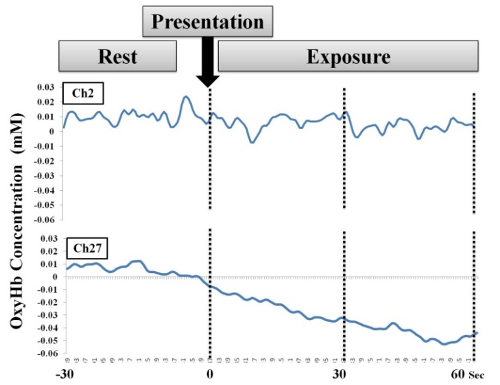 | Figure 11. Time-series changes in oxygenated hemoglobin (oxyHb) concentration during exposure to non- favorable stimuli |
3.3.3. Male participants
- When the participants who preferred the green plant were viewing it, the CBF significantly increased in the occipital lobe, which is especially involved in visual and perceptual functions (Ch43,44,46), as found by Simonov [28]. Moreover, the results showed that the brain activity decreased in the prefrontal lobe of the brain, represented as which constitutes feeling area (Ch1) (Fig. 10). These findings suggest that the experience of relaxation was associated with the sedation at this area of the brain which controls the emotion. In contrast, when the participants who did not prefer the green plant were viewing it, it was directly associated with CBF sedation in the visual area. In the case of the participants who preferred the green-red plant, the CBF was significantly increased in the visual area, which stimulates the visual function. On the other hand, for the group who did not prefer it, CBF was significantly decreased in the judgment area (Ch2). These results suggest that a decrease in the concentration and the creativity were associated with the lower activity in this part of the brain, which controls attention and interest. In addition, it was clearly observed that CBF was sedated in the somatosensory area (Ch27), indicating that the green-red plant could result in diminishing the sensation of the body; it means that participants did not care for the vision of this plant color (Fig 11).Concerning the participants who preferred the green-white plant, CBF was sedated in the visual and somatosensory areas, while, for the group who did not prefer the green-white plant, the CBF significantly decreased in the judgment and somatosensory areas. These findings suggest that viewing the green-white plant decreased the concentration, and diminished the sensation of the skin and visual function.
3.3.4. Female participants
- Responses of the female participants who preferred the green plant the results were similar to males’ responses. The cerebral response indicates that the green plant provides a relaxing effect through the sedation of the feeling area and stimulated visual function (fig 10).In the case of the participants who did not prefer the green-red plant, CBF was significantly decreased in the somatosensory area. This cerebral response indicates that the green-red plant diminishes the skin and body sensation.While, the participants who preferred the green-white plant were viewed it, the CBF indicates that the plant led to increase sensation of the skin and body, because of the significant increment of CBF in the somatosensory area, furthermore, it stimulated the visual function evidently by the significant increment of cerebral activity in the vision area.The present study has shown the possibility of quantifying the human psycho-physiological responses to plant variegation based on eye movement and brain activity. The particular outcomes in this study support the findings of previous studies and confirm that plant color variations can be used to stimulate different responses as well as, plants might well be able to stimulate desire responses such as promoting calm and relaxation. Based on the results of eye movement, color preference and brain activity derived from the experience of viewing plants with three different foliage colors. It can be concluded that green plants can be used to promote a feeling of relaxation and stimulate pleasant and high elated emotion. The results of this study are in harmony with those of a previous study that showed that green plants are useful for creating a comfortable environment [9, 29, 30]. Even more relevant, was the observation that when the participants were viewing the green and green-red plants, there was a strong association that those colors received longer duration and higher numbers of eye fixation (Figs 5 and 6). It has concluded that the CBF increment was correlated with the degree of attention settled by the participants to the visual [31]. Furthermore, it has been reported that the occipital visual cortex is activated by visually evocative stimuli, whereas the activation in this area is independent of emotional state [32, 33]. In the case of participants who preferred a particular color, it was clear that the CBF significantly increased in the visual area, while, it was significantly decreased in the case of the participants who did not. A study by Hoshi [34], distinguished pleasant and unpleasant human emotions based on cerebral blood flow. The study showed that unpleasant emotion was followed by increases in oxy-Hb at the right prefrontal cortex, while a pleasant and high elated emotion was accompanied by a decrease in oxy-Hb. This results are in full agreement with those of the current study, our results showed that the participants who preferred the green plant color had a significantly decrease of cerebral blood flow in the feeling area (Ch1). This indicates that the participants felt pleasure when they were viewed the green plant. On contrary, the CBF decreased in the judgment and somatosensory areas, when the participants did not prefer a particular color, which indicates that they were showing less concentration and attention. There are a similarity between the attitude expressed by the participants in our study and the study described by Suzuki [35] who examined brain activity during semantic differential rating of drawing stimuli containing different affective polarities. They reported that oxy-Hb concentration was lower around the right superior temporal and partial regions and especially in the somatosensory and auditory areas when subjects were viewing a noisy or temperamental picture. Recent studies have demonstrated that the brain is predominantly activated by unfavorite colors over favorite colors [36]. Although, positive and negative emotions appear to be associated with different styles of processing information, it is apparent that, in this study, the brain activation patterns in response to visual stimulation with favorite and unfavorite colors were different from each other. These results support other studies that suggested that the brain has a limited capacity for attention, therefore attention to one cue will decrease the brain’s focus on other environment cues. In words, Eye movement and brain activity can be used as indicators to detect human psycho-physiological responses. Different results in eye movement indicate that color variations make some view interesting and others not. As well as, brain activity reflects the human emotional status. Furthermore, increasing cerebral blood flow at the occipital lobe was correlated with the degree of attention to the visual stimuli. Variegated plants might well be able to stimulate desire responses such as promoting calming, pleasure and relaxation. Limitation of the study. To reduce confounding variables, this experiment controlled the age, and nationality of participants as well as environmental conditions such as noise, temperature, and humidity [37, 38]. Accordingly, the effect found in this study might not be generalizable to certain populations or different environment conditions. Further studies are needed to test outcome with a wider range of plant colors, participants and methodology. As well as, the findings of this study have a number of important implications for future practice. More broadly, research can shed light on the discussed issues by determining the psychological and physiological responses of participants of other culture using the same foliage colours to show how ethnic differences vary in the relationship between psycho-physiological responses and colors.
4. Conclusions
- The results provide important scientific evidence of the health benefits of the presence of indoor variegated plants. We suggest that each color is recommended for a specific situation. The green plant is useful for creating a comfortable environment. Moreover, this study has a potential understanding of the different neural mechanism on the subjective emotion state in viewing the favorite and unfavorite colors. The brain activation in response to visual stimuli with favorite and unfavorite colors was different from each others. In conclusion, carful choices of plant colors should be made during decorating the indoor environment. Green plant could be incorporated into parks or hospitals in order to provide relaxing environment. Different color variations can be used according to different environmental demands to create healthy and livable indoor environments.
ACKNOWLEDGMENTS
- The authors wish to express a deep gratitude to all the students who agreed to participate in the survey as volunteers. This research was funded with a grant from the Egyptian Ministry of Higher Education.
References
| [1] | C. Marcus and B. Marni, “Healing gardens: Therapeutic benefits and design recommendations”, John Wiley & Sons. New York; (1999). |
| [2] | P.D. Relf and V.I. Lohr, “Human issues in horticulture”, Hort Science. 38, 984 (2003). |
| [3] | T. Fjeld, B. Veiersted, L. Sandvik, et al., “The effect of indoor foliage plants on health and discomfort symptoms among office workers”, Indoor and Built Environment. 7(4), 204 (2002). |
| [4] | B. Butterfield and D. Relf, “National survey of attitudes towards plants and gardening. The role of horticulture in human well-being and social development”, A national symposium (proceedings). Timer press, Portland, OR, p. 211-212 (1992). |
| [5] | P. Grahn, and U.A. Stigsdotter, “Landscape planting and stress”, Urban forestry urban greening. 2, 1(2003). |
| [6] | S., Simson and M. C. Straus. Horticulture as theraby: Principale and practice. The Haworth Press, New York, NY, (1998). |
| [7] | A.C. Hurlbert and Y. Ling, “Biological components of sex differences in color preferences”, Current Biology. 17, 623 (2007). |
| [8] | M. Saito, “Comparative studies on color preference in Japan and other Asian regions, with special emphasis on the preference for white”, Color Research and Application 21(1),35 (1996). |
| [9] | X. Li, Z. Zhang, M. Gu, D-Y. Jiang, Q-X. Zhang, and H-T. Pan, “Effects of plantscape colors on psycho-physiological responses of university students”, J. Food Agri. Environ. 10,702 (2012). |
| [10] | JE. Song, “Effects of interior plantscape in office on psycho-physiological improvement and stress alleviation of indoor workers”, Konkuk University, Seoul, South Korea, PhD thesis.(2004). |
| [11] | K. Arai and K. Hasegawa, “Method for psychological status monitoring with Line of sight vector changes (Human Eye Movements) detected with wearing glass International”, Journal of Advanced Research in Artificial Intelligence. 2(6),65 (2013). |
| [12] | W. J. Thomas and G. Isabel, “Brain areas engaged during visual judgments by involuntary access to novel semantic information”, Vision Research 44, 429 (2004). |
| [13] | M. Davis and PJ. Whalen, “The amygdala: Vigilance and emotion”, Mol Psychiatry 6, 13 (2001). |
| [14] | L. Pessoa, S. Kastner and LG. Ungerleider, “Attentional control of the processing of neural and emotional stimuli”, Brain Res Cogn Brain Res. 15, 31 (2002). |
| [15] | KL. Phan, T. Wager, SF. Taylor and I. Liberzon, “Functional neuroanatomy of emotion: a meta-analysis of emotion activation studies in PET and fMRI”, Neuroimge. 16, 331 (2002). |
| [16] | A.Villringer, and U. Dirnafl. Coupling of brain activity and cerebral blood flow: Basis of functional neuroimaging. Cerebrovasc. Brain Metab. Rev. 7:240–276 (1995). |
| [17] | E.K. Silberman, and H. Weingartner, “Hemispheric lateralization of functions related to emotion”, Brain Cogn. 5, 322 (1986). |
| [18] | D.M. Tucker, “Lateral brain functions, emotion, and conceptualization”, Psychol. Bull. 89,19 (1981). |
| [19] | Jo, H., Rodiek, S., Fujii, E., Miyazaki, Y., Park, B-J. & Ann S-W. Physiological and psychological response to floral scent. HortScience, 48(1), 1-7 (2013). |
| [20] | Bach, D.R., et al. Altered lateralisation of emotional prosody processing in schizophrenia. Schizophrenia Research. 110: 180–187, (2009). |
| [21] | Alfano, K.M., and C.R. Cimino. Alteration of expected hemispheric asymmetries: Valence and arousal effects in neuropsychological models of emotion. Brain and Cognition. 66: 213–220, (2008). |
| [22] | L.R. Caplan, “Brain-stem localization and function”, Springer New York (1993). |
| [23] | A.L. Yarbus, “Eye Movements and Vision”, New York: Plenum (Originally published in Russian) (1962). |
| [24] | N.H. Mackworth, and A.J. Morandi, “The gaze selects informative details within pictures”, Perception & Psychophysics. 7,173 (1967). |
| [25] | M.A. Baker and M. Loeb, “Implications of measurement of eye fixations for a psychophysics of form perception”, Perception & Psychophysics. 13,185 (1973). |
| [26] | R. Kuller, B. Mikellides and J. Janssens, “Color, arousaland performance-A comparison of three experiments”, Col. Res. Appl. 34,141 (2009). |
| [27] | M.T. FitzGerald, “Neuroanatomy: Basic and Clinical”, London, W.B. Saunders (1996). |
| [28] | PV. Simonov, “Neurobiological basis of creativity”, Neurosci Behav Physiol. 27,585 (1997). |
| [29] | C-Y. Chang, and P-K. Chen, “Human response to window views and indoor plants in the work place”, HortScience. 40, 1354 (2005). |
| [30] | BK. Kim, “Study regarding visual preference anger analysis of an indoor landscape architecture plant”, Kyunghee Univ. Suwon. South Korea, PhD. thesis (1997). |
| [31] | S C. Bondy, R. A. Lehman, and J. L. Purdy, “Visual attention affects brain blood flow”, Nature. 248,440 (1974). |
| [32] | KL. Phan, T. Wager, SF. Taylor, et al. “Functional neuroanatomy of emotion: a meta-analysis of emotion activation studies in PET and fMRI”, Neuroimge. 16, 331(2002). |
| [33] | EM. Reiman, R. Lane, GL. Aher, et al. “Neuroanatomy correlates of externally and internally generated human emotion”, Am J Psychiatry. 154, 918 (1997). |
| [34] | Y. Hoshi, N. Kobyashi, and M. Tamura, “Interpretation of near-infrared spectroscopy signals, a study with a newly developed perfuse rat brain model”, Journal of Applied Physiology. 90(5),1657 (2001). |
| [35] | M. Suzuki, J. Gyoba, and Y. Sakuta, “Multichannel NIRS analysis of brain activity during semantic differential rating of drawing stimuli containing different effective polarities”, Neurosci. Lett. 375(1), 53 (2005). |
| [36] | T.H. Kim, J.K. Song, and G.W. Jeong, “Neural Responses to the human color preference for assessment of eco-friendliness: a functional magnetic resonance imaging study”, Int. J. Environ. Res. 6(4),953(2012). |
| [37] | Bocca, E. & Battiston, MN. (1964). Odour Perception and Environment Conditions. Acta Oto-Laryngologica 57: 391-400. |
| [38] | Brand, G & Millot, J. (2001). Sex differences in human olfaction: Between evidence and enigma. J. Exp. Psychol 54:259–270. |