
-
Paper Information
- Paper Submission
-
Journal Information
- About This Journal
- Editorial Board
- Current Issue
- Archive
- Author Guidelines
- Contact Us
International Journal of Materials and Chemistry
p-ISSN: 2166-5346 e-ISSN: 2166-5354
2026; 16(5): 103-115
doi:10.5923/j.ijmc.20261605.03
Received: Aug. 24; Accepted: Oct. 15, 2026; Published: Oct. 17, 2026

Synthesis and Structural and Microstructural Characterization of Chromium(III) Benzoate Coordination Polymer
 Abstract
 Reference
 Full-Text PDF
 Full-text HTML
Ferland Ngoro-Elenga1, 2, Hubert Makomo3, Narcisse Nicaise Obaya3, Grâce Jeanci Viboudoulou1, Ibrahima Elhadji Thiam4, Timothée Nsongo2
1Laboratory of Inorganic Chemistry and Applied, Marien Ngouabi University, Brazzaville, Congo
2Geological and Mining Research Centre (CRGM), Ministry of Mining Industries and Geology, Brazzaville, Congo
3Team of Organic and Analytical Chemistry of Bioactive Substances, Marien Ngouabi University, Brazaaville, Congo
4Laboratory of Organic Coordination Chemistry, Cheikh Anta Diop University, Dakar, Sénégal
Correspondence to: Ferland Ngoro-Elenga, Laboratory of Inorganic Chemistry and Applied, Marien Ngouabi University, Brazzaville, Congo.
| Email: |  |
Copyright © 2026 The Author(s). Published by Scientific & Academic Publishing.
This work is licensed under the Creative Commons Attribution International License (CC BY).
http://creativecommons.org/licenses/by/4.0/

A chromium (III) benzoate coordination polymer was synthesized and characterized to elucidate its structural and microstructural properties. The crystal structure, chemical functionalities, morphology, and elemental composition were investigated using powder X-ray diffraction (PXRD), Fourier-Transform infrared (FTIR) spectroscopy, scanning electron microscopy coupled with energy-dispersive X-ray spectroscopy (SEM-EDS), and X-ray fluorescence (XRF) spectroscopy. The PXRD pattern reveals a highly crystalline phase with numerous reflections, particularly in the low-angle region, indicating a well-organized crystal structure with relatively large interplanar spacings. The absence of a reference phase indicates that the obtained material may correspond to a crystalline Cr(III) benzoate phase for which no corresponding crystallographic structure has been idnetified in the database consulted. FTIR analysis confirms the deprotonation of the lignad carboxylic groups and their involvement in coordination with the Cr(III) centers. SEM observations reveal predominantly prismatic crystal morphologies, with particle sizes ranging from 2.26 to 17.21 µm (mean size: 8.15 µm), while EDS and XRF analysis confirm the elemental composition of the material. The microstructural parameters were evaluated using the Scherrer, Monshi-Scherrer, Williamson-Hall (UDM), Size-Strain Plot (SSP), and Halder-Wagner methods. The apparent coherent domain sizes obtained ranged from 49.17 to 550.99 nm, depending on the approach employed, highlighting the influence of the underlying assumptions associated with each diffraction profile analysis method. The SSP and Halder-Wagner methods provides provided the best statistical fits (R2 = 0.823) and yielded consistent estimates of the coherent domain size (49.17 nm), together with low microstrain values (2.05×10⁻³ and 3.33×10⁻³, respectively). Overall, the structural and microstructural results confirm the formation of a crystalline Cr(III)-benzoate material and indicate that the SSP and Halder-Wagner approaches provide the most consistent estimates of the microstructural parameters for this system.
Keywords: Chromium(III) benzoate coordination polymer, Benzoic acid, Powder X-ray diffraction, Diffraction peak broadening analysis, Microstructural parameters
Cite this paper: Ferland Ngoro-Elenga, Hubert Makomo, Narcisse Nicaise Obaya, Grâce Jeanci Viboudoulou, Ibrahima Elhadji Thiam, Timothée Nsongo, Synthesis and Structural and Microstructural Characterization of Chromium(III) Benzoate Coordination Polymer, International Journal of Materials and Chemistry, Vol. 16 No. 5, 2026, pp. 103-115. doi: 10.5923/j.ijmc.20261605.03.
Article Outline
1. Introduction
- Coordination polymers are currently among the most extensively studies classes of crystalline materials in coordination chemistry and materials science, owing to their remarkable structural diversity and outstanding functional properties. The diversity of coordination geometries of metal centers, combined with the wide variety of available ligands, gives rise to a remarkable range of structural architectures, which can extend from one dimensional (1D) chains to two dimensional (2D) layers and three dimensional (3D) frameworks. This modularity endows coordination polymers with a broad range of physicochemical properties, making them particularly attractive for numerous applications, including adsorption, catalysis, molecular separation, chemical sensing, gas storage, molecular magnetism, and materials science [1-6]. Among the transition metals used in the design of these materials, chromium(III) is of particular interest owing to its d3 electronic configuration, high thermodynamic stability, and strong tendency to from octahedral complexes. Cr(III)-based coordination polymers are generally distinguished by their excellent chemical and thermal stability, as well as their electronic and magnetic properties, making them promising candidates for the development of functional materials [7,8]. Carboxylate ligands occupy a prominent position in this field of coordination chemistry because they exhibit multiple coordination modes, including monodentate, chelating, and bridging modes, thereby enabling the construction of extended networks diverse geometries [9,10]. In particular, the benzoate ion is a simple aromatic ligand capable of bridging multiple metal centers and promoting the formation of crystalline coordination networks [11,12]. The rational design of such materials relies not only on the determination of their crystal structure but also on a thorough understanding of their microstructural organization, as both aspects directly govern their physicochemical properties. Powder X-ray diffraction (PXRD) remains the reference technique for identifying crystalline phases and assessing long-range structural order. However, when the crystallographic structure is unavailable or when no suitable single crystal can be obtained, the interpretation of diffraction patterns should be complemented by spectroscopic and microscopic techniques to establish the nature of the synthesized material [4,13]. In this context, FTIR spectroscopy provides essential information on the coordination modes of the ligands, whereas SEM-EDS and XRF analyses enable the characterization of the solid in terms of its morphology and elemental composition, respectively. Beyond structural identification, diffraction peak broadening contains valuable information on the microstructure of crystalline materials. The sizes of coherently diffracting crystallite domains, microstarin, and crystal defect density directly influence numerous material properties, including stability, reactivity, and functional performance. Several approaches based on diffraction peak-broadening analysis have been de developed to estimate these parameters, includind the Scherrer, Monshi-Scherrer, Williamson-Hall, Size-STrain Plot, and Halder-Wagner methods. However, the reliable assessment of these microstructural parameters remains remains challenging because the different analytical methods are based on distinct physical assumptions and may therefore yield divergent results. Despite their widespread application to oxides, nanoparticles, and ceramic materials, comparative studies addressing their applicability to coordination polymers remain relatively scarce. To the best of our knowledge, a comprehensive literature search revealed no reported crystal structure describing a coordination polymer composed exclusively of Cr(III) centers interconnected by benzoate ligands. The absence of a crystallographic reference structure is therefore of particular significance for the characterization of the material obtained and supports the use of a multi-technique analytical approach combining several complementary methods [11]. Beyond the synthesis of the Cr(III)-benzoate coordination polymer, this study also aims to provide a critical assessment of the principal diffraction peak-broadening analysis methods in order to identify those that offer the most representative description of its crystalline microstrure.
2. Experimental
2.1. Hydrothermal Synthesis of the Cr(III)-Benzoate Coordination Polymer
- Benzoic acid (1.50 g) was dissolved in 30 mL of distilled water. In parallel, chromium(III) nitrate nonahydrate (1.56 g) was dissolved in 30 mL of distilled water. The resulting aqueous solution of chromium(III) nitrate nonahydrate was then added to the benzoic acid solution under continuous stirring for 30 min at room temperature. The resulting susspension was transferred to a stainless-steel autoclave equipped with a polytetrafluoroethylene (PTFE) inner liner and heated at 120 °C in an oven for 4 days. After the hydrothermal treatment, the autoclave was allowed to cool naturally to room temperature. The resulting solid was recovered by filtration and washed with distilled water. The material was subsequently dried at 100 °C for 30 min. The resulting green powder was collected and stored in a glass vial.
2.2. Structural Characterization Method for the Synthesized Powder
- The synthesized product was characterized using several complementary techniques. Powder X-ray diffraction (PXRD) measurements were performed using an X’Pert3 Panalytical powder diffractometer with CuKα radiation, with wavelengths of λKα1 = 1.54059 Å and λKα2 = 1.54441 Å. The diffraction data were collected over the 2θ rage of 5 - 50°, with a step size of Δ2θ = 0.02°, at an accelating voltage of 45 kV and a current of 100 mA. A systemetic search for crystallographic structures of Cr(III) benzoate compounds was conducted in the CCDC and COD databases using the keywords ‘’chromium(III) benzoate’’, ‘’chromium benzoate’’, ‘’Cr(III) benzoate’’, ‘’chromium(III) benzoate complex’’, ‘’chromium(III) benzoate coordination compound’’, ‘’chromium(III) benzoate coordination polymer’’ and ‘’chromium(III) benzoate crystal structure’’, supplemented by searches based on the chemical formulas Cr(C6H5COO)3 et Cr(O2CPh)3. Fourier-transform infrared spectroscopy was performed using a Thermo Scientific Nicolet iS50 FTIR spectrometer over wavenumber range of 500 – 4000 cm-1. The morphology and elemental composition of the synthesized material were investigated by scanning electron microscopy coupled with energy-dispersive X-ray spectroscopy (SEM-EDS). The analyses were carried out using an SU8100 Regulus scanning electron microscope equiped with an energy-dispersive X-ray spectroscopy detector. SEM observations were performed at an accelerating voltage of 2 kV, using magnifications of ×5000, ×2000, ×1000, and ×500, with a working distance of 7.8 mm. Elemental analysis by EDS was conducted at a magnification of ×10000, with an acquisition time of 7.68 µs, a real analysis time of 47.4 s, and an energy resolution of 123.8 eV. The elemental composition was further determined using a Thermo Fisher Scientific X-ray fluorescence (XRF) spectrometer. The coherent diifraction domain size and lattice microstrain were estimated using the Scherrer, Monshi-Scherrer, Williamson-Hall, Halder-Wagner, and Size-Strain Plot methods. Only the reflections attributed to the coordination complex were considered for the determination of the coherent crystallite domain size and microstrain. The instrumental broadening of the XRD peaks was determined using a crystalline silicon standard, for which an instrumental FWHM, βinstrumental, of 0.071° was measured for the (1 1 1) reflection at 2θ = 28.4°. The instrumental contribution was corrected using a Gaussian deconvolution procedure according to
 , where βobs is the observed FWHM . The profiles of the individual XRD reflections were fitted in OriginPro 2024 software using a Voigt function to determine the peak positions and FWHM values. Only well-resolved reflections without significant overlap with neighboring peak were considered for the microstructural analysis and subsequent fitting procedures.
2.3. Microstructural Characterization Methods of the Synthesized Powder
2.3.1. Scherrer et de Monshi-Scherrer Methods
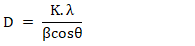
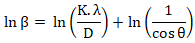
- The Scherrer method is used to estimate the average coherent crystallite domain size from XRD peak broadening. This approach assumes that peak broadening arises exclusively from the finite size of the coherent crystallite domains and does not account for the contribuation of crystal lattice microstrain. The coherent crystallite domain size is determined using Equation (1) [14,15].
 | (1) |
 | (2) |
2.3.2. Williamson-Hall Method: Uniform Deformation Model (UDM)
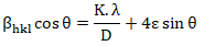
- The UDM model was employed to distinguish the contribution of the coherent crystallite domain size and microstrain (ε) to XRD peak broadening. This approach assumes that the microstrain is uniform along all crystallographic directions, allowing the total peak broadening to be resolved into a size-related contribution associated with lattice distorsion (Equation 3) [18].
 | (3) |
 | (4) |
2.3.3. Size-Strain Plot Method
- The Size-Strain Plot (SSP) method was employed to simultaneously estimate the coherent crystallite domain size and microstrain by accounting for their respective contributions to XRD peak broadening (Equation 5). This approach assumes that size-induced broadening follows a Lorentzian distribution, whereas strain-induced broadening is Gaussian in nature [15,21,22].
 | (5) |
 | (6) |
2.3.4. Halder-Wagner Method
- This method was applied to simultaneously estimate the coherent crystallite domain size and crystal lattice microstrain from X-ray diffraction peak broadening. The approach is based on a Voigt function, assuming that the diffraction profiles result from the convolution of Lorentzian and Gaussian components, which are generally attributed to the effects of coherent crystallite domain and microstrain, respectively (Equation 7) [21,24-26].
 | (7) |
 | (8) |
3. Results and Discussion
3.1. XRD Data Analysis
- The powder X-ray diffraction pattern of the synthesized solid (Figure 1) reveals a highly crystalline compound, with the main intense reflections distributed over the 5 – 30° (2θ) range. The principal reflections are located at approximately 2θ(°) = 8.08, 8.22, 8.42, 8.6, 8.88, 9.56, 10.16, 10.84, 11.7, 11.86, 15.16, 16.24, 16.52, 16.74, 17.28, 17.84, 18.46, 18.96, 19.82, 20.26, 20.54, 22.44, 22.9, 23.66, 23.8, 24.92, 25.66, 26.80, 28.00. The presence of numerous well-defined and well-resolved reflections is consistent with a highly ordered crystalline phase exhibiting long-range periodicity. This structural organization is consistent with the nature of coordination polymers, in which metal centers and organic ligands are assembled in a repetitive manner to form periodic crystalline networks [28-30]. In particular, carboxylate ligands can act as coordinating and bridging groups between metal centers, thereby promoting the propagation of structural order throughout the coordination network [28,29,31]. The reflections observed at low angles correspond, according to Bragg’s law, to relatively large interplanar spacings. They therefore indicate the presence of crystallographic planes characterized by signicant interplanar distances [32]. In the context of the coordination polymer investigated, these results are consistent with an extended structural organization involving coordination units interconnected through Cr-O-carboxylate interactions. In its worth nothing that the reflections at 2θ (°) = 8.08, 16.24, 17.28, 18.96, and 26.8° also coincide with reflections attributed to residaul benzoic acid (CCDC 2533705). The experimental diffraction pattern does not reproduce the entire diffraction pattern of benzoic acid. Several major reflections of the synthesized material occur at diffent positions or exhibit markedly altered relative intensities, particularly around 2θ(°) = 8.22, 8.42, 8.60, 8.88, 9.56, 10.16, 10.84, 11.7, 11.86, 15.16, 16.52, 16.74, 18.46, 19.82, 20.26, 20.54, 22.44, 22.90, 23.66, 23.80, 24.92, 25.66, and 28.00. These differences indicate that the crystalline structure of the synthesized material does not correspond to that of either the free ligand or chromium(III) nitrate nonahydrate, whose principal (-111) reflection occurs at 2θ = 13.40° (JCPDS card n°31-0409). These changes are consistent with deprotonation of the carboxylic group followed by its coordination to the Cr(III) centers, resulting in a new structural arrangement. A comprehensive literature search did not identify any CIF file corresponding to a coordination polymer composed exclusively of Cr(III) centers and benzoate ligands. Consequently, direct indexing of the experimental diffraction pattern could not be performed by comparison with a reference crysatl structure. The absence of a reference phase suggests that the obtained material may correspond to a crystalline Cr(III)-benzoate phase for which no corresponding crystallographic strucutre was identified in the databases consulted. The PXRD analysis therefore suggests the formation of a crystalline Cr(III)-benzoate coordination network; however, the lack of an available CIF structure necessitates an interpretation based on the overall diffraction signature of the material rather than on an unambiguous crystallographic identification.
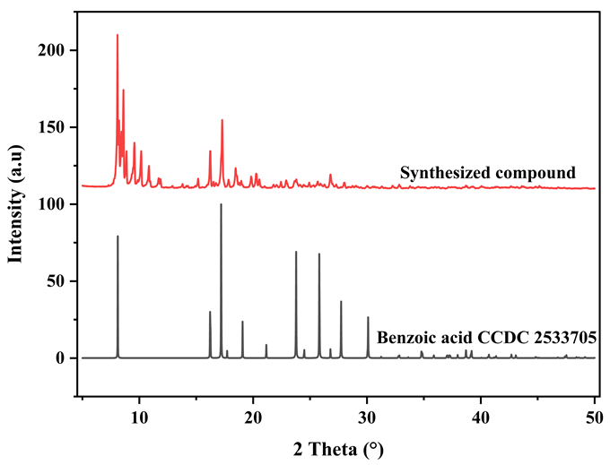 | Figure 1. XRD pattern of synthetized compound |
3.2. Fourier –Transform Infrared Spectroscopy
- The FTIR spectrum of the synthesized material (Figure 2) exhibits several characteristic bands consistent the presence of benzoate ligands coordinated to chromium(III). The band located at around 3605 cm-1 is assigned to ν(O-H) vibrations, suggesting the presence of hydroxyl groups weakly involved in hydrogen bonding within the material. The band observed at 3066 cm-1 is attributed to ν(C-H), stretching vibrations, of the aromatic hydrogens atoms of the benzene ring of the benzoate ligand. The weak band observed at 1710 cm-1 is assigned to the stretching vbration, of residual carboxylic acid groups, suggesting that a small fraction of benzoic acid may remain in the non-deprotanated or uncoordinated form. The band observed around 1610 cm-1 is primarity assigned to the stretching vibration of the aromatic C=C bonds of the benzoate ring; a contribution from the asymmetric stretching vibration of the carboxylate group, νasym(COO-), cannot be excluded because of the possible averlap of vibrational bands in this spectral region. The bands observed at 1572 and 1529 cm-1 are assigned to the asymmetric stretching vibrations, νasym(COO-), whereas those at 1410 and 1332 cm-1 correspond to the symmetric stretching vibrations, νsym(COO-), of the carboxylate groups [33,34]. The appearance of these carboxylate bands, together with the decrease in the contribution associated with the the protonated carboxylic group, is consistent with the deprotonation of benzoic acid and the formation of benzoate groups associated with the Cr(III) centers. The differences in wavenumber, Δν, are 162 and 197 cm-1, obtained from (1572 – 1410 cm-1) and (1529 – 1332 cm-1), respectively. The Δν values obtained indicate that the carboxylate groups are involved in the coordination environment of chromium. This interpretation is consistent the coordination modes reported for certain Cr(III)-carboxylate complexes. Sydora and collaborators notably demonstrated that benzoate ligands can adopt different coordination modes around Cr centers, including a bridging mode in the complex Cr2(µ-O2CPh)4(THF)2, in which the benzoate groups bridge two Cr(III) centers [34]. Likewise, Vlachos and collaborators reported, by single crystal X-ray diffraction, the trinuclear oxo-carboxylate complex Cr3O(O2CPh)6(MeOH)3(NO3).2MeOH, in which the benzoate ligands participate in the connection of the Cr(III) centers through a µ2-η1:η bridging mode [35]. Thus, these values are consistent with the presence of coordinated benzoate groups involved in an extended coordination framework. However, in the absence of direct structural determination, the Δν values alone do not allow the coordination mode (monodentate, bridging, or chelating) to be unambiguously established. The bands at 1218, 1179, 1115, 1072, 1025, 1002, 975 cm-1 are mainly assigned to vibrations of the aromatic ring and C-H deformation modes of the benzoate ligand [36]. The bands observed at 937, 817, and 713 cm-1 are attributed to the out-of-plane C-H bending vibrations, δ(C-H), of the substituted aromatic ring, with are characteristic of benzoate ligand. The bands located at 659 and 510 cm-1 are consistent with metal-oxygen stretching vibrations, ν(Cr-O), and may therefore be associated with Cr-O interaction involving the oxygen atom of the carboxylate groups. These low-wavenumber vibrational features provide additional spectroscopic support for the interaction between the Cr(III) centers and the oxygen donor atoms of the carboxylate groups.
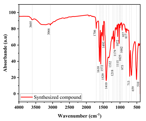 | Figure 2. FTIR spectrum of the synthesized product |
3.3. Scanning Electron Microscopy Coupled with Energy Dispersive X-Ray Spectroscoy (SEM-EDS)
- The SEM micrographs acquired at different magnifications (figure 3) show that the material consists of an assembly of micrometre-size crystalline particles exhibiting predominantly a prismatic morphology. The image acquired at ×500 highlights the overall organization of the synthesized powder, whereas the ×1000 magnification reveals faceted particles with planar surfaces and clearly defined edges. The particle size ranges from 2.26 to 17.21 µm, with an average size of 8.15 µm (Figure 4). These observations also reveal partial agglomeration of the crystals, with several particles being in direct contact with one another. The particle surfaces appear generally compact, and no clearly discernible open macroporosity is observed at the scale investigated. However, this observation does not rule out the presenec of microporosity or mesoporosity, the assessment of which requires specific techniques, such as nitogen adsorption-desorption measurements [37]. The presence of planar faces and well-defined edges is consistent with relatively ordered crystal growth and the preferential development of specific crystallographic faces [38,39]. However, crystal morphology results from the interplay of several factors associated with nucleation and growth processes, as well as the physicochemical conditions prevailing during crystallization [38,39]. In carboxylate-based coordination polymer systems, the synthesis conditions can notably affect the crystal growth kinetics, resulting in variations in particle morphology and size [40]. Therefore, the observed morphology should be regarded as the outcome of the specific synthetic conditions employed in the study [38,39,40].
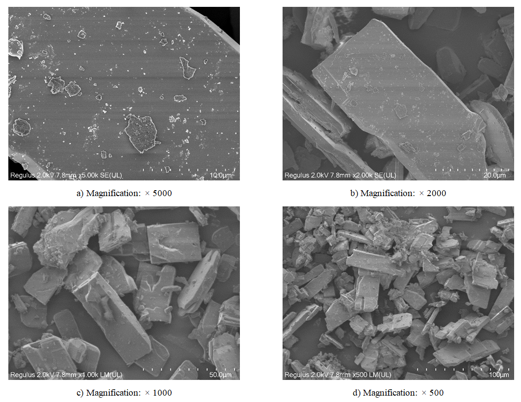 | Figure 3. SEM of synthetized compound |
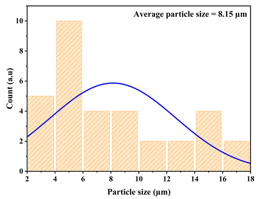 | Figure 4. Particle size of the synthesized compound |
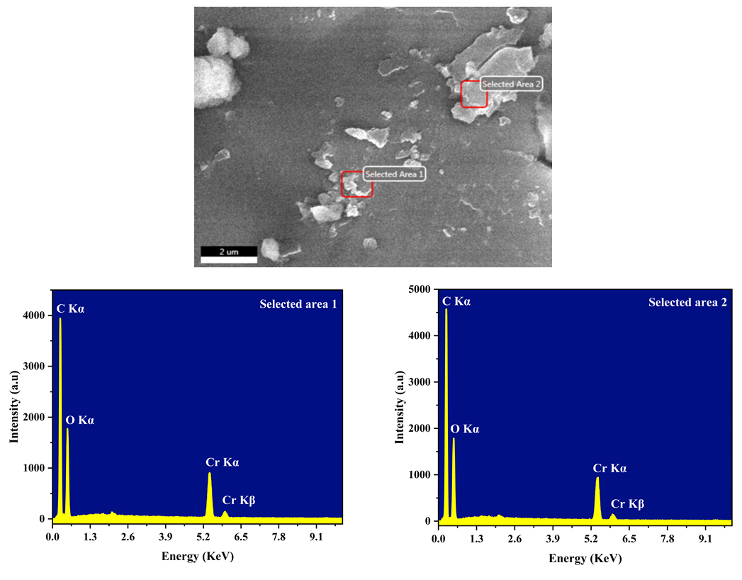 | Figure 5. SEM-EDS analysis of the synthesized compound |
3.4. X-ray Fluorescence (XRF)
- XRF analysis revealed the presence of Cr as the metallic center, whereas the elements constituting the ligand (C, O, and H) were not detected owing to the intrinsic limitations of XRF for thr analysis of low atomic number elements [41,42]. The apparent Cr conent was 97.97%. This values does not directly represent the actual chromium content of the material, as the contribution of light elements is not adequately accounted for during quantitative analysis. Accordingly, the remaining 2.03% cannot be considered representative of the actual C, O, and H content, but rather corresponds to the unsassigned fraction under the analytical conditions employed. XRF therefore provided only partial information on the composition of the synthesized material. In contrast, the presence of the organic ligand is corroborated by EDS analysis, which detected carbon and oxygen, as well as by FTIR spectroscopy, whose characteristic bands are consistent with the coordination of carboxylate groups to metal center, in agreement with the behavior reported for Cr(III) benzoate compleces [11].
3.5. Microstructural Study on the Synthesized Cr-Benzoate Coordination Polymer
- The angular positions and corrected FWHM values of the Ce-MOF diffraction peaks used to estimate the coherent crystallite domain size and lattice microstrain. are listed in Table 1.
|

3.5.1. Scherrer and Monschi-Scherrer Methods
- Figure 6 represents the coherent crystallite domain sizes determined using the Scherrer equation for the different crystallographic reflections, together with their average value. The calculated coherent crystallite domain size exhibit significant variation, ranging from 39.66 to 297.57 nm, indicating a dependence on the crystallographic direction considered. The average size, estimated at 120.23 nm, therefore provides a representative overall value for the material. In contrast, Figure 7 presents the experimental data and the corresponding linear fit obtained from the plot of lnβ versus de ln(1/cosθ) according to the Monshi-Scherrer method. This fitting procedure yielded an estimated coherent crystallite domain size of 168.54 nm.
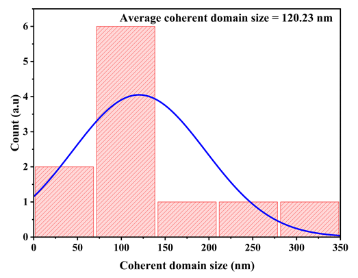 | Figure 6. Scherrer method |
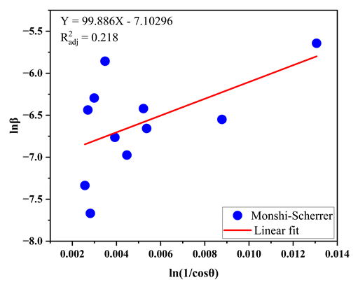 | Figure 7. Monshi-Scherrer method |
3.5.2. Williamson-Hall Method: Uniform Deformation Model (UDM)
- Figure 8 presents the plot of βcosθ versus 4sinθ according to the unifprm deformation model (UDM) of Williamson-Hall. The experimental data were fitted by linear regression, with the intercept providing the coherent crystallite domain size and slope yielding the microstrain of the material. The resulting values were 550.99 nm for the coherent crystallite domain size and 4.73×10⁻³ for the microstrain.
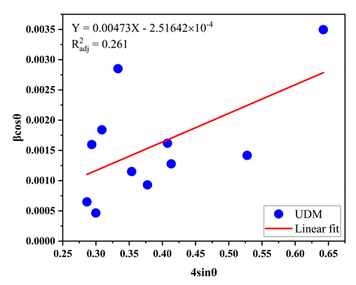 | Figure 8. UDM |
3.5.3. Size-Strain Plot Method
- Figure 9 presents the experimental data and the corresponding linear fit obtained using the SSP method, based on the plot of
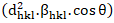 versus
 . The linear fitting yielded an estimated coherent crystallite domain size of 49.17 nm and a microstrain of 2.053×10⁻³.
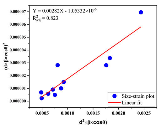 | Figure 9. Size-strain Plot method |
3.5.4. Halder-Wagner Method
- Figure 10 presents the Halder-Wagner plot and the corresponding linear of the experimental data, obtained by plotting
 versus
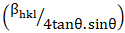 . The regression line parameters enabled the simultaneous determination of the coherent crystallite domain size and microstrain. The resulting values were 49.17 nm for the coherent crystallite domain size and 3.331×10⁻³ for the microstrain.
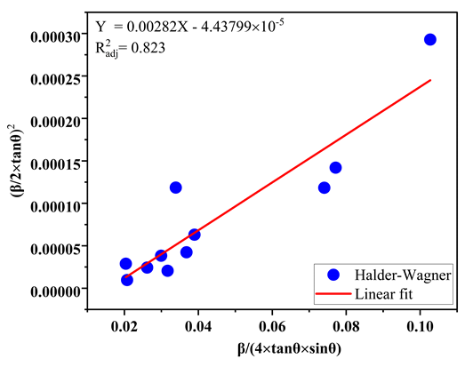 | Figure 10. Halder – Wagner method |
|

|

4. Conclusions
- In this this study, a chromium(III)benzoate coordination polymer was synthesized and characterized using a multi technique approach combining XRD, FTIR, SEM-EDS, and XRF. The XRD results reveal the formation of a highly crystalline phase exhibiting numerous low angle reflections, indicative of an extended periodic organization. Although no crystallographic reference structure could be identified, the structura signatures obtained suggest the formation of a crystalline Cr(III)-benzoate coordination network distinct from both the benzoate ligand and the chromium precursor. FTIR analysis confirms the transformation of the benzoic acid ligand into carboxylate groups coordinated to the Cr(III) centers, with a coordination mode consistent with bridging carboxylates, while the bands assigned to Cr-O vibrations further support the establishment of metal-ligand interactions. SEM-EDS observations revealed a prismatic crystalline morphology with micrometre-size particles, while EDS and XRF analyses confirmed the presence of chromium and an elemental composition consistent with the formation of the target material. The convergence of the results obtained from these complementary techniques therefore provides consistent experimental evidence for the formation of a Cr(III)-benzoate coordination polymer. Analysis of diffraction peak broadening using the Scherrer, Monshi-Scherrer, Williamson-Hall, Size-Strain Plot, and Halder-Wagner methods highlighted the influence of the underlying assumptions of each model on the estimation of microstructural parameters. Among thses approaches, the SSP and Halder-Wagner methods exhibited the best agreement with the experimental data (R2 = 0.823), yielded consistent values for the coherent crystallite domain size (49.17 nm) and microstrain. Thus, this study demonstrates the value of combining structural and microstructural characterization techniques for the identification of poorly documented crystalline coordination phases. It also highlights the suitability of the Size-Strain Plot and Halder-Wagner models for evaluating the microstructural parameters of metal carboxylate-based coordination polymers, particularly when the contributions of coherent crystallite size and crystal defects need to be distinguished.
Author Contributions
- Ferland Ngoro-Elenga: conceptualisation, data curation, methodology, writing-original draft and writing-reviews and editing; Hubert Makomo: Methodology, writing-reviews and editing; Nicaisse Narcisse Obaya: methodology, writing-review and editing; Grâce Jeanci Viboudoulou: methodology; Ibrahima Elhadji Thiam: supersivion and validation; Timothée Nsongo: supersivion and validation. All authors have red and approved the final version of the manuscrit.
Data Availability Statement
- The authors confirm that all data supporting the finding of this study are included in the article.
Conflict of Interest
- The authors declare that they have no conflict of interest.
ACKNOWLEDGEMENTS
- The authors gratefully acknowledge the Director General of the Geological and Mining Research Centre (CRGM) of the Republic of Congo for providing technical and administrative support.