-
Paper Information
- Paper Submission
-
Journal Information
- About This Journal
- Editorial Board
- Current Issue
- Archive
- Author Guidelines
- Contact Us
International Journal of Modern Botany
p-ISSN: 2166-5206 e-ISSN: 2166-5214
2016; 6(3): 37-40
doi:10.5923/j.ijmb.20160603.01

New Record of Phytoparasitic Alga, Cephaleuros diffusus Thomson & Wujekin (Trentepohliaceae, Chlorophyta) on Artocarpus incisus (Thunb.) L.f., Kerala, India
 Abstract
 Reference
 Full-Text PDF
 Full-text HTML
Binoy T. Thomas, V. P. Thomas, M. V.Bhagya, Swathi S. Nair, Reshma Rajan, S. T. Saranyamol
Phycotechnology Centre, Post Graduate & Research Department of Botany, Catholicate College, Pathanamthitta, India
Correspondence to: Binoy T. Thomas, Phycotechnology Centre, Post Graduate & Research Department of Botany, Catholicate College, Pathanamthitta, India.
| Email: |  |
Copyright © 2016 Scientific & Academic Publishing. All Rights Reserved.
This work is licensed under the Creative Commons Attribution International License (CC BY).
http://creativecommons.org/licenses/by/4.0/

No other pathological symptoms have been identified so far except root and fruit rot caused by fungi in Artocarpus incisus. Algal leaf spot by Cephaleuros species associated with Artocarpus incisus (Breadfruit tree) Kerala, India of the Western Ghats region has not been reported yet. The aim of the study was to explore the alga which causes leaf spot in Bread fruit tree, Kerala. Leaves of the breadfruit tree were found to be damaged internally by the algal growth and found externally as powdery form. Early fruit drop of the plant is a common symptom observed in all parts of Kerala conspicuously during the southwest monsoon season by the infection of alga. The causative algal species was identified as Cephaleuros diffusus (Trentepohliaceae, Chlorophyta) on the basis of morphological and taxonomical characteristics. The species is resembles Cephaleuros virescens, but can be readily differentiated by filaments size and nature, length and width of sporangia, sporangiophores and gametangia. The present study examined that infection by Cephaleuros diffusus in Artocarpus incisus caused necrosis in the internal tissues and leading to early fruit drop and falling of leaves. The present study would help to understand the various pathogenic algae which cause leaves and fruits spot in diverse crop plants.
Keywords: Bread fruit tree, Pathogen, Southwest Monsoon Season
Cite this paper: Binoy T. Thomas, V. P. Thomas, M. V.Bhagya, Swathi S. Nair, Reshma Rajan, S. T. Saranyamol, New Record of Phytoparasitic Alga, Cephaleuros diffusus Thomson & Wujekin (Trentepohliaceae, Chlorophyta) on Artocarpus incisus (Thunb.) L.f., Kerala, India, International Journal of Modern Botany, Vol. 6 No. 3, 2016, pp. 37-40. doi: 10.5923/j.ijmb.20160603.01.
Article Outline
1. Introduction
- Artocarpus incisus (Thunb.) L.f. (1782:411) commonly known as bread fruit tree, belongs to the family Moraceae which can be grown under wide range of ecological conditions (Ragone, 1997; Murai et al., 1958). It is pantropical in distribution (Dignan et al., 2004). It is an important staple food crop and a primary component of traditional agro forestry (Ragone, 1997). The bread fruit is high in carbohydrates, minerals and vitamins. The fruit is rich in fibre and so good to control blood sugar in diabetics, reduce blood lipids and control weight (Graham & Bravo, 1981). The tree provides medicine, construction materials and animal feed. It is relatively free of diseases and pests. Root rot and fruit rot of the tree are caused by the fungus, Phellinusnoxius and Phytopthora respectively (Ragone, 1997). No other pathological cause has been identified so far. However, algal leaf spot is a common disease in the breadfruit tree and most severe in Kerala during the southwest monsoon season. Algal leaf spot is also called green scurf (Alfieri, 1969) because the spots have a crusty or flaky appearance. In general, algal leaf spot is characterized by greyish, green, brown or orange cushion like marks on the leaf surface. The spot is most noticeable when the algae affect leaves, but also seasonally affects twigs, branches and fruits. Severe infection might lead to localized leaf yellowing and premature drop. Cephaleuros species is known to be parasitic on several plants in the tropics and subtropics regions (Suto et al., 2014; Pitaloka et al., 2015). It belongs to the division Chlorophyta, class Ulvophyceae, order Trentepohliales and family Trentepohliaceae (Guiry & Guiry, 2016). The disease appeared on the leaf surface as burnt orange to brown spots. Stems, fruits, leaves of the tree are infected by the Cephaleuros species, which produces a thallus on the leaf surface which bearing sporangiophores and sporangia (Pitaloka et al., 2015).Cephaleuros species has a wide range of host plants (Sinclair and Lyon, 2005). La Rue (1923) reported that algal leaf spot of rubber plants is caused by Cephaleuros parasiticus. La et al. (2009) observed that Cephaleuros virescens infected on Camellia japonica in Korea. Han et al. (2011) firstly described that Cephaleuros virescens diseased on Ficus benghalensis. Suto et al. (2014) stated that C. virescens and C. parasiticus affected the different host plant such as Polyalthia longifolia and Syzygium aromatium respectively. Pitaloka et al. (2015) examined that algal leaf spot of Para rubber is caused by Cephaleuros virescens. Sunpapao et al. (2016) and Pitaloka et al. (2015) surveyed the occurrence of C. virescens in Nephelium lappaceum and Durian plants respectively in Thailand. Sunpapao et al. (2015) surveyed the occurrence of phytoparasitic algae, C. diffusus on the leaves of Acasia in Thailand. The disease caused by the Cephaleuoros has been reported from various places such as Hawaii, America, Japan, Africa, Panama and Florida (Rindi et al., 2006; Suto and Ohtani, 2009; Marlatt and Campbell, 1980). However the report on the pathogenic algae was scarce in India, particularly from Kerala. So the present study fills the vacuum.
2. Materials and Methods
- Twenty eight specimens of algal spot bearing leaves from the different breadfruit trees were collected from the different parts of Kerala (10° 00’N- 76° 25 E) during all the three seasons such as southwest monsoon, northeast monsoon and summer seasons. Macroscopic features of lesions and thallii of algae were observed under stereomicroscope. Sections were made by hand with a razor blade, placed in a drop of Shear’s fluid on a glass slide with a cover glass sealed to observe through the Trinocular microscope (Suto et al., 2014). Microscopic features of thalli and the associated parts were observed by using the Olympus LX 400 Trinocular microscope and photographs were taken by using BioLinkz Cmos Cam (3.0m pixels) attached to the microscope. Sporangiophore, sporangia, zoospore etc. were observed and took measurements and compared with that of the most related Cephaleuros species. A voucher specimen of algal leaf spot affected twig was deposited in the Catholicate College Herbarium (CATH), Pathanamthitta, Kerala, India (Accession no. 12001). The permanent slides of the identified alga, Cephaleuros diffusus was also kept in the Phycotechnology centre Herbarium (CAPH), Department of Botany, Catholicate College, Pathanamthitta, Kerala, India with accession number (CAPH 118 & 119).
3. Result
- Leaf spot by phytoparasitic alga, C. diffusus was the firstly noted algal disease in Artocarpus incisus from Kerala, India. A preliminary investigation of the lesions of the leaf was associated with pathogenic algal growth. The leaf spots on the upper leaf surface seemed as orange areas, approximately 1-4 mm diameter (Fig. 1 A). Plant tissue of the affected regions withers and dies beneath the algal spots. Twigs and fruits (Fig. 1 C) of the tree were also affected.Transverse sectioning revealed that thalli were sub cuticular or sub epidermal growth through leaf tissue and caused necrosis of cells (Fig. 1D). Filaments are short and cylindrical with irregularly shaped, 17- 41 µm long and 4-15 µm width. Disk of thalii are composed of open filamentous. Setae rarely found as cylindrical filaments. No thalii grown on the lower leaf surface. Sporangiophores forming head cell with sporangia on the top (Fig. 1F). Also produce sporangiate laterals, zoosporangia and suffultory cells. Sporangia were spherical in shape, dark orange, 12.5 – 30.5 µm long and 12.5 – 22.5 µm width (Fig. 1D & F). The sporangiophores were sparsely produced on the upper leaf surface, solitary or in a tuft of 250- 439 µm long and 10-12.5 µm width.
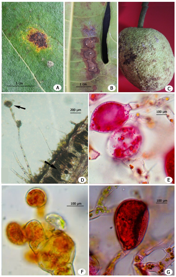 | Figure 1. A. Affected leaf of Artocarpus incisus (Adaxial). B. Affected leaf (abaxial). C. Affected fruit. D. Anatomy of affected leaf with sporangiophore and sporangium of Cephaleuros diffusus. E. Gametangium. F. Sporangium. G. Gametangium (enlarged view) |
4. Discussion
- Spreading of algal disease is due to various environmental conditions such as frequent rains and warm weather (Han et al., 2011; Sunpapao et al., 2016). The present findings agreed by the report of Sinclair and Lyon (2005) that unusual environmental conditions might promote the viability of the pathogens in the host plants. Southwest monsoon of Kerala, followed by extreme summer promoted rapid infection in all parts of the Artocarpus particularly on leaves. The pathogen has the ability to withstand in the infected plant debris also. The pathogen was identified as C. diffusus because of the morphological features and it has been agreed with the description of Sunpapao et al. (2015) and Thompson and Wujek (1997). It is seen that lesions produced by C. parasiticus are appeared on both leaf surfaces. While in C. diffusus and C. virescens, lesions are appeared on upper leaf surface only. In the present report also infection by C. diffusus observed on the upper surfaces of leaves. C. parasiticus infects the leaf sub epidermally and intramatrically only (Sunpapao et al., 2015). C. diffusus caused necrosis of epidermis, mesophyll cells and palisade tissue of the leaves.Krishnamurthy (2000) reported that there is only one species of Cepahaleuros in India and neighboring countries. According to him C. parasiticus is not distinct from C. diffusus by the morphological features. But Guiry and Guiry (2016) reported that C. diffusus and C. parasiticus are separate species. Sunpapao et al. (2015) reported the main differences of C. diffusus from that of closely related species, C. virescens. In the present investigation also, similar features of the C. diffusus was observed and recorded.
5. Conclusions
- The present study witnessed the infection of Cephaleuros diffusus in Artocarpus incisus caused necrosis in the internal tissues of breadfruit tree and consequently caused early fruit drop and falling of leaves leading to crop loss mainly in southwest monsoon season, Kerala, India.
ACKNOWLEDGEMENTS
- The authors gratefully acknowledged Kerala State Council for Science, Technology and Environment (KSCSTE) for providing financial assistance through the Science Research Scheme (No.030/SRSLS/2014/CSTE).