
-
Paper Information
- Next Paper
- Previous Paper
- Paper Submission
-
Journal Information
- About This Journal
- Editorial Board
- Current Issue
- Archive
- Author Guidelines
- Contact Us
International Journal of Genetic Engineering
p-ISSN: 2167-7239 e-ISSN: 2167-7220
2026; 14(11): 364-372
doi:10.5923/j.ijge.20261411.06
Received: Oct. 15; Accepted: Nov. 3, 2026; Published: Nov. 12, 2026

Time-Dependent Changes in the Activity of the Mitochondrial ATP-Dependent Potassium Channel of Rat Liver in CCl4-Induced Toxic Hepatitis and Their Correction by Polyphenols
 Abstract
 Reference
 Full-Text PDF
 Full-text HTML
Odinakhon M. Turdiyeva, Ma’murjon K. Pozilov
Department of Zoology and General Biology, Fergana State University, Fergana, Uzbekistan
Correspondence to: Odinakhon M. Turdiyeva, Department of Zoology and General Biology, Fergana State University, Fergana, Uzbekistan.
| Email: |  |
Copyright © 2026 The Author(s). Published by Scientific & Academic Publishing.
This work is licensed under the Creative Commons Attribution International License (CC BY).
http://creativecommons.org/licenses/by/4.0/

Mitochondrial ATP-dependent potassium channels (mitoKATP channels) take part in maintaining matrix volume and membrane potential, but data on the activity of this channel in toxic hepatitis and on the effect of biologically active substances on it are scarce. The aim of this study was to determine the activity of the mitoKATP channel of rat liver mitochondria in toxic hepatitis induced by carbon tetrachloride (CCl4), its time-dependent changes, and the effect of the polyphenols gossitan and getasan on it, in comparison with the flavonoid quercetin. Toxic hepatitis was induced in outbred white male rats by injection of 50% CCl4 (0.5 ml/kg) every 3 days for 3 weeks. Channel activity was assessed by the change in optical density of the mitochondrial suspension at 540 nm in the presence of 200 µM ATP; the activity of the control group was taken as 100%. In toxic hepatitis the activity of the mitoKATP channel was inhibited by 29.5±2.6% relative to the control. After 20 days of treatment, quercetin, gossitan and getasan (50 mg/kg) increased the channel activity by 13.0±1.1%, 19.3±1.6% and 14.0±1.2% relative to the hepatitis group. In the 6-week dynamic study the activity of the channel in toxic hepatitis decreased by 34.5±2.1% relative to the control by week 6; when the compounds were administered from week 3, the inhibition at week 6 was 18.5±2.0%, 9.4±1.8% and 14.6±1.5%, respectively, and the activity was higher than at week 3. The results show that gossitan and getasan, like quercetin, may activate K+ flux through the mitochondrial membrane in toxic hepatitis.
Keywords: Toxic hepatitis, Carbon tetrachloride, Rat liver mitochondria, Mitochondrial ATP-dependent potassium channel, Gossitan, Getasan, Quercetin
Cite this paper: Odinakhon M. Turdiyeva, Ma’murjon K. Pozilov, Time-Dependent Changes in the Activity of the Mitochondrial ATP-Dependent Potassium Channel of Rat Liver in CCl4-Induced Toxic Hepatitis and Their Correction by Polyphenols, International Journal of Genetic Engineering, Vol. 14 No. 11, 2026, pp. 364-372. doi: 10.5923/j.ijge.20261411.06.
Article Outline
1. Introduction
- Hepatocytes are the specific functional cells of the liver and make up 70–85% of its mass. They perform metabolic, secretory and storage functions, and the liver carries out the metabolism of lipids and carbohydrates and regulates the level of proteins and amino acids in the blood [1]. Liver cells are targets of various toxic substances, in which their mitochondria are damaged. Under the action of various xenobiotics the amount of free radicals in liver mitochondria may increase, the antioxidant defense system may change and membrane disturbances may occur. Mitochondria occupy about 20–25% of the cytoplasm of liver cells, and each cell contains 1000–2000 mitochondria [2]. The ion-transport systems located in the liver mitochondrial membrane, the passive ion permeability, ATP synthesis and the antioxidant defense systems determine the physiological regulation of hepatocytes, and in various pathological processes these systems become dysfunctional.Carbon tetrachloride (CCl4) is a hepatotoxic substance that is well soluble in fats. The oxidative stress caused by CCl4 leads to cell damage through the oxidation of cell components such as lipids, proteins and DNA [3], and the binding of CCl4 to fats and proteins causes peroxidative degeneration of many tissues [4]. Various pharmacological agents can trigger the opening of the mitochondrial permeability transition pore (mPTP) in liver mitochondria [5,6]. The opening of the mPTP is defined as a sudden increase in the permeability of the inner mitochondrial membrane to low-molecular-weight solutes (up to 1.5 kDa) in response to stimuli including high Ca2+ and oxidative stress [7]. In toxic hepatitis a sharp increase in the permeability of liver mitochondria to water and water-soluble molecules has been observed, which may lead to a drop of the membrane potential.Mitochondrial potassium channels have been described as important factors of cell life and death. The activation of mitochondrial K+ channels, such as the ATP-regulated or Ca2+-activated large-conductance channels, may have a cytoprotective effect in cardiac or neuronal tissue, while the inhibition of the mitochondrial Kv1.3 channel may lead to the death of cancer cells [8]. The field began in the early 1990s with the first publication describing a K+ channel sensitive to ATP and to the antidiabetic sulfonylurea glibenclamide (the mitoKATP channel) [9]. The discovery that the activation of a mitochondrial K+ channel by channel openers triggers protective mechanisms in cardiomyocytes showed that these proteins are important in ischemia–reperfusion [10]. This was the point at which trials of directing drugs that affect cell function to various mitochondrial K+ channels began.Various K+ channels are present in the inner mitochondrial membrane [11]: the ATP-regulated channel (mitoKATP), which was the first K+ channel described in the inner mitochondrial membrane, the large-, intermediate- and small-conductance Ca2+-activated channels (mitoBKCa, mitoIKCa, mitoSKCa), the voltage-gated channels mitoKv1.3, mitoKv1.5 and mitoKv7.4, the two-pore-domain mitoTASK3 channel, the Na+-activated mitoSLO2 channel and the mitoHCN channel. Most probably, the mitoKATP channel is formed by ROMK-type channels [12] or by the recently described CCDC51 protein, and the ABCB8 protein (the mitochondrial sulfonylurea receptor) is a part of the channel [13].Different tissues contain different sets of mitochondrial K+ channels [14]. For example, five types of K+ channels were described in cardiomyocyte mitochondria, but only two types in the mitochondria of skin fibroblasts [15]; the mitoKATP, mitoBKCa and mitoKv1.3 channels are the most widespread in mammalian mitochondria [8]. The number of mitoKATP channels is higher in the brain than in heart mitochondria [16]. Therefore, before targeting specific mitochondrial K+ channels with drugs, the type of the channel, its presence in the particular tissue and its abundance in the target tissue should be considered.The following mitochondrial K+ channels have been described [11]: mitoKATP (the first channel described in the inner mitochondrial membrane); mitoBKCa (large-conductance, encoded by the KCNMA1 gene, a splice isoform of the BKCa channel of plasma membranes); mitoIKCa (intermediate-conductance, KCNN4); mitoSKCa (small-conductance, KCNN1–KCNN3); the voltage-gated mitoKv1.3, mitoKv1.5 and mitoKv7.4 (KCNQ4); the acid-sensitive two-pore-domain mitoTASK3 (KCNK9); the Na+-activated mitoSLO2 (KCNT2 in mammals); and the hypopolarization-activated cyclic-nucleotide-gated mitoHCN channel (HCN1–HCN4). Thus, three parameters should be defined before mitochondrial K+ channels are considered as pharmacological targets: the molecular identity of the channel, its tissue profile, and its level in the particular tissue (the so-called “3D mitoK channel mapping”).Another aspect of this problem is the presence of the channels in different cell compartments. The BKCa channels, which are abundant in the membranes of many cells, are present in cardiomyocytes only in the inner mitochondrial membrane and not in the plasma membrane [17]; this is unique for mitochondrial K+ channels and supports the view that these channels and cardiac tissue are targets for pharmacological agents. The functional consequences of the uneven distribution of the channels and of the intracellular heterogeneity of mitochondria with respect to K+ channels have not yet been determined.Before the identification of mitochondrial K+ channels, the electrogenic flow of K+ ions into the mitochondrial matrix was described as “potassium uniport” [18]. The mitoKATP channel takes part in maintaining the matrix volume and in the formation of the membrane potential, and it has a great significance in the formation of adaptation processes of cells in many pathological conditions [19]. The proteins forming this channel are inhibited by physiological concentrations of ATP and take part in the K+ cycle [20]; the regulation of the channel plays an important role in cell adaptation to hypoxia [21, 22], and the molecular structures of mitochondria may be specific targets for pharmacological agents with antihypoxant properties [23].In Uzbekistan, the dysfunction of liver mitochondria in CCl4-induced toxic hepatitis and the effect of bioactive substances on it have been studied by Asrarov and Abdullaeva [24]. In a complex study of the dynamics of mitochondrial functional indices in CCl4-induced toxic hepatitis it was found that the damaging effect is associated with the transition of the cyclosporin A-sensitive pore into the open conformation and with the activation of endogenous phospholipase A2, and that these changes intensify as the pathological process develops [25]. The corrective effect of the alkaloid protopine on the functional indices of mitochondria in toxic hepatitis has been found [26]; the effect of flavonoid compounds on mitochondrial enzymes in heliotrine-induced hepatitis [27] and the effect of polyphenol extracts of Helichrysum maracandicum on mitochondrial antioxidant enzymes in toxic hepatitis [28] have also been studied.The search for new agents that prevent liver damage in hepatitis, the identification of bioactive substances in local raw materials with hepatoprotective properties, their isolation from plants in pure form and the study of the mechanisms of their corrective action at the molecular, cellular and tissue levels are among the topical tasks of modern physiology, pharmacology and medicine. Hepatoprotective compounds isolated from plants differ from chemical synthetic preparations in the absence of toxic properties and in having a physiological mechanism of action and pharmacological properties. Therefore, in this work the membrane-active properties of the polyphenols gossitan and getasan were evaluated with the aim of determining membrane disturbances of mitochondria in toxic hepatitis and correcting them with natural substances.However, data on the activity of the rat liver mitoKATP channel in toxic hepatitis and on the effect of biologically active substances on it are scarce. In toxic hepatitis the volume of the mitochondrial matrix changes and ion homeostasis may be disturbed, and the K+-dependent permeability of the inner membrane is regulated by the mitoKATP channel. The aim of this work was to determine the activity of the mitoKATP channel of rat liver mitochondria in CCl4-induced toxic hepatitis, to study its time-dependent changes, and to determine the effect of the polyphenols gossitan and getasan on it, in comparison with the flavonoid quercetin.
2. Materials and Methods
2.1. Animals
- The study was carried out on outbred white male rats weighing 180–200 g, kept on a standard ration in vivarium conditions, in the Laboratory of Molecular Biophysics of the Institute of Biophysics and Biochemistry at the National University of Uzbekistan. The research on experimental animals was carried out in accordance with the Declaration of Helsinki, the guidelines of the Council for International Organizations of Medical Sciences (CIOMS, 1985) and the bioethical regulation of the Institute on the use of laboratory animals in scientific research (2019).
2.2. Toxic Hepatitis Model and Experimental Groups
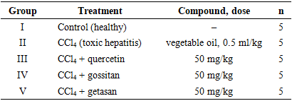
- Toxic hepatitis was induced by injection of 50% CCl4 dissolved in vegetable oil (0.5 ml/kg) under the abdominal skin once every 3 days [29]. Blood was taken from the animals every 3 days and the activities of ALT and AST were determined; 21 days after the beginning of CCl4 administration the plasma ALT (normal value 70±1 U/l) and AST (normal value 58±3 U/l) were increased. The animals were divided into five groups (n=5 in each), as shown in Table 1.
|
2.3. Test Compounds and Reagents
- Gossitan (C177H154O85) isolated from Gossypium hirsutum [30] and getasan (C55H36O34) isolated from Geranium sanguineum were provided by scientists of the Institute of Bioorganic Chemistry of the Academy of Sciences of the Republic of Uzbekistan. Gossitan and getasan were compared with the existing hepatoprotective flavonoid quercetin (C15H10O7, M=302 g/mol, isolated from Plantago major L.), which was used as a standard prototype [31]. Quercetin is widely found in the vegetative and generative organs of tea (Camellia sinensis), apple (Malus domestica), onion (Allium), grape (Vitis vinifera), citrus fruits and tomato (Solanum lycopersicum), and anti-inflammatory, antiviral, antiallergic, anticarcinogenic and hypotensive effects of quercetin have been found in in vitro experiments. Among the plant substances tested (the diterpenoid alkaloids zongorine and zeravshanizine, the flavonoids isorhamnetin and norwogonin, and the polyphenols gossitan and getasan), only those with hepatoprotective activity in toxic hepatitis were selected by screening; the corrective effect of gossitan and getasan on the disturbances of liver cell and mitochondrial functions was more pronounced than that of the other plant substances. The molar mass of getasan is 1240 g/mol. The structural formulas of the compounds are shown in Figure 1. The following reagents were used: tetrachloromethane (Reakhim, Russia), Tris-HCl, EDTA, EGTA, KH2PO4, HEPES, KCl, succinate (Sandoz, Switzerland), oligomycin, rotenone, ATP (Serva, Germany) and sucrose. All reagents had the chemical purity required for the experiments.
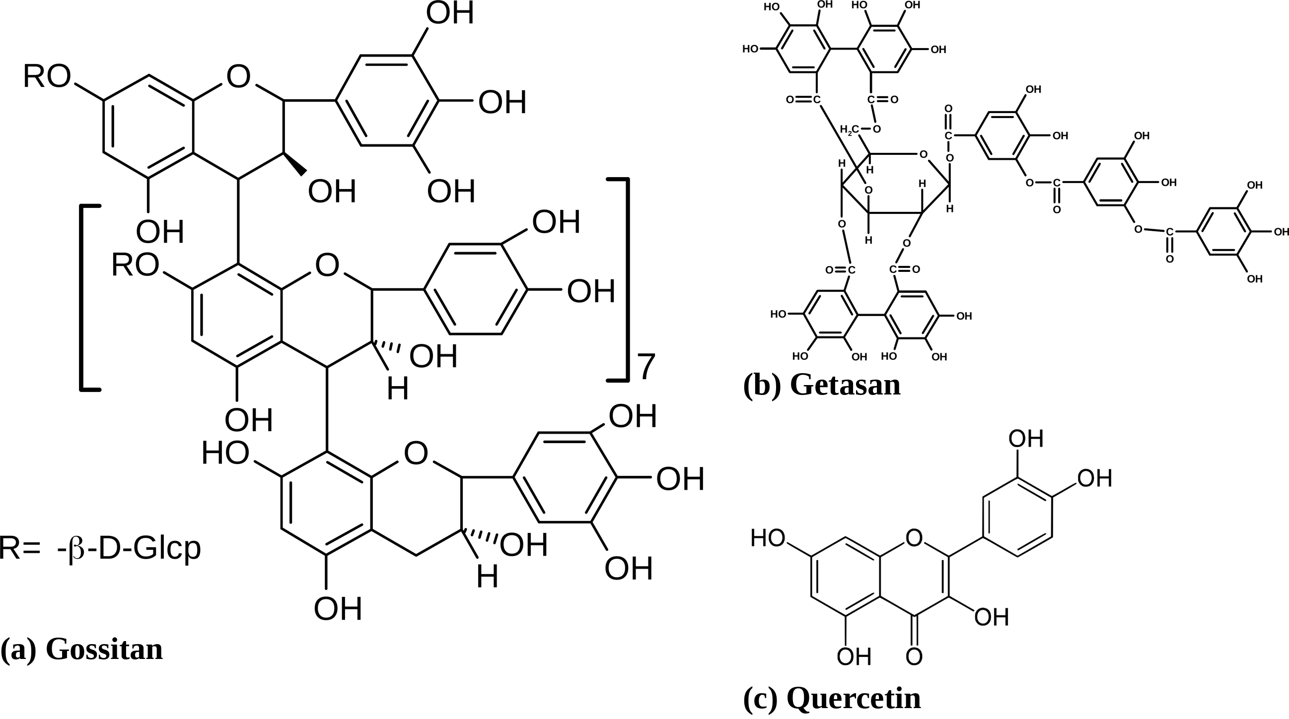 | Figure 1. Structural formulas of the compounds used: (a) gossitan (C177H154O85) isolated from Gossypium hirsutum; (b) getasan isolated from Geranium sanguineum; (c) quercetin (C15H10O7) isolated from Plantago major L. |
2.4. Isolation of Liver Mitochondria
- Rat liver mitochondria were isolated by the differential centrifugation method of Schneider [32]. After decapitation the liver was placed in a semi-ice isolation medium (250 mM sucrose, 10 mM Tris-HCl, 1 mM EGTA, pH 7.4), minced, and homogenized in a ratio of 1:6 (liver weight in g to ml of medium) with a Teflon pestle at 6000–8000 rpm. Centrifugation was carried out at 0–2°C in two steps: first at 1500 rpm for 7–8 min to sediment heavy cell aggregates, then the supernatant was centrifuged at 6000 rpm (6000 g) for 17 min. The mitochondrial pellet was washed in an EDTA-free medium (1:5 by volume) and stored in a vessel with ice. Mitochondrial protein was determined by the biuret method.
2.5. Determination of mitoKATP Channel Activity
- The permeability of the mitoKATP channel was determined by the change in the optical density of the mitochondrial suspension (0.3–0.4 mg/ml) at 540 nm in an open 3-ml cell at 26°C with constant stirring. The incubation medium contained 125 mM KCl, 10 mM HEPES, 5 mM succinate, 1 mM MgCl2, 2.5 mM K2HPO4, 2.5 mM KH2PO4, 0.005 mM rotenone and 0.001 mM oligomycin, pH 7.4 [33]. The physiological concentration of ATP (200 µM) was present in the incubation medium; the channel activity of the control group under these conditions was taken as 100%.
2.6. Statistical Analysis
- Statistical processing and plotting were performed with OriginPro 8.6 (USA). The values were calculated as the arithmetic mean of 4–7 independent experiments. The differences between the control, experimental and experimental + test compound values were calculated by the t-test; P<0.05, P<0.01 and P<0.001 were taken as statistically significant. The results are presented as M±m (n=5).
3. Results
3.1. Body and Liver Weights
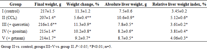
- In the hepatitis group (II) the change in the body weight of the rats between the beginning and the end of the experiment was lower (5.6±0.4%) than in the control (group I, 11.3±1.2%). The absolute liver weight of the rats with toxic hepatitis was 10.6±0.9 g, which was 41.3±3.4% higher than in the control, and the relative liver weight index was 5.12±0.6%, compared with 3.45±0.2% in the control (Table 2).When the animals with toxic hepatitis were treated with quercetin (group III) for 20 days, the change in body weight and the relative liver weight index were 11.3±0.9% and 3.61±0.2%, i.e. the body and liver weights were restored to the control level in comparison with group II. After the administration of gossitan (group IV) and getasan (group V), the change in body weight was 10.2±0.8% and 9.2±0.7%, and the relative liver weight index decreased relative to the pathological group and was slightly higher than the control (3.81±0.4% and 4.06±0.3%, respectively; Table 2). Thus, the selected compounds effectively restore the body and liver weights of rats in toxic hepatitis.
|
3.2. Transaminases and Bilirubin in Toxic Hepatitis
- After 21 days of CCl4 administration, the plasma ALT, AST and total bilirubin of the hepatitis group (II) were 203.4±20.5 U/l, 178.6±15.5 U/l and 18.9±1.6 µmol/l, i.e. 2.6, 3.3 and 1.7 times higher than in the control, which confirmed the development of toxic hepatitis (Table 3). In group III, treated with quercetin for 20 days, the plasma ALT (86.6±9.4), AST (68.2±6.6) and bilirubin (12.5±1.4) were lower than in group II by 2.3, 2.61 and 1.5 times, respectively. After 20 days of treatment with gossitan (group IV), ALT, AST and bilirubin were 97.8±10.6 U/l, 82.6±7.5 U/l and 13.4±1.2 µmol/l; with getasan (group V) they were 122.2±11.3 U/l, 94.3±6.2 U/l and 14.8±1.3 µmol/l, i.e. lower than in the hepatitis group by 2.08, 2.16 and 1.41 times for gossitan and by 1.66, 1.90 and 1.27 times for getasan. The effect of gossitan on the increased enzymes and bilirubin was more pronounced than that of getasan.
|
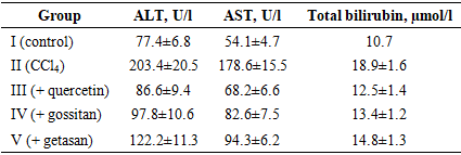
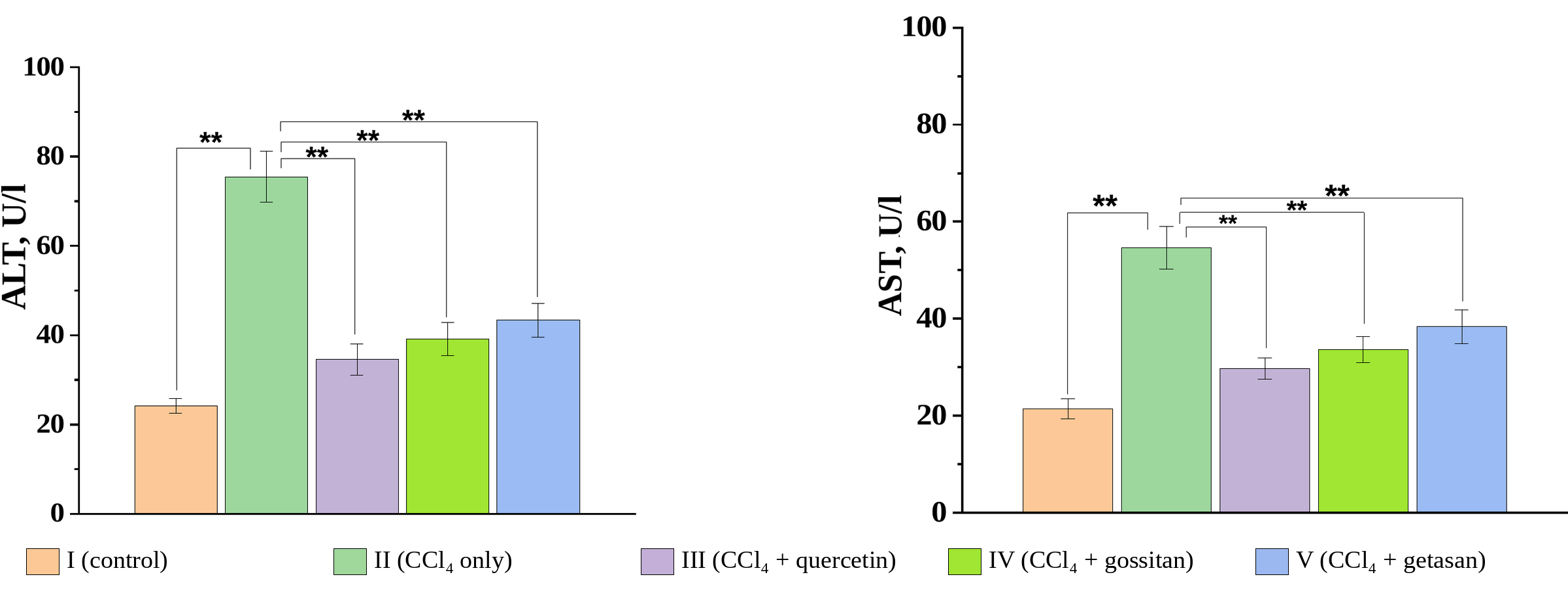 | Figure 2. Effect of quercetin, gossitan and getasan on the activity of ALT (left) and AST (right) in the liver of rats with CCl4-induced toxic hepatitis (**P<0.05; group II relative to the control, groups III–V relative to group II; n=5) |
3.3. mitoKATP Channel Activity After 20 Days of Treatment
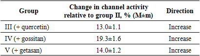
- In the hepatitis group (II) the permeability of the mitoKATP channel in the presence of 200 µM ATP in the incubation medium was inhibited by 29.5±2.6% relative to the control (Figure 3). In the hepatitis group treated with quercetin (III) the channel permeability was increased by 13.0±1.1% relative to group II. In groups IV and V, treated with gossitan and getasan for 20 days, the channel permeability was activated by 19.3±1.6% and 14.0±1.2% relative to group II, respectively (Figure 3, Table 4).
|
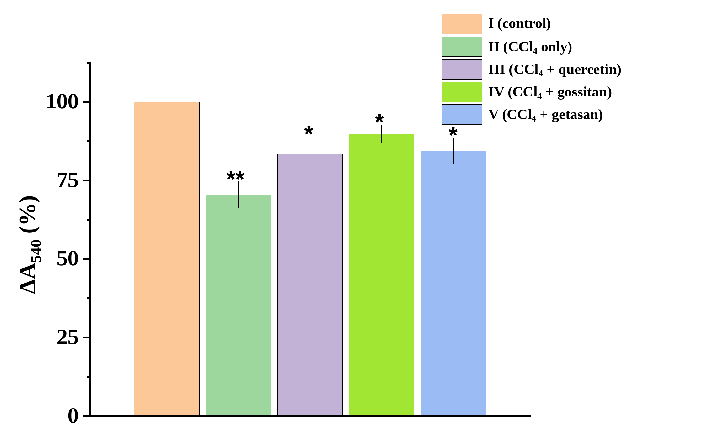 | Figure 3. The effect of quercetin, gossitan and getasan on the activity of the mitoKATP channel of rat liver mitochondria in CCl4-induced toxic hepatitis after 20 days of treatment (activity of the control group in the presence of 200 µM ATP is taken as 100%; *P<0.05 relative to the control, P<0.05 relative to group II; n=5) |
3.4. Time-Dependent Changes in mitoKATP Channel Activity
- To determine how the activity of the channel changes with the development of the pathological process and how it depends on the administration of the compounds, the activity of the mitoKATP channel of the liver mitochondria was followed for 6 weeks (Figure 4). In the presence of 200 µM ATP the activity of the channel in the control (group I) was taken as 100%. In the hepatitis group (II) the activity of the channel decreased with time relative to the control, and by week 6 the decrease was 34.5±2.1%.Quercetin, gossitan and getasan were administered to the animals with toxic hepatitis (groups III, IV and V) from week 3, and mitochondria were isolated at weeks 4, 5 and 6. In group III the activity of the channel was inhibited up to week 3; after the administration of quercetin, from week 4 the permeability was higher than at week 3, and at week 6 the activity of the mitoKATP channel was inhibited by 18.5±2.0% relative to the control, i.e. it was activated under the action of quercetin compared with week 3.In groups IV and V the activity of the mitoKATP channel was inhibited at week 3 by 25.0±3.0% and 24.0±2.0% relative to the control, respectively. When gossitan and getasan were administered from week 3, at week 6 the inhibition relative to the control was 9.4±1.8% and 14.6±1.5%, respectively. When these week 6 values were compared with the values at week 3, the activity of the mitoKATP channel was found to be increased (Figure 4, Table 5).
|

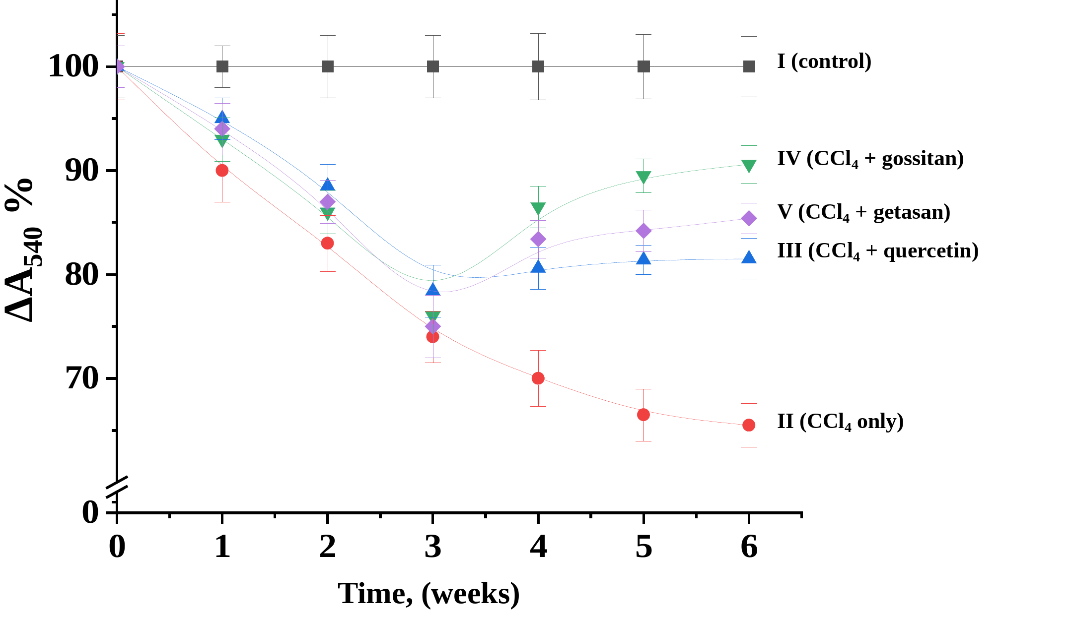 | Figure 4. Time-dependent changes in the activity of the mitoKATP channel of rat liver mitochondria in CCl4-induced toxic hepatitis and the effect of quercetin, gossitan and getasan (all points P<0.05; n=5). The activity of the control group in the presence of 200 µM ATP was taken as 100%; treatment of groups III–V was started at week 3 |
4. Discussion
- The results show that in CCl4-induced toxic hepatitis the activity of the mitoKATP channel of rat liver mitochondria is inhibited (by 29.5±2.6% in the first experiment) and that the inhibition increases with the development of the pathological process (up to 34.5±2.1% by week 6). The mitoKATP channel takes part in maintaining the matrix volume and in the formation of the membrane potential, and the K+-dependent permeability of the inner membrane is regulated by this channel. In toxic hepatitis the matrix volume changes and ion homeostasis may be disturbed. Because of the inhibition of the channel, the flow of K+ ions into the matrix sharply decreases and the volume of the mitochondria changes; as a result, the K+ cycle in the mitochondria may be disturbed and the membrane potential may decrease.Quercetin, gossitan and getasan increased the activity of the channel in toxic hepatitis. After 20 days of treatment the increase relative to the hepatitis group was 13.0±1.1%, 19.3±1.6% and 14.0±1.2%, respectively. In the dynamic experiment, when the compounds were administered from week 3, the activity of the channel at week 6 was higher than at week 3, although the inhibition relative to the control remained (18.5±2.0%, 9.4±1.8% and 14.6±1.5% for quercetin, gossitan and getasan, respectively); the smallest inhibition at week 6 was observed in the gossitan group. Thus, the polyphenols gossitan and getasan may activate the flow of K+ ions through the mitochondrial membrane in toxic hepatitis induced by CCl4.The development of toxic hepatitis in this model was confirmed by the decrease in the increase of body weight, the increase in the absolute liver weight and the relative liver weight index, and by the increase of ALT, AST and total bilirubin. Aminotransferases catalyze the transfer of amino groups from amino acids to keto acids, and pyridoxal phosphate, the active form of vitamin B6, serves as their coenzyme. Their activity in the blood plasma is very low, and it increases considerably when the membranes of liver cells are damaged and their integrity is disturbed; this is why the determination of the activities of both ALT and AST is important in toxic hepatitis. Gossitan and getasan decreased the activities of these enzymes in the plasma and liver and reduced the increased bilirubin, and the effect of gossitan was more pronounced than that of getasan, which indicates their hepatoprotective properties.The corrective effect of the polyphenols on the mitoKATP channel is consistent with the other findings obtained in the same model: in toxic hepatitis the permeability of liver mitochondria for K+, H+, Ca2+ and Mg2+ ions and the opening of the mPTP increased, and gossitan, getasan and quercetin inhibited the permeabilization of the membrane; they also reduced the content of the lipid peroxidation product MDA and restored the activity of the antioxidant enzymes SOD, catalase and glutathione peroxidase, which indicates their antioxidant properties. In addition, the polyphenols decreased the activity of ALT and AST and the content of bilirubin, i.e. they showed a hepatoprotective effect.The experiments were carried out in two complementary stages. In the first stage the polyphenols corrected the changes of body and liver weights, the plasma and liver transferases and bilirubin; in the second stage they corrected the dysfunction of liver mitochondria, including the activity of the mitoKATP channel. The mechanisms of the corrective action of the compounds on the dysfunction of mitochondria in toxic hepatitis were determined at the mitochondrial level, which may serve as a theoretical and practical basis for the creation of new preparations against liver diseases on the basis of the revealed properties of these compounds.Therefore, the study of disturbances of organ, tissue and cell functions in toxic hepatitis at the mitochondrial level, of the corrective role of the KATP channel, and the identification of new promising hepatoprotective substances and their mechanisms of action remain relevant problems. Biologically active substances that correct the dysfunction of liver homogenate and mitochondria in hepatitis may serve as a theoretical and practical basis for the development of new hepatoprotective drugs.
5. Conclusions
- (1) In CCl4-induced toxic hepatitis the plasma ALT, AST and total bilirubin increased by 2.6, 3.3 and 1.7 times, and gossitan and getasan decreased them, like quercetin; the body and liver weight indices were also restored. The permeability of the mitoKATP channel of rat liver mitochondria in the presence of 200 µM ATP is inhibited by 29.5±2.6% relative to the control.(2) After 20 days of treatment, quercetin, gossitan and getasan (50 mg/kg) increased the activity of the channel by 13.0±1.1%, 19.3±1.6% and 14.0±1.2% relative to the hepatitis group.(3) With the development of toxic hepatitis the activity of the mitoKATP channel decreases with time and by week 6 is 34.5±2.1% lower than in the control. When quercetin, gossitan and getasan were administered from week 3, the activity of the channel at week 6 was higher than at week 3; the inhibition relative to the control was 18.5±2.0%, 9.4±1.8% and 14.6±1.5%, respectively.(4) The polyphenols gossitan and getasan may activate K+ flux through the mitochondrial membrane in toxic hepatitis, and their effect is comparable to that of quercetin.