
-
Paper Information
- Paper Submission
-
Journal Information
- About This Journal
- Editorial Board
- Current Issue
- Archive
- Author Guidelines
- Contact Us
International Journal of Genetic Engineering
p-ISSN: 2167-7239 e-ISSN: 2167-7220
2026; 14(10): 287-290
doi:10.5923/j.ijge.20261410.05
Received: Sep. 22; Accepted: Oct. 19, 2026; Published: Oct. 23, 2026

Morphofunctional Changes in the Stomach Under the Influence of Food Colorants E-171 and E-173 and Their Assessment
 Abstract
 Reference
 Full-Text PDF
 Full-text HTML
Oltiboyeva Mavsuma Gulom qizi1, Oripov Firdavs Suratovich2, Khalikov Qahhor Mirzayevich3
1Assistant, Samarkand State Medical University, Samarkand, Uzbekistan
2Doctor of Medical Sciences, Professor, Samarkand State Medical University, Samarkand, Uzbekistan
3Candidate of Medical Sciences, Samarkand State Medical University, Professor, Samarkand, Uzbekistan
Correspondence to: Oltiboyeva Mavsuma Gulom qizi, Assistant, Samarkand State Medical University, Samarkand, Uzbekistan.
| Email: |  |
Copyright © 2026 The Author(s). Published by Scientific & Academic Publishing.
This work is licensed under the Creative Commons Attribution International License (CC BY).
http://creativecommons.org/licenses/by/4.0/

This article examines the potential effects of the food colorants E-171 (titanium dioxide) and E-173 (aluminium), used in the food industry, on gastric tissues and considers the assessment of possible morphofunctional changes caused by their exposure. The literature review indicates that titanium dioxide particles may interact with biological barriers of the gastrointestinal tract and may be associated with local inflammatory processes and cellular changes. At the same time, the effects observed in experimental studies depend on particle size, dose, duration of exposure, and biological properties; the available data are insufficient to draw a uniform conclusion. The effects of E-173 on gastric tissues also require separate experimental assessment. A comprehensive study of the gastric mucosa, glandular structure, vascular changes, and functional parameters may help clarify potential mechanisms of tissue injury. These data are important for the scientific assessment of the biological safety of food colorants and for expanding current knowledge of their effects on the stomach.
Keywords: Food additives, Food colorants, E-171, Titanium dioxide, E-173, Aluminium, Gastric mucosa, Morphofunctional changes, Histopathological alterations, Oxidative stress, Sesame oil, Biological safety
Cite this paper: Oltiboyeva Mavsuma Gulom qizi, Oripov Firdavs Suratovich, Khalikov Qahhor Mirzayevich, Morphofunctional Changes in the Stomach Under the Influence of Food Colorants E-171 and E-173 and Their Assessment, International Journal of Genetic Engineering, Vol. 14 No. 10, 2026, pp. 287-290. doi: 10.5923/j.ijge.20261410.05.
1. Introduction
- Various food additives are used to improve the appearance, colour, and consumer properties of food products. Assessing their biological safety, particularly their potential effects on the digestive system under conditions of long-term intake, is an important task in modern experimental medicine and toxicology.E-171 is titanium dioxide, which was widely used as a white colouring additive in food products. Its particle size and physicochemical properties may influence its interactions with biological systems. In 2021, the EFSA expert panel reassessed the available evidence and concluded that E-171 could no longer be considered safe as a food additive because concerns regarding genotoxicity could not be ruled out [1].In an experimental study, Bettini and colleagues investigated the passage of food-grade titanium dioxide particles across the intestinal barrier, their effects on immune homeostasis, and their association with certain pathological changes in the rat colon [2]. However, findings on the effects of E-171 on the stomach are not uniform. In an experiment by Han and colleagues, titanium dioxide particles were detected in gastric tissues, and changes in certain immunological indicators were reported; nevertheless, a dose-dependent relationship was not observed for all toxicological parameters [3]. E-173 is known as an aluminium colourant, and assessment of its biological effects requires clear specification of the composition, form, dose, and route of exposure. It is scientifically inappropriate to assume that E-171 and E-173 have identical effects. The gastric mucosa comes into direct contact with substances entering the body; therefore, studying changes in its epithelium, glands, stroma, and microcirculatory system is important for identifying possible mechanisms of toxic effects [6,7,8,9].Aim of the study: to investigate the separate and combined effects of food colorants E-171 and E-173 on the morphofunctional status of the stomach and to assess the effectiveness of correction with sesame oil.
2. Materials and Methods
- Adult white outbred male and female laboratory rats were selected for the study. The rats were divided into control, E-171, E-173, E-171 + E-173, and correction groups. The E-171 and E-173 colorants were administered for three months. Gastric tissue specimens, blood, and blood serum were selected as the study materials.The study used biochemical methods, histological staining methods (Masson's trichrome, haematoxylin and eosin, and Van Gieson staining), morphometry, biochemical analysis, biological correction methods, and statistical methods.
3. Results
- We investigated the effects of titanium dioxide (E-171) and aluminium (E-173) food colorants, as well as morphofunctional changes arising from their combined effects on the structural and functional state of the rat stomach. The widespread use of inorganic compounds, nanoparticles, and metal compounds in the food industry may lead to their accumulation in the body and may adversely affect various organs and systems, particularly the digestive system, potentially causing pathomorphological changes.Titanium dioxide had long been considered an inert substance. However, recent biomedical studies indicate that some food additives may cross mucosal barriers and have the potential to induce oxidative stress. The E-173 food colorant (aluminium metal) may affect microcirculation in small blood vessels and the enzymatic structures of epithelial cells. Particular attention is paid to their combined effects because, under real dietary conditions, people may consume these chemical substances together.The fundic and pyloric regions of the stomach were the principal objects of morphological analysis because, owing to their anatomical and physiological characteristics, these regions may respond differently to chemical exposures. Morphological examination of parietal cells, chief cells, and mucosal cells (mucocytes) under experimental intoxication makes it possible to investigate tissue injury and potential mechanisms underlying disruption of the organ’s secretory and protective functions. An accurate description of the normal morphology of the gastric fundus is an important step in understanding xenobiotic-induced changes, as it provides a reference for comparative assessment of destructive alterations (Figure 1).
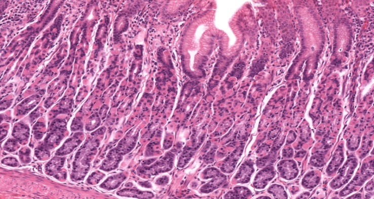 | Figure 1. Structure of the fundic gastric mucosa in rats from the control group. Haematoxylin and eosin staining. Magnification: 10×10 |
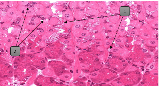 | Figure 2. Morphological changes in epithelial cells of the fundic gastric mucosa under the influence of E-171. 1 — hydropic change in the cytoplasm; 2 — changes in the continuity and thickness of the nuclear envelope. Haematoxylin and eosin staining. Magnification: 10×20 |
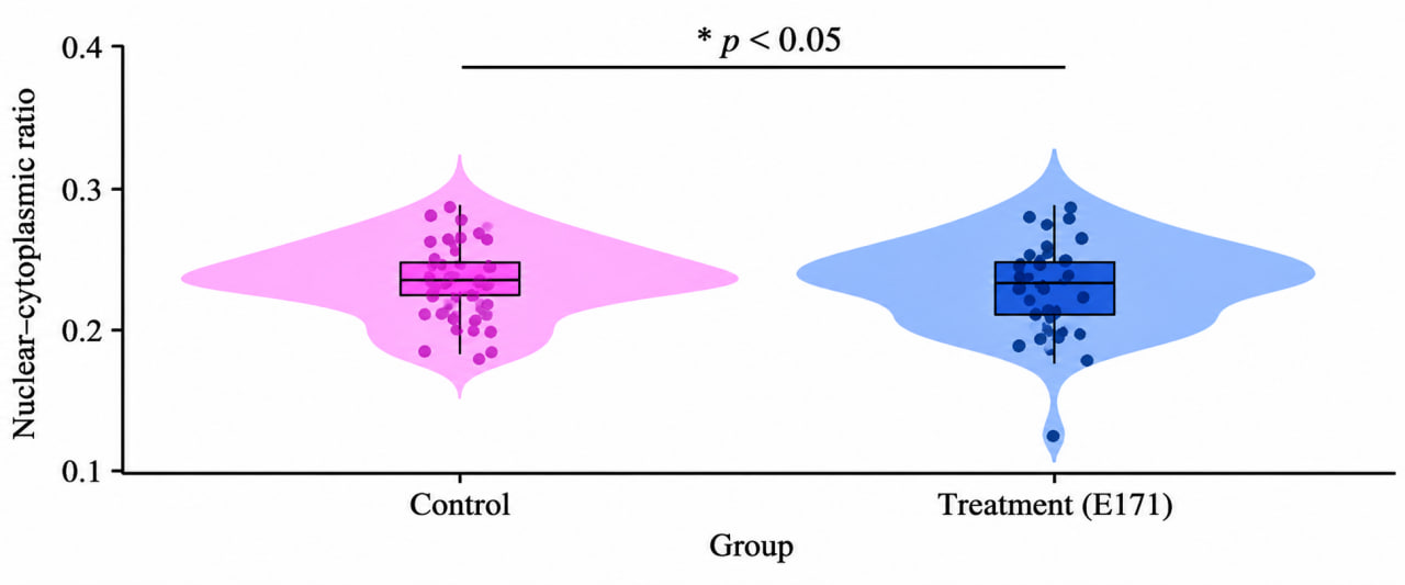 | Figure 3. Comparative data on cellular parameters. RStudio |
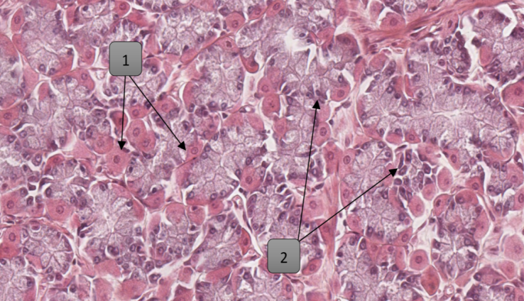 | Figure 4. Fundic region of the stomach in rats from the experimental group. 1 — parietal cells; 2 — chief cells. Haematoxylin and eosin staining. Magnification: 10×20 |
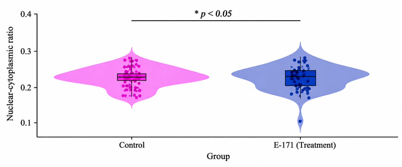 | Figure 5. Comparative analysis of the nuclear-to-cytoplasmic ratio in parietal cells of the gastric fundus. RStudio |
4. Discussion
- This study focused on a comparative assessment of the morphometric parameters of glands, parietal cells, and chief cells in the fundic and antral mucosa of rat stomachs exposed to the food colorants E-171 and E-173. Cytoplasmic and nuclear parameters, the nuclear-to-cytoplasmic ratio, and the morphological state of glandular structures are important criteria for interpreting the findings. Their integrated assessment helps characterise the potential cytotoxic effects of food colorants on gastric tissues. The literature contains differing findings on the biological effects of titanium dioxide. Bettini and colleagues reported changes related to immune homeostasis and pathological findings in the rat colon following exposure to E-171 [2]. In contrast, Blevins and colleagues found no treatment-related significant changes in the histopathology of the small and large intestines or in certain immunological parameters in rats fed an E-171-containing diet for 7 or 100 days [4,5]. These differences highlight the importance of experimental design, route of administration, dose, duration of exposure, and tissue type assessed. Therefore, findings from intestinal studies cannot be directly applied to parietal or chief cells of the stomach.
5. Conclusions
- Investigating the effects of the food colorants E-171 (titanium dioxide) and E-173 (aluminium) on the body, particularly assessing possible morphofunctional changes in gastric tissues, is an important area of modern toxicology and experimental morphology.Assessing the structure of the gastric mucosa, glandular activity, cellular condition, and signs of local inflammation together with functional and biochemical parameters may provide a deeper understanding of the potential mechanisms of action of these colorants. Comparing exposed groups with the control group and analysing the findings using appropriate statistical methods, including assessment of dose-related changes where applicable, improves the scientific reliability of the study.Thus, comprehensive morphofunctional assessment of the effects of E-171 and E-173 on the stomach is important for expanding scientific knowledge of their biological safety, identifying potential risk factors, and developing future preventive and practical recommendations. Final conclusions should be based only on experimental findings and reliable scientific evidence.