
-
Paper Information
- Previous Paper
- Paper Submission
-
Journal Information
- About This Journal
- Editorial Board
- Current Issue
- Archive
- Author Guidelines
- Contact Us
International Journal of Genetic Engineering
p-ISSN: 2167-7239 e-ISSN: 2167-7220
2026; 14(9): 237-245
doi:10.5923/j.ijge.20261409.03
Received: Aug. 16, 2026; Accepted: Sep. 6, 2026; Published: Sep. 9, 2026

GC–MS Profiling of Lipid and Sterol Metabolites in the Biomass of Inonotus hispidus (Bull.) P. Karst.
 Abstract
 Reference
 Full-Text PDF
 Full-text HTML
Xumoyun N. Quziboyev
Department of Microbiology and Biotechnology, Karshi State University, Karshi, Uzbekistan
Correspondence to: Xumoyun N. Quziboyev, Department of Microbiology and Biotechnology, Karshi State University, Karshi, Uzbekistan.
| Email: |  |
Copyright © 2026 The Author(s). Published by Scientific & Academic Publishing.
This work is licensed under the Creative Commons Attribution International License (CC BY).
http://creativecommons.org/licenses/by/4.0/

This study aimed to characterize the gas chromatography–mass spectrometry (GC–MS) profile of a lipophilic extract obtained from naturally grown fruiting bodies of Inonotus hispidus and to identify putative lipid- and sterol-related metabolites. Dried and milled biomass (54 g) was extracted with 1 L of 96% ethanol for 5–7 days. The concentrated extract was fractionated with hexane and chloroform and subsequently subjected to GC–MS analysis. A total of 28 chromatographic peaks were detected. Comparison with the NIST mass spectral library showed that the predominant peak, observed at 10.752 min, matched methyl linoleate with 99% similarity and accounted for 27.86% of the total peak area. Methyl palmitate (12.62%), ethyl linoleate (8.46%), methyl stearate (7.75%), palmitic acid (5.16%), and linoleic acid (5.35%) were also major constituents. Squalene represented 1.61% of the total peak area, whereas peaks at 20.282 and 20.995 min showed putative matches to an ergosta-type sterol and ergosterol, respectively. A phthalate derivative was interpreted as a possible laboratory contaminant. These findings indicate that the lipophilic fraction of I. hispidus fruiting bodies may be rich in fatty-acid derivatives and compounds associated with sterol biosynthesis. Because compound assignments were based on library matching, they should be regarded as tentative and confirmed using authentic standards, retention indices, and complementary LC–MS and/or NMR analyses.
Keywords: Inonotus hispidus, GC–MS, Lipid metabolites, Fatty acids, Methyl linoleate, Palmitic acid, Squalene, Ergosterol, Sterols, Bioactive compounds
Cite this paper: Xumoyun N. Quziboyev, GC–MS Profiling of Lipid and Sterol Metabolites in the Biomass of Inonotus hispidus (Bull.) P. Karst., International Journal of Genetic Engineering, Vol. 14 No. 9, 2026, pp. 237-245. doi: 10.5923/j.ijge.20261409.03.
Article Outline
1. Introduction
- Medicinal and biologically active fungi are important sources of natural metabolites, including polysaccharides, phenolic compounds, terpenoids, sterols, fatty acids, organic acids, and numerous other secondary metabolites. These compounds may exhibit antioxidant, anti-inflammatory, antimicrobial, immunomodulatory, and, in some cases, cytotoxic activities. Therefore, detailed characterization of the chemical composition of macrofungi is of considerable scientific and practical relevance to pharmacology, food biotechnology, nutraceutical development, and the cosmetics industry.Inonotus hispidus (Bull.) P. Karst. is a wood-decaying basidiomycete belonging to the family Hymenochaetaceae and occurs predominantly on the trunks of deciduous trees. Its fruiting bodies are typically yellowish brown to dark brown, with a distinctly hispid surface, and may develop as either parasites or saprotrophs. In recent years, the bioactive constituents, pharmacological properties, and potential food and health-related applications of I. hispidus have attracted increasing scientific attention [11,12].Previous studies have reported phenolics, flavonoids, polysaccharides, terpenoids, fatty acids, and sterol-type compounds in I. hispidus. Ergosterol and its derivatives are particularly important because they are major structural components of fungal cell membranes. Ergosterol can therefore serve as a chemical marker of fungal biomass and sterol metabolism. Variations in ergosterol peroxide and other ergostane derivatives have also been associated with the physiological state and developmental stage of I. hispidus.The metabolite profile of fungi may vary markedly with growth substrate, host-tree species, geographic origin, sampling season, developmental stage of the fruiting body, extraction procedure, and solvent system. Consequently, profiling naturally occurring I. hispidus collected from local habitats is scientifically relevant. Such analyses can help assess its value as a biological resource, identify promising metabolites, and provide a basis for subsequent bioactivity studies.Gas chromatography–mass spectrometry is a powerful analytical technique for detecting volatile and semivolatile organic compounds and for the preliminary identification of fatty-acid derivatives, aliphatic hydrocarbons, terpenoids, and sterols. Library-based spectral matching enables tentative assignment of extract constituents; however, particularly in complex natural matrices, GC–MS identifications should be verified using authentic standards or complementary analytical approaches.Accordingly, this study aimed to characterize lipid and sterol metabolites in an extract prepared from naturally occurring I. hispidus fruiting bodies by GC–MS and to assess their potential biotechnological relevance.To the best of our knowledge, this work provides the first systematic GC–MS description of a lipophilic extract obtained from naturally grown I. hispidus fruiting bodies collected in Uzbekistan [13-16]. Major fatty-acid derivatives, squalene, and putative ergosta-type sterols were evaluated together on the basis of their relative peak areas.The specific objectives of this study were to: (i) prepare dried biomass from I. hispidus fruiting bodies and obtain a 96% ethanolic extract; (ii) partially fractionate the extract using hexane, chloroform, and ethanol while reducing pigmented impurities; (iii) determine the GC–MS chemical profile of the resulting extract; (iv) evaluate the major fatty acids, aliphatic hydrocarbons, squalene, and ergosterol-like compounds detected; and (v) assess the potential biological and biotechnological significance of the identified metabolites.
2. Materials and Methods
2.1. Collection and Preparation of Fungal Material
- Fruiting bodies of naturally occurring I. hispidus were collected from a tree trunk and transported to the laboratory. Visible debris and remnants of tree bark were removed. The material was then dried, milled, and homogenized to obtain a uniform biomass fraction. The prepared dry biomass was weighed on an electronic balance and had a total mass of 54 g.
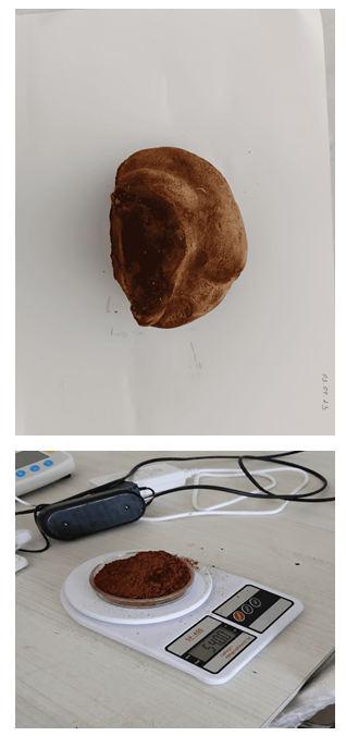 | Figure 1. Dried and milled biomass prepared from an I. hispidus fruiting body collected from a tree trunk |
2.2. Preparation of the Ethanolic Extract
- Milled dry I. hispidus biomass (54 g) was placed in a glass vessel and mixed with 1 L of 96% ethanol. Extraction was carried out in the dark under laboratory conditions for 5–7 days. The mixture was agitated periodically to improve the transfer of extractable constituents into the solvent.At the end of extraction, the dark-brown suspension was filtered through filter paper. The filtrate was separated from the solid residue and concentrated for subsequent processing.
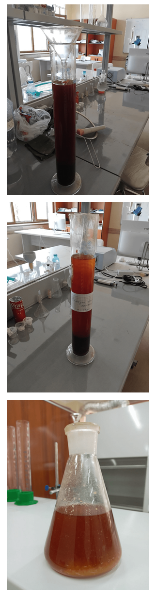 | Figure 2. Filtration of the 96% ethanolic extract prepared from 54 g of dried I. hispidus biomass |
2.3. Partial Purification of the Extract with Organic Solvents
- To reduce co-extracted compounds and obtain a cleaner fraction for GC–MS analysis, the concentrated extract was sequentially treated with organic solvents. First, the viscous extract was washed with hexane to separate part of the nonpolar and strongly lipophilic constituents.The sample was subsequently treated with chloroform to recover a lipophilic metabolite fraction. To reduce pigments and other strongly colored compounds, the resulting fraction was washed again with 96% ethanol. This procedure yielded a relatively purified, dark-colored extract fraction suitable for GC–MS analysis.
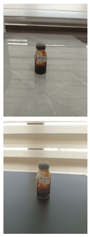 | Figure 3. Concentrated fraction of the I. hispidus extract after sequential solvent treatment |
2.4. GC–MS Analysis
- The prepared extract fraction was analyzed by gas chromatography–mass spectrometry to obtain a preliminary profile of lipids, sterols, and other semivolatile organic constituents. Chromatographic peaks were evaluated according to retention time and mass-spectral characteristics. Preliminary compound identification was performed by comparison with a mass-spectral library; consequently, all reported assignments were regarded as tentative, and definitive identification of selected compounds requires authentic standards and/or complementary spectroscopic analyses.GC–MS analysis was performed in full-scan mode using an automated sample-introduction system. Data were acquired with the instrumental method "Drugs_SCAN_MED_22_757_auto.M". The mass spectrometer was operated under electron ionization at 70 eV. Peaks were evaluated on the basis of retention time, integrated peak area, and diagnostic mass fragments. Experimental spectra were compared with the NIST17.L mass-spectral library using a minimum match threshold of 80%. In total, 28 chromatographic peaks were recorded between 4.277 and 20.995 min.
2.5. Criteria for Compound Identification
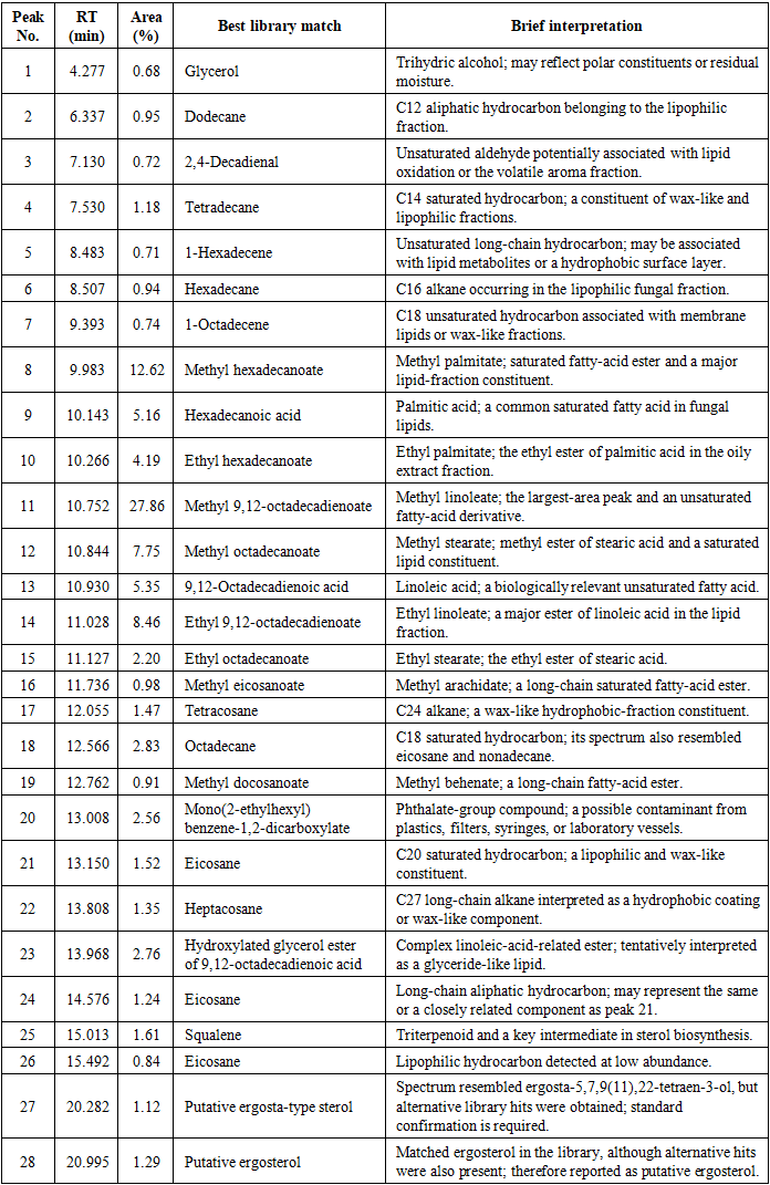
- Preliminary identification was based on comparison of experimental mass spectra with the NIST library. Table 1 reports the library match, retention time, and relative peak area for each peak. Identification confidence should be strengthened in future work by calculating linear retention indices using an n-alkane series, confirming major compounds with authentic standards, and examining the sterol fraction by LC–MS or NMR. Names based solely on spectral-library similarity are therefore designated as tentative or putative.
|
2.6. Experimental Design and Statistical Analysis
- The GC–MS data presented here represent a descriptive chemical profile of a single extract; therefore, inferential statistics were not applied. Relative abundance was calculated as the percentage contribution of each integrated peak to the total area of all detected peaks in the total-ion chromatogram. At least three independent biological replicates, together with technical replicates for each sample, should be analyzed in follow-up work, with results reported as mean ± standard deviation.
2.7. Control of Contamination and Analytical Artifacts
- To distinguish phthalate derivatives and solvent-related artifacts from genuine sample constituents, solvent blanks, extraction blanks, and predominantly glass laboratory ware with minimal plastic contact are recommended. Interpretation of methyl and ethyl esters should explicitly document the solvents used, any derivatization step, and the complete sample-preparation workflow. Because no separate derivatization was performed in this experiment, methyl- and ethyl-ester assignments should be treated as tentative library-based identifications.
3. Results and Discussion
3.1. GC–MS Profile of the Inonotus hispidus Extract
- GC–MS analysis of the I. hispidus extract revealed 28 chromatographic peaks. Spectral-library comparison indicated the presence of aliphatic hydrocarbons, aldehydes, fatty acids and their methyl and ethyl esters, terpenoids, sterol-like compounds, and several possible exogenous contaminants.The largest peak was detected at 10.752 min and showed 99% similarity to methyl 9,12-octadecadienoate (methyl linoleate), accounting for 27.86% of the total peak area. The 99% spectral match is a very high similarity score, indicating a high level of confidence in the tentative library identification of this peak; nevertheless, because no authentic standard or retention-index confirmation was performed, the assignment is still reported as tentative. Other abundant constituents included methyl hexadecanoate, ethyl 9,12-octadecadienoate, methyl stearate, palmitic acid, and linoleic acid.
3.2. Chemical Classes of the Detected Compounds
- Fatty acids and their esters constituted the largest chemical class in the extract. This group included methyl palmitate, palmitic acid, ethyl palmitate, methyl linoleate, methyl stearate, linoleic acid, ethyl linoleate, ethyl stearate, methyl arachidate, and methyl behenate, demonstrating the predominance of lipid-related metabolites. This predominance likely reflects both a biological and a methodological factor: fatty-acid esters are typical storage and membrane-associated lipids of basidiomycete fruiting bodies, and the sequential hexane–chloroform–ethanol treatment used here was specifically designed to enrich nonpolar, lipophilic constituents while removing polar pigments and other co-extracted compounds, so the resulting fraction was expected to be dominated by this chemical class.Methyl linoleate was the dominant component, representing 27.86% of the total peak area. Methyl palmitate, ethyl linoleate, and methyl stearate contributed 12.62%, 8.46%, and 7.75%, respectively. These compounds may be major constituents of the oily extractive fraction of the fruiting body.Long-chain hydrocarbons, including dodecane, tetradecane, hexadecane, octadecane, eicosane, tetracosane, and heptacosane, were also detected, suggesting the presence of hydrophobic and wax-like components. Such compounds may be associated with the protective surface layer, intracellular lipid droplets, or the natural wax fraction of the fruiting body. This interpretation is supported by their chain lengths (C12–C27) and saturation pattern, which are typical of cuticular wax hydrocarbons that form a hydrophobic barrier on fungal fruiting-body surfaces, protecting against desiccation and microbial colonization, as well as of hydrocarbons stored within intracellular lipid droplets.Squalene, detected at 15.013 min, is an important intermediate in triterpenoid and sterol biosynthesis. Its occurrence supports the possible presence of lipophilic metabolites associated with fungal sterol-forming pathways.Peaks at 20.282 and 20.995 min produced spectra resembling an ergosta-type compound and ergosterol, respectively. Because alternative library hits were also obtained, these compounds are more appropriately described as a putative ergosta-type sterol and putative ergosterol; confirmation requires comparative analysis with an authentic ergosterol standard and/or LC–MS analysis.The phthalate-related compound detected at 13.008 min was interpreted as a likely laboratory contaminant rather than a natural fungal metabolite, because phthalates may leach from plastic containers, caps, syringes, filters, or other solvent-contact materials.
3.3. Relative Abundance and Biological Relevance of the Major Components
- The 28 detected peaks were classified, on the basis of library matches, as aliphatic hydrocarbons, fatty acids and their esters, terpenoids, sterol-like compounds, and possible exogenous contaminants. Fatty acids and their derivatives clearly predominated, confirming that the analyzed fraction was enriched in lipophilic metabolites.
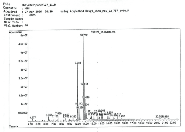 | Figure 4. Total-ion chromatogram of the I. hispidus extract obtained by GC–MS |
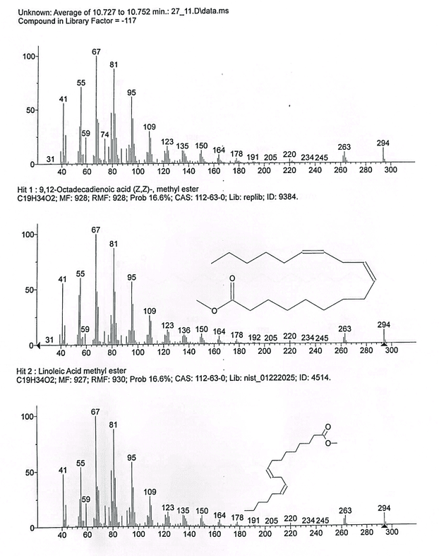 | Figure 5. Mass spectrum of the peak detected at 10.752 min and its library match to methyl 9,12-octadecadienoate |
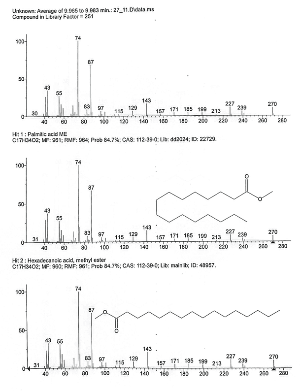 | Figure 6. Experimental mass spectrum of palmitic acid detected in the I. hispidus extract and comparison with the library spectrum |
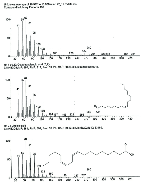 | Figure 7. Experimental mass spectrum of linoleic acid detected in the I. hispidus extract and comparison with the library spectrum |
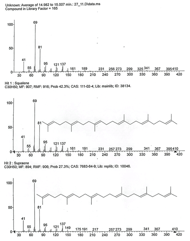 | Figure 8. Mass spectrum of squalene detected in the I. hispidus extract and comparison with library data |
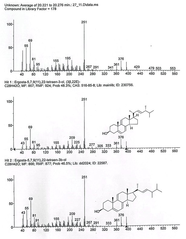 | Figure 9. Mass spectrum of the putative ergosta-type sterol detected at 20.282 min |
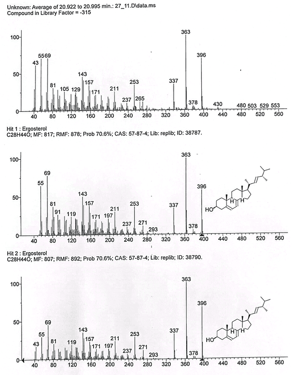 | Figure 10. Mass spectrum of the putative ergosterol detected at 20.995 min and comparison with the library spectrum |
3.4. Comparison with Previous Studies
- The GC–MS profile obtained in this study was dominated by fatty acids and their methyl and ethyl esters, together with long-chain hydrocarbons, squalene, and sterol-like compounds. The predominance of the methyl-linoleate-like peak and the detection of palmitic-, stearic-, and linoleic-acid derivatives indicate that lipid metabolism makes a substantial contribution to the chemical composition of I. hispidus biomass.These observations are broadly consistent with earlier studies of wood-decaying basidiomycetes, in which palmitic, stearic, and linoleic acids have frequently been reported among the major fatty acids; in some fungal species, linoleic acid represents a substantial proportion of the total fatty-acid pool [1,2,9].The chemical diversity of I. hispidus has also been documented in previous experimental and review articles. Polyphenols, triterpenoids, and other bioactive secondary metabolites have been reported, and their abundance may vary substantially with geographic origin, developmental stage, host-tree species, and extraction procedure [3–5].A squalene-like peak was detected at 15.013 min in the present study. Because squalene is a key intermediate in sterol biosynthesis, its occurrence indicates the presence of lipophilic metabolites associated with terpenoid and sterol metabolism. Previous reports describing the concurrent detection of palmitic and stearic acids, long-chain hydrocarbons, and squalene in selected wood-decaying basidiomycetes support this interpretation [6].The peaks observed at 20.282 and 20.995 min showed library similarity to an ergosta-type compound and ergosterol, respectively. Ergosterol is one of the principal sterols in fungal membranes and is widely applied as a biomarker of fungal biomass; it has been reported as the predominant sterol in several edible and medicinal mushrooms [2,7,8,10].However, the ergosterol and ergosta-type assignments in this study were based exclusively on spectral-library similarity. They should therefore be interpreted as putative ergosterol and a putative ergosta-type sterol. Definitive identification requires comparative GC–MS analysis with authentic standards and confirmation by LC–MS or NMR.The results suggest that unsaturated fatty-acid derivatives may predominate in the lipophilic fraction of I. hispidus, particularly methyl linoleate, ethyl linoleate, and linoleic-acid-like compounds. Nevertheless, GC–MS profiling alone cannot establish pharmacological or cosmetic efficacy; antioxidant, antimicrobial, cytotoxicity, and safety assays are required before any functional claims can be made.An important contribution of this study is the preliminary lipid and sterol profile generated from naturally grown I. hispidus collected under local conditions. Future comparative studies should include samples from different host trees, developmental periods, and solvent systems to clarify the variability of the chemical composition of this fungus.
3.5. Study Limitations
- The principal limitations were the use of a single biological sample, the absence of technical replication and inferential statistics, reliance primarily on NIST library matching, lack of retention-index data and authentic-standard confirmation, and the absence of experimental antioxidant, antimicrobial, or cytotoxicity assays. The results should therefore be interpreted as a preliminary chemical profile.
4. Conclusions
- This study provides a preliminary GC–MS assessment of lipid and sterol metabolites in an extract prepared from naturally occurring I. hispidus fruiting bodies. Dried biomass (54 g) was extracted with 1 L of 96% ethanol for 5–7 days, and the concentrated extract was sequentially treated with hexane, chloroform, and ethanol before GC–MS analysis.A total of 28 chromatographic peaks were recorded. Library-based assignments indicated that the extract contained mainly aliphatic hydrocarbons, fatty acids and their esters, triterpenoids, and sterol-type compounds. The major conclusions are as follows: (i) fatty acids and their esters were the dominant chemical group, with palmitic, linoleic, and stearic acids and their methyl and ethyl esters indicating that products of lipid metabolism are prominent in I. hispidus biomass; (ii) the most intense peak, at 10.752 min, showed a high library match to methyl linoleate, suggesting that unsaturated fatty-acid derivatives are important constituents of the extract; (iii) detection of long-chain hydrocarbons confirmed the hydrophobic and lipophilic character of the extract; (iv) the detection of squalene suggests the presence of metabolites associated with terpenoid and sterol biosynthesis; and (v) peaks at 20.282 and 20.995 min showed library similarity to an ergosta-type sterol and ergosterol, respectively, supporting the possible occurrence of characteristic fungal sterol metabolism, although definitive identification requires authentic-standard comparison and complementary LC–MS or NMR analyses. The phthalate-related compound was considered a likely laboratory contaminant and was therefore excluded from the natural metabolites of I. hispidus.Taken together, the results indicate that the lipophilic fraction of the analyzed I. hispidus sample may contain fatty-acid derivatives, squalene, and sterol-like compounds. Because this conclusion is based on a descriptive, library-assisted profile of a single sample, species-level generalization requires biological replication, quantitative confirmation with authentic standards, and biological-activity testing.
ACKNOWLEDGEMENTS
- The author thanks the Department of Microbiology and Biotechnology, Karshi State University, for laboratory support.
Funding
- This research received no external funding.
Conflict of Interest
- The author declares no conflict of interest.
Data Availability
- The data presented in this study are available from the corresponding author upon reasonable request.