
-
Paper Information
- Previous Paper
- Paper Submission
-
Journal Information
- About This Journal
- Editorial Board
- Current Issue
- Archive
- Author Guidelines
- Contact Us
International Journal of Genetic Engineering
p-ISSN: 2167-7239 e-ISSN: 2167-7220
2026; 14(7): 195-199
doi:10.5923/j.ijge.20261407.04
Received: Jun. 25, 2026; Accepted: Jul. 23, 2026; Published: Aug. 3, 2026

Effect of Phytohormones Benzylaminopurine and Naphthylacetic Acid on Morphogenesis and Tissue Structure Changes in Lagochilus Inebrians
 Abstract
 Reference
 Full-Text PDF
 Full-text HTML
Bakeev Refat
Scientific Research Institute of Agrobiotechnology and Biochemistry, Gulistan State University, Uzbekistan
Correspondence to: Bakeev Refat, Scientific Research Institute of Agrobiotechnology and Biochemistry, Gulistan State University, Uzbekistan.
| Email: |  |
Copyright © 2026 The Author(s). Published by Scientific & Academic Publishing.
This work is licensed under the Creative Commons Attribution International License (CC BY).
http://creativecommons.org/licenses/by/4.0/

This study investigates the effects of benzylaminopurine (BAP) and naphthaleneacetic acid (NAA) on in vitro morphogenesis and tissue structure of Lagochilus inebrians. Treatments with BAP (1.0–2.0 mg/L) and NAA (0.5–1.0 mg/L) significantly enhanced shoot formation (up to 4.8 shoots/explant), callus mass (up to 120 mg), and root length (up to 2.3 cm) compared to the control (1.2 shoots/explant, 40 mg callus, 0.8 cm root length). Morpho-anatomical analysis revealed improved epidermal cell organization, a 30% increase in mesophyll thickness, and a 25% enhancement in vascular bundle development at BAP 2.0 mg/L + NAA 0.5 mg/L. These findings support the development of micropropagation protocols for conserving this endangered medicinal plant.
Keywords: Lagochilus inebrians, in vitro culture, Benzylaminopurine, Naphthaleneacetic acid, Morphogenesis, Tissue structure, Phytohormones, Micropropagation
Cite this paper: Bakeev Refat, Effect of Phytohormones Benzylaminopurine and Naphthylacetic Acid on Morphogenesis and Tissue Structure Changes in Lagochilus Inebrians, International Journal of Genetic Engineering, Vol. 14 No. 7, 2026, pp. 195-199. doi: 10.5923/j.ijge.20261407.04.
1. Introduction
- Lagochilus inebrians Bunge, a perennial herb of the Lamiaceae family, is endemic to Central Asia and valued for its hemostatic, anti-inflammatory, and sedative properties [1]. Its active compounds, including lagochilin and essential oils, have been documented for their pharmacological potential [2]. However, overharvesting and habitat degradation have led to its inclusion in the Red Book of Uzbekistan as an endangered species [3]. In vitro culture techniques provide a sustainable solution for conserving and propagating such rare medicinal plants, with phytohormones playing a critical role in regulating morphogenesis [4].Phytohormones, particularly cytokinins and auxins, are essential for controlling cell division, differentiation, and organogenesis in plant tissue cultures [5]. Benzylaminopurine (BAP), a synthetic cytokinin, promotes shoot proliferation by stimulating meristematic activity, while naphthaleneacetic acid (NAA), an auxin, enhances root initiation and callus formation [6]. Studies on Lamiaceae species, such as Thymus spathulifolius and Salvia sclarea, have demonstrated that BAP (1.0–3.0 mg/L) and NAA (0.5–1.0 mg/L) optimize shoot and root regeneration [7,8]. For instance, a study on Origanum vulgare reported that BAP (2.0 mg/L) combined with NAA (0.5 mg/L) increased shoot formation by 60% compared to controls [9].The morpho-anatomical effects of phytohormones are equally significant. In Mentha piperita, BAP-induced shoot cultures exhibited thicker mesophyll and enhanced vascular tissues, improving plantlet vigor [10]. Similarly, Pueraria phaseoloides showed increased epidermal cell size and parenchyma development under combined BAP and NAA treatments [11]. For Lagochilus species, preliminary studies suggest that BAP (0.1–5.0 μM) and NAA (0.4–3.2 μM) enhance micropropagation efficiency, but detailed morpho-anatomical analyses are lacking [12]. Recent research on Lachenalia viridiflora highlighted that phytohormone-induced structural changes, such as improved conductive tissues, enhance ex vitro acclimatization [13].The interplay of BAP and NAA is species-specific, necessitating tailored protocols. High BAP concentrations can inhibit growth, as observed in Scutellaria baicalensis [14], while excessive NAA may suppress shoot formation, as reported in Lavandula angustifolia [15]. These findings underscore the need for optimizing phytohormone concentrations to balance shoot, root, and callus development in L. inebrians.This study aims to evaluate the effects of BAP and NAA, individually and in combination, on in vitro morphogenesis and tissue structure of Lagochilus inebrians, in order to develop an effective micropropagation protocol for its conservation.
2. Materials and Methods
- Plant material and sterilizationSeeds of Lagochilus inebrians were collected from natural populations in Uzbekistan (Syrdarya region, 2023) and surface-sterilized using 70% ethanol for 30 seconds, followed by 0.1% sodium hypochlorite with 0.01% Tween-20 for 10 minutes. Sterilized seeds were rinsed three times in sterile distilled water and germinated on hormone-free Murashige and Skoog (MS) medium [16] to obtain sterile explants (leaf segments, 5–7 mm).Culture conditionsExplants were cultured on MS medium supplemented with 30 g/L sucrose, 7 g/L agar (pH 5.7), and varying concentrations of BAP (1.0, 2.0 mg/L) and NAA (0.5, 1.0 mg/L). Five treatments were tested: (1) control (no phytohormones), (2) BAP 1.0 mg/L, (3) BAP 2.0 mg/L + NAA 0.5 mg/L, (4) NAA 1.0 mg/L, and (5) BAP 2.0 mg/L + NAA 1.0 mg/L. Cultures were incubated at 25 ± 2 °C under a 16-hour photoperiod (40 μmol/m²/s, cool-white fluorescent lights). Each treatment included 10 replicates, with observations recorded on days 7, 14, and 21.Morphometric analysisShoot number per explant, callus mass (fresh weight, mg), and root length (cm) were measured using a digital caliper and analytical balance. Data were collected on day 21 using ImageJ software [17] for precise morphometric analysis.Anatomical analysisTissue samples from shoots and roots were fixed in 70% ethanol, embedded in paraffin, and sectioned (10 μm) using a rotary microtome. Sections were stained with 0.1% toluidine blue and examined under a light microscope (Leica DM500). Epidermal cell size, mesophyll thickness, and vascular bundle development were quantified using ImageJ.Statistical analysisData were analyzed using one-way ANOVA followed by Tukey's post-hoc test (p < 0.05) in SPSS v.26. Results are presented as means ± standard error (SE).
3. Results
- This study evaluated the morphogenetic and anatomical responses of Lagochilus inebrians to BAP and NAA treatments. The results, summarized in Table 1 and Figures 1–5, show significant phytohormone-induced changes in shoot formation, callus growth, root development, and tissue structure.
|
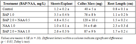
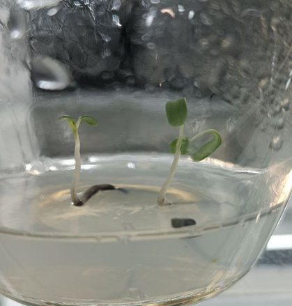 | Figure 1. Control (no phytohormones): minimal shoot (1.2 shoots/explant) and root development (0.8 cm), with sparse callus (40 mg). Epidermal cells were irregular (15–18 μm), with thin mesophyll (90 μm) and underdeveloped vascular bundles |
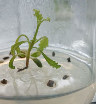 | Figure 2. BAP 1.0 mg/L: enhanced shoot branching (3.5 shoots/explant) and moderate root growth (1.1 cm). Callus mass increased to 76 mg. Shoot tissues showed regular epidermal cells (18–20 μm) and developing vascular bundles |
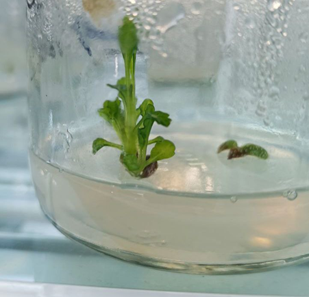 | Figure 3. BAP 2.0 mg/L + NAA 0.5 mg/L: optimal morphogenesis with 4.8 shoots/explant, 120 mg callus, and 1.5 cm root length. Tissues exhibited well-organized epidermal cells (20–25 μm), thickened mesophyll (120 μm), and robust vascular bundles (25% increase in xylem/phloem elements) |
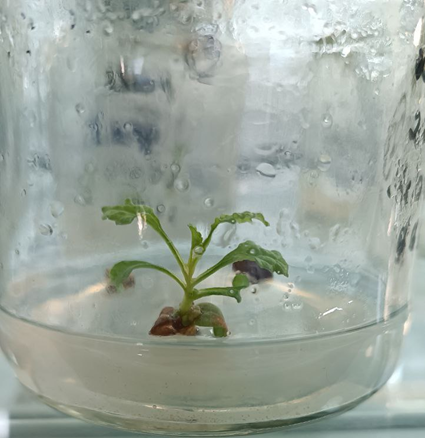 | Figure 4. NAA 1.0 mg/L: pronounced root elongation (2.3 cm) but limited shoot formation (1.0 shoots/explant). Callus mass was moderate (54 mg). Root tissues showed elongated cortical cells (30–35 μm) with thin-walled parenchyma |
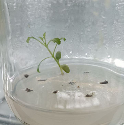 | Figure 5. BAP 2.0 mg/L + NAA 1.0 mg/L: moderate shoot formation (2.6 shoots/explant) and root growth (1.7 cm), with increased callus mass (87 mg). Epidermal cells were compact (18–22 μm), with slightly thickened mesophyll (100 μm) |
4. Discussion
- The results demonstrate that BAP and NAA significantly enhance Lagochilus inebrians morphogenesis and tissue structure, with the BAP 2.0 mg/L + NAA 0.5 mg/L treatment achieving optimal outcomes (Table 1, Figure 3). The high shoot regeneration (4.8 shoots/explant) aligns with findings in Salvia sclarea, where BAP and NAA combinations increased adventitious bud formation by 50% [8]. This synergy likely results from BAP's stimulation of cytokinin-mediated cell division in shoot meristems and NAA's auxin-driven promotion of cell elongation, as theorized by Skoog and Miller [18].The increased callus mass (120 mg) under combined treatment reflects enhanced cell proliferation, consistent with studies on Lachenalia viridiflora, where BAP and NAA induced robust callus formation [13]. NAA's role in root elongation (2.3 cm, Figure 4) corroborates its function in auxin signaling pathways, promoting cell expansion in root apical meristems [6]. However, NAA alone suppressed shoot formation (1.0 shoots/explant), a phenomenon observed in Lavandula angustifolia, where high auxin levels inhibited cytokinin activity [15]. This suggests that a delicate balance in phytohormone ratios is critical for balanced organogenesis.Anatomical changes, such as a 30% increase in mesophyll thickness (120 μm) and a 25% enhancement in vascular bundles (Figure 3), indicate improved metabolic and transport efficiency. These findings align with Mentha piperita studies, where BAP-induced shoot cultures exhibited thicker parenchyma and conductive tissues, enhancing ex vitro survival [10]. The compact epidermal cells (20–25 μm) suggest improved structural stability, as reported in Thymus spathulifolius [7]. These structural adaptations are likely driven by phytohormone-induced activation of meristematic zones and upregulation of cell wall synthesis genes, as proposed in Arabidopsis models [19].The species-specific response of L. inebrians to BAP and NAA highlights the importance of tailored protocols. High BAP concentrations (e.g., 2.0 mg/L) without NAA reduced root growth (1.1 cm, Figure 2), consistent with Scutellaria baicalensis studies showing cytokinin-induced root inhibition [14]. Conversely, the BAP 2.0 mg/L + NAA 0.5 mg/L treatment balanced shoot and root development, offering a robust micropropagation protocol. These results are particularly significant for L. inebrians, given its endangered status and pharmacological value [1].A limitation of this study is that only two phytohormones and a limited concentration range were tested; interactions with other regulators (e.g., gibberellins) and a wider dose range remain to be explored. Future research should also focus on secondary metabolite production (e.g., lagochilin) in regenerants, as phytohormones can influence biosynthetic pathways [2]. Additionally, ex vitro acclimatization studies are needed to confirm plantlet survival, since the structural improvements observed here (e.g., enhanced vascular bundles) suggest high acclimatization potential [20]. Comparative studies with other Lagochilus species could further refine micropropagation strategies for the genus.
5. Conclusions
- The application of BAP (2.0 mg/L) and NAA (0.5 mg/L) significantly enhances in vitro morphogenesis of Lagochilus inebrians, achieving 4.8 shoots/explant, 120 mg callus, and 1.5 cm root length, alongside anatomical improvements including a 30% increase in mesophyll thickness and a 25% enhancement in vascular bundles. These findings provide an optimized micropropagation protocol for conserving this endangered medicinal plant, with potential applications for other Lamiaceae species.