D. U. Mamadiyarova1, S. B. Azimova2, M. U. Mamadiyarov3
1Department of Physiology, Samarkand State Medical University, Uzbekistan
2Department of Normal and Pathological Physiology, Tashkent State Medical University, Uzbekistan
3Department of Biology, Samarkand State Pedagogical Institute, Uzbekistan
Copyright © 2026 The Author(s). Published by Scientific & Academic Publishing.
This work is licensed under the Creative Commons Attribution International License (CC BY).
http://creativecommons.org/licenses/by/4.0/

Abstract
This article examines the role of clinical and demographic characteristics, age, gender, and medical history in the development and progression of angiopathy in 94 patients with type 2 diabetes mellitus. The article is based on a detailed, multivariate, and systematic analysis of clinical data obtained during the patient examination, including age, gender, and disease duration.
Keywords:
Diabetic angiopathy, Oxidative system, Antioxidant system, Vascular damage, Endothelial dysfunction, Visceral condition, Endothelin-1, NO, VEGF-A
Cite this paper: D. U. Mamadiyarova, S. B. Azimova, M. U. Mamadiyarov, Clinical and Demographic Characteristics of Diabetic Angiopathy Development, Molecular Markers of Endothelial Dysfunction, International Journal of Genetic Engineering, Vol. 14 No. 7, 2026, pp. 186-190. doi: 10.5923/j.ijge.20261407.02.
1. Introduction
This study examines the scientific basis for elucidating the role of genetic polymorphisms in the development of endothelial dysfunction, which occurs in the presence of hyperglycemia in the blood, leading to a decrease in the amount of NO, a vasodilator, and an increase in the amount of endothelin-1, a vasoconstrictor [3,4,10]. The article uses the results of molecular genetic studies to conduct genetic tests at the first signs of angiopathy, rather than for its diagnosis and treatment, and to carry out preventive work, which is more cost-effective than treatment [1,2,7]. Particular attention is paid to the evolution of the classification system developed under the auspices of the World Health Organization (1965, 1980, 1999, 2019), which emphasizes the prognostic, pathogenetic and practical significance for paramedical work based on molecular genetic and clinical phenotypic characteristics [5,6,8,9].This article is based on a comprehensive, multifaceted, and systematic analysis of clinical data obtained during a prospective screening of 94 residents of the Samarkand region. The research methodology was developed using the principles of evidence-based medicine and mathematical modeling, which enabled us to assess the risk of developing metabolic disorders leading to irreversible structural changes in blood vessels. The clinical phase included a detailed medical history, symptom assessment, physical examination, and medical record review. This enabled the formation of representative samples for subsequent molecular genetic analysis. To ensure the reliability of biochemical and genetic comparisons and minimize the influence of random factors, the first group (n=51) was formed from patients with clearly defined diabetic angiopathy of the lower extremities, corresponding to stages IIB, III, and IV according to the Fontaine-Pokrovsky classification. These patients had hemodynamically significant arterial stenosis, severe intermittent claudication (less than 200 meters without pain), or trophic ulcers and necrosis. The second group (n=43) included patients with long-standing type 2 diabetes mellitus but with preserved hemodynamics of the main arteries (ankle-brachial index >0.9) and no signs of stress ischemia. The control group (n=83) consisted of apparently healthy volunteers from the same region, matched for demographic characteristics, but without carbohydrate metabolism disorders or cardiovascular disease.Objective of the study: To study the clinical and demographic characteristics of the development of diabetic angiopathy, the importance of molecular markers of endothelial dysfunction.
2. Study Materials and Methods
The study included 94 patients with type 2 diabetes mellitus registered at the Samarkand Endocrinology Dispensary, including those assigned to the study group based on clinical and laboratory tests. All patients were citizens of Uzbekistan and resided in Uzbekistan.The diagnosis was based on a combination of clinical, laboratory, and instrumental data and confirmed according to the international classification. The main group was divided into two subgroups: Group 1 – patients with uncomplicated diabetes (n=43), Group 2 – patients with diabetes complicated by diabetic angiopathy (n=51).
3. Study Results
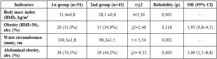
Evaluation of endothelial dysfunction in the development of angiopathy begins with an analysis of the internal organs and lipotoxicity. In this study, we examined the accumulation of visceral adipose tissue as one of the main causes, examining it not only as a manifestation of excess body weight but also as an independent etiological factor in pathogenesis. Anthropometric parameters such as body mass index (BMI) and waist circumference (WC) were used to quantitatively analyze abdominal obesity and the development of angiopathy. Analysis of the obtained data (Table 1) revealed a statistically significant predominance of the visceral fat phenotype in patients with diabetic angiopathy. The average intima-media thickness index in the first group was 31.4 ± 0.8 kg/m², which corresponds to stage 1 obesity and shows a significant difference with diabetic patients without angiopathy (28.1 ± 0.6 kg/m²; p = 0.001).Table 1. Comparative characteristics of anthropometric indicators and the risk of metabolic syndrome
 |
| |
|
Analysis of the topography of inflamed tissue is primarily informative for diagnostic purposes. In our group, 74.5% of participants had a specific residential area, while in the control group, this percentage was significantly lower—44.2%.Statistically significant differences (χ² = 9.12; p = 0.003 by Pearson's test) indicate a significant impact of abdominal obesity. The odds ratio (OR = 3.69) demonstrates that abdominal obesity increases the risk of developing diabetic angiopathy in patients with diabetes mellitus by almost fourfold. Pathogenetically, this is associated with excessive production of proinflammatory adipokines (TNF-α, IL-6, leptin) by visceral adipocytes. These mediators cause systemic endothelial inflammation, leading to the expression of adhesion molecules (ICAM-1, VCAM-1) and subsequent adhesion of leukocytes to the vascular wall, which is a key initial step in atherogenesis.Let's consider the stratification of metabolic diseases by gender and age. To exclude the influence of gender or age on the identified metabolic changes, we also analyzed the glycemic profile in the group of patients with diabetes and severe angiopathy and divided the patients into subgroups.The main intragroup comparative analysis (Table 2) did not reveal statistically significant differences in carbohydrate metabolism parameters depending on gender (p = 0.351) and age (p = 0.145). Table 2. Metabolic parameters in gender-age subgroups of patients with diabetes mellitus with developed angiopathy (n=51)
 |
| |
|
The study's results have significant clinical value, as they confirm the ubiquity of common metabolic pathways leading to vascular damage. Glucotoxicity and lipotoxicity have been found to have equally pronounced pathogenic effects on both men and women, as well as across age groups. This, in turn, underscores the importance of developing unified yet individualized metabolic correction strategies for all patients with diabetic angiopathy, regardless of their demographic characteristics.We will also examine molecular markers of endothelial dysfunction in combination with clinical dermatographic signs of angiopathy development in diabetes. Clinical manifestations of vascular complications in diabetes mellitus are the culmination of a long-term, latent process of cellular dysfunction. Modern pathological physiology views diabetic angiopathy as a panendotheliopathy—a complete disruption of the endothelial regulatory function.To investigate the underlying mechanisms that block natural tissue repair processes in patients with diabetes, we conducted a comparative quantitative analysis of two main systems:1. Angiogenesis systems, in which vascular endothelial growth factor A (VEGF-A), responsible for the formation of new capillaries, served as a marker.2. Vasomotor control systems, in which endothelin-1 (ET-1), the most potent known vasoconstrictor, served as a marker.We put forward and tested a scientific hypothesis that the terminal stage of angiopathy in patients of the first group is characterized not by excessive vascular spasm, as previously thought, but, on the contrary, by profound insensitivity and suppression of functional reserve.An assessment of angiogenesis in the examined patients revealed that soft tissue resistance to ischemic injury correlates with the effectiveness of collateral circulation. A decrease in partial oxygen pressure induces stabilization of the transcription factor HIF-1α. This factor, translocating to the nucleus, initiates expression of the VEGFA gene, which promotes proliferation of vascular collaterals. "Angiogenic deficiency," defined as a VEGF-A concentration below 1000 pg/ml, was adopted as a criterion for high ischemic risk (see Table 3).Table 3. Vascular endothelial growth factor (VEGF-A) concentration and risk assessment of angiogenic deficiency
 |
| |
|
An analysis of experimental data revealed a paradoxical phenomenon in the functioning of the vascular system. In patients in the first group, experiencing severe chronic ischemia, a multiple increase in growth factor levels was theoretically expected as a compensatory mechanism. However, the opposite trend was observed: the average VEGF-A level in the study group was 966.45 pg/ml, which is statistically significantly lower (p = 0.043) compared to the comparison group (1149.66 pg/ml) and even to healthy control values. This phenomenon was designated "angiogenic depression," since tissues experiencing hypoxia were unable to adequately produce signaling molecules. Further research showed that profound angiogenic deficiency is diagnosed in 66.7% of patients with critical ischemia. Calculation of the odds ratio (OR = 5.82) with a high degree of statistical significance (χ2 = 15.8; p < 0.001) indicates that a deficiency in basal VEGF levels increases the risk of irreversible necrotic changes and amputation by almost sixfold. The pathophysiological cause of this condition is a disruption of adaptive mechanisms under the influence of glucose toxicity. Hyperglycemia induces glycation of HIF-1α and the destruction of protein growth factors by free radicals, which impedes angiogenesis and leads to ischemia without alternative.When assessing vascular tone and endothelial dysfunction in patients with diabetes, endothelin-1 (ET-1) is considered a marker of early-stage endothelial dysfunction, causing vasospasm. However, our data showed that with long-term diabetes (more than 10 years), the dynamics of this indicator invert. We analyzed ET-1 levels, using a decrease in its concentration below the physiological optimum (<210 pg/ml) as a marker of maladaptation (see Table 4).Table 4. Endothelin-1 (ET-1) levels and endothelial secretory function assessment
 |
| |
|
Our data show that all patients with diabetes mellitus exhibit decreased endothelin-1 levels compared to healthy individuals. Peptide concentrations in groups 1 and 2 (198.66 and 193.69 pg/ml) were more than 20% lower than control values (253.75 pg/ml; p < 0.001).Moreover, no statistically significant difference was recorded between the groups of diabetics (with and without angiopathy) (p = 0.661).Interpretation of the obtained data indicates complete exhaustion of the endothelial secretory function. The pathogenesis of vascular changes in diabetes mellitus includes two phases: 1. The activation phase (the first 5-7 years), characterized by endothelial hyperactivity and increased production of endothelin-1 (ET-1), which causes vasospasm and arterial hypertension. 2. The exhaustion phase (disease duration >10 years), accompanied by apoptosis of endothelial cells and their replacement with connective tissue. Transformation of the vascular wall into a rigid structure devoid of active regulation of tone leads to the development of a condition that we propose to designate "vasomotor paralysis" or atony. This condition is a clinically unfavorable prognostic factor. Loss of basal vascular tone initiates a slowdown in linear blood flow velocity, venous stasis, an increase in intracapillary pressure, and plasma extravasation. Therefore, the low level of endothelin detected in our patients does not reflect the preservation of vascular function, but its pathological impairment.The VEGF/ET-1 index was used to assess the dynamic balance between angiogenesis and vascular tone. In the control group, reflecting physiological norms, this index was 4.5. In the second group, demonstrating compensatory capabilities, it increased to 5.9. This increase was achieved by reducing ET-1 levels while maintaining normal VEGF concentrations. Thus, the body actively engages a compensatory mechanism: when vascular atony increases, the angiogenic signal is enhanced, which is presumably a key factor in preventing the development of angiopathy in this group of patients.In the decompensated group, a significant decrease in the index to 4.8 is observed. Despite the apparent recovery to pseudo-normal values, this level is pathological, as confirmed by low concentrations of VEGF and ET-1. This condition can be interpreted as a "functional collapse," characterized by complete exhaustion of both the tonic and reparative regulatory systems.Our studies revealed significant differences in the mechanisms of angiopathy development. Instead of the general diabetes-related decrease in endothelin-1 (a marker of vascular atony in patients with long-standing diabetes), the key predictor of adverse outcomes is angiogenic depression specific to the first group—a decrease in VEGF-A below 1000 pg/ml (OR=5.82). This deficiency blocks the formation of collaterals, leading to necrosis of ischemic limbs.
4. Conclusions
The following key findings were reached during the clinical phase of the study. It was established that the traditional indicator of diabetes compensation, glycated hemoglobin (HbA1c), does not accurately predict the development of vascular complications of diabetes (AUC=0.54). Postprandial hyperglycemia, characterized by high variability in blood glucose levels, is a true factor in vascular damage. An increase in glucose levels to 11.1 mmol/L or higher significantly increases the risk of developing angiopathy by 8.4 times. This glucose surge is a key factor triggering mitochondrial oxidative stress. This aggravating factor is compounded by visceral obesity (OR=3.7), which contributes to the overall background of systemic inflammation.A key factor in the development of diabetic angiopathy is the disruption of adaptive antioxidant defense mechanisms. Patients with angiopathy experience a sharp decrease in superoxide dismutase activity, which is 35.2 times more frequent than in healthy individuals. This results in an uncontrolled accumulation of malondialdehyde, with levels increasing 192 times above normal. High concentrations of malondialdehyde act as a toxic agent, damaging endothelial cell membranes and causing vascular hardening.We have described a specific molecular profile of end-stage diabetic angiopathy, characterized by "angiogenic depression" (VEGF-A <1000 pg/ml; OR=5.8) and "vasomotor atony" (ET-1 reduction). Pathogenesis involves the loss of the ability of ischemic tissues to synthesize growth factors and vasoconstrictors, which prevents the formation of collaterals and autoregulation of blood flow.The identified biochemical abnormalities directly correlate with vascular structural changes. Oxidative stress and VEGF deficiency manifest as intima-media thickening (OR=21.1), hemodynamically significant stenosis, and a critical decrease in perfusion pressure (ABI < 0.9; OR=89.1). All of this leads to the development of tissue hypoxia (TcPO2 < 30 mmHg), which is incompatible with the vital functions of cellular structures.Summarizing the obtained data, we are forced to conclude that the identified profound disturbances in the antioxidant defense system and angiogenesis regulation in patients with long-standing diabetes are systemic in nature. However, the fact that some patients with similar diabetes history and glycemic levels maintain intact blood flow and high enzyme activity cannot be explained solely by external factors.
References
| [1] | Efremova L.S., and Vasilyeva L.V. "The role of biomarkers in the pathogenesis of chronic heart failure in patients with diabetes mellitus" Practical Medicine, vol. 20, no. 7, 2022, pp. 117-121 (in Russian). |
| [2] | Zakharyan E.A., Ageeva E.S., Shramko Yu.I., Maly K.D., Gurtovaya A.K., and Ibragimova R.E. "Modern concepts of the diagnostic role of biomarkers of endothelial dysfunction and the possibilities of its correction" Complex Issues of Cardiovascular Diseases, vol. 11, no. S4, 2022, pp. 194-207 (in Russian). |
| [3] | Litvinov A.S., Krasnorutskaya O.N., Dugusheva V.A., Kotova Yu.A., Bugrimov D.Yu., A.E. Morozova, and S.A. Strelnikova. "Markers of cellular stress and endothelial dysfunction as components of the diagnosis of type 2 diabetes mellitus and obesity." Scientific and Medical Bulletin of the Central Black Earth Region, vol. 26, no. 1, 2025, pp. 110-114. doi:10.18499/1990-472X-2025-26-1-111-115 (in Russian). |
| [4] | Ugleva S. V. et al. Molecular genetic monitoring and digital transformation technologies in modern epidemiology // Bulletin of the Russian Academy of Medical Sciences. - 2023. - Vol. 78. - No. 4. - P. 363-369. Aliyu U. et al. Genetic risk and polygenic risk score assessment of prediabetes and progression to type 2 diabetes // Diabetes, Obesity and Metabolism. – 2025, р. 43-56 (in Russian). |
| [5] | Schnurr T. M. et al. Obesity, unfavourable lifestyle and genetic risk of type 2 diabetes: a case-cohort study // Diabetologia. – 2020. – Т. 63. – С. 1324-1332. |
| [6] | Izzo C. et al. The role of oxidative stress in cardiovascular aging and cardiovascular diseases // Life. – 2021. – Т. 11. – №. 1. – С. 60. |
| [7] | Giglio R. V. et al. Novel molecular markers of cardiovascular disease risk in type 2 diabetes mellitus // Biochimica et Biophysica Acta (BBA)-Molecular Basis of Disease. – 2021. – Т. 1867. – №. 8. – С. 166148. |
| [8] | Lyssenko V., Vaag A. Genetics of diabetes-associated microvascular complications // Diabetologia. – 2023. – Т. 66. – №. 9. – С. 1601-1613. |
| [9] | Yang J., Liu Z. Mechanistic pathogenesis of endothelial dysfunction in diabetic nephropathy and retinopathy // Frontiers in endocrinology. – 2022. – Т. 13. – С. 816400. |
| [10] | Zhang Z. et al. The impact of oxidative stress-induced mitochondrial dysfunction on diabetic microvascular complications // Frontiers in endocrinology. – 2023. – Т. 14. – С. 1112363. |


 Abstract
 Reference
 Full-Text PDF
 Full-text HTML