-
Paper Information
- Paper Submission
-
Journal Information
- About This Journal
- Editorial Board
- Current Issue
- Archive
- Author Guidelines
- Contact Us
International Journal of Genetic Engineering
p-ISSN: 2167-7239 e-ISSN: 2167-7220
2026; 14(5): 151-154
doi:10.5923/j.ijge.20261405.03
Received: May 8, 2026; Accepted: May 25, 2026; Published: May 30, 2026

Morphological Features of Rat Kidneys in Ontogenesis
 Abstract
 Reference
 Full-Text PDF
 Full-text HTML
Ilyasov A. S.1, Mamarajabova B. A.2
1Doctor of Biological Sciences, Professor, Navoi Innovation University, Navoi, Uzbekistan
2Independent Researcher, Berdakh Karakalpak University, Nukus, Uzbekistan
Copyright © 2026 The Author(s). Published by Scientific & Academic Publishing.
This work is licensed under the Creative Commons Attribution International License (CC BY).
http://creativecommons.org/licenses/by/4.0/

The kidney are bilateral bean-shaped organs, reddish-brown in colour and located in the posterior abdomen. Their main function is to filter and excrete waste products from the blood. They are also responsible for water and electrolyte balance in the body. Background: The present study investigated the morphofunctional characteristics of the rat kidney structure during postnatal development. Method: The experiment was performed on outbred white rats. A total of 48 animals were divided into four age groups: newborns (n=11), 3-month-old (reproductive young age, capable of breeding, n=12), 6-month-old (active reproductive period, n=13), and 12-month-old (reproductive mature age / period of decline, n=12). The age groups were established to assess the dynamics of morphometric changes in rat kidney structural elements during postnatal development. All animals were kept under standard vivarium conditions with a regular diet. Rats were euthanized under ether anesthesia at newborn, 3-, 6-, and 12-month ages. After laparotomy, kidneys were removed. Body weight was recorded, and absolute and relative kidney masses were determined. Kidney samples were fixed in 10% neutral buffered formalin, dehydrated through graded alcohols, and embedded in paraffin. Paraffin sections (5–8 μm thick) were stained with hematoxylin and eosin (H&E) and by the van Gieson method. Results: To assess the results obtained, the dynamics of postnatal development of macro- and microanatomical parameters in ontogenesis were examined in a control group comprising newborn, 3-month-old, 6-month-old, and 12-month-old outbred white rats. Based on the age period subdivision used to identify changes in morphometric parameters of structural elements of the rat kidney in postnatal development, the morphometric parameters of rat kidneys were analyzed. Result: During the postnatal period, the highest growth rates were observed by 6 months of age relative to 3-month-old animals: body weight, kidney weight, and cortical thickness increased by up to 55.7%. In contrast, the increase in renal capsule thickness was observed at 12 months of age compared to 6-month-old animals, reaching 33.0%.
Keywords: Rat, Kidney, Morphology, Physiology, Ontogenesis
Cite this paper: Ilyasov A. S., Mamarajabova B. A., Morphological Features of Rat Kidneys in Ontogenesis, International Journal of Genetic Engineering, Vol. 14 No. 5, 2026, pp. 151-154. doi: 10.5923/j.ijge.20261405.03.
Article Outline
1. Introduction
- The morphological and physiological features of the urinary organs are of significant importance for the functional state of vital organs and body systems. These features should be considered when implementing preventive and diagnostic interventions aimed at the prevention of diseases. Complete prevention and effective treatment are unattainable without a thorough understanding of the anatomical and physiological characteristics of the organism, as well as the histological structure of each organ within this system. In outbred white rats, the kidneys are located in the retroperitoneal space on both sides of the vertebral column and demonstrate minor variations in their topographical anatomy [1].The kidneys of mammals possess a bean-like shape, exhibit a reddish-brown coloration, and contain numerous glandular excretory tubules [2].According to certain authors, two poles (proximal and distal), two surfaces (anterior and posterior), and two borders (medial and lateral) can be distinguished in the kidney. The lateral border is more convex and descends inferiorly, while the medial border is slightly concave or straight and is directed dorsally [3].Externally, the rat kidney is covered by a capsule loosely connected to the parenchyma of the organ. The kidney is externally surrounded by adipose tissue and is covered anteriorly by a serous membrane. On the medial surface of the kidney, the renal hilum is present, through which nerves and vessels enter the kidney, while veins and the ureter exit. Deep to the hilum is located the renal cavity (sinus), which contains the renal pelvis [4].The renal cortex is composed of alternating dark and light regions. The light-colored regions are conical in shape and constitute medullary rays radiating from the renal medulla into the cortex [5].The renal pyramid is composed of straight tubules forming the nephron loop (loop of Henle) and of collecting tubules that traverse the renal medulla. The proximal convoluted tubules are situated surrounding the renal corpuscles and account for the greatest area. The distal tubules exhibit a smaller diameter and a rounded lumen [6,7].According to published data, the structure of the rat kidney has been adequately characterized; nevertheless, the morphofunctional features of the rat kidney during postnatal ontogenesis have been insufficiently studied, and contradictory views exist regarding this issue.
2. Aim of the Study
- To investigate the morphofunctional features of the rat kidney structure during the postnatal period of development.
3. Study Design and Animals
- The experiment was conducted on outbred white rats. A total of 48 rats were used, including: 11 newborns; 12 rats at 3 months of age (reproductive young age, capable of reproduction); 13 rats at 6 months of age (active reproductive period); and 12 rats at 12 months of age (reproductively mature, or aging period).The subdivision of age periods was performed to identify the dynamics of changes in morphometric parameters of structural elements of the rat kidney during postnatal development. All laboratory animals were kept under standard vivarium conditions and received a standard diet.
4. Animal Euthanasia and Organ Collection
- Animals were sacrificed at newborn, 3month, 6month, and 12month ages using ether anesthesia. After opening the abdominal cavity, the kidneys were excised. Body weight was recorded, and the kidneys were weighed to determine absolute and relative kidney masses.
5. Histological Processing
- Kidney specimens were fixed in 10% neutral buffered formalin, subsequently dehydrated through ascending concentrations of alcohol, and embedded in paraffin wax. Paraffin sections of 5–8 μm thickness were cut and stained with hematoxylin and eosin (H&E) as well as with van Gieson's stain.
6. Statistical Analysis
- The results are expressed as mean standard deviation (M ± SD). Intergroup differences were assessed using Student's t-test. A p-value of less than 0.05 was considered statistically significant.
7. Results and Discussion
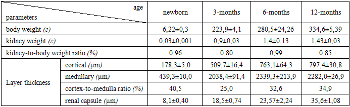
- The kidneys are situated between the lumbar muscles and the parietal layer of the peritoneum, thus exhibiting a retroperitoneal topography. Anteriorly, structures such as the duodenum, the celiac trunk, adipose tissue, mesenteric vessels, and the descending portion of the small intestine are present, while posteriorly (caudally) only adipose tissue is found. Both kidneys are enveloped by layers of adipose tissue overlying the fibrous capsule. Such structural organization and topographical arrangement create distinctive barriers that impede the dissemination of pathological processes, including infection or bleeding.On the medial surface of the kidneys, the renal hilum can be observed. Taking into account that the mass and size of the right kidney are somewhat larger compared to the left kidney, the right kidneys were used for subsequent organometric studies.In newborn rats, the body weight varies between 5.0 and 8.0 g, while the kidney weight ranges from 0.024 to 0.036 g. The relative kidney weight (kidney-to-body weight ratio) is 0.96%. At 3 months of age (sexually mature juvenile), the body weight ranges from 200.0 to 250.0 g, and kidney weight ranges from 0.7 to 1.1 g.At this age, the relative kidney weight (kidney-to-body weight ratio) equals 0.80%. At 6 months of age (reproductive young period), the body weight of the animals ranges from 260.0 to 300.0 g, while the kidney weight ranges from 1.2 to 1.6 g.At this stage, the relative kidney weight (kidney-to-body weight ratio) equals 0.99%. At 12 months of development (reproductively mature age), the body weight of the animals varies between 310.0 and 350.0 g, while the kidney weight ranges from 1.3 to 1.7 g. In the reproductively mature age group, the kidney-to-body weight ratio is 0.85%. In the control group of 3monthold rats, the dynamics of renal organometric parameters were as follows: with an increase in body weight of the white rats throughout the observation period, the examined organometric indices of the kidney showed a corresponding increase. It was found that the capsule thickness varies along the surface of the kidney and is not uniform. In newborn rats, the renal capsule thickness ranges between 6.0 and 10.0 μm.The kidneys of rats are enveloped by a dense connective tissue capsule, which consists of bundles of collagen fibers together with elastic and reticular fibers. In 3-month-old animals, the thickness of the renal capsule ranges from 14.0 to 23.0 μm. The microscopic architecture of the kidney at 3 months of age is presented in Figure 1. By the age of 6 months, the capsule thickness varies between 19.0 and 28.0 μm. The structure of the renal cortex in 6-month-old rats from the control group is illustrated in Figure 2.
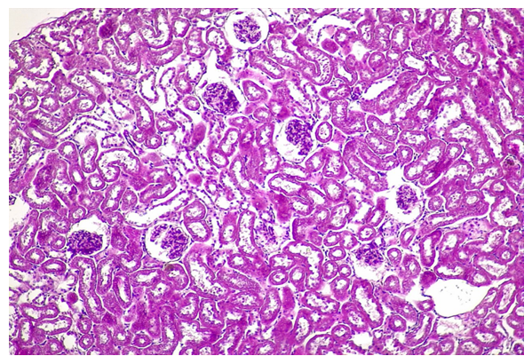 | Figure 1. Light micrograph of the kidney structure in a 3-month-old rat. 1. Renal corpuscle; 2. Proximal convoluted tubule; 3. Collecting ducts; 4. Renal capsule. Hematoxylin and eosin (H&E) staining. Magnification: ocular 10×, objective 20× (total magnification 200×) |
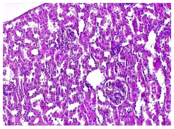 | Figure 2. Histological structure of the renal cortex in a 6-month-old control rat. 1. Renal capsule; 2. Cortex (cortical substance); 3. Medulla (medullary substance); 4. Renal corpuscle. Hematoxylin and eosin (H&E) staining. Magnification: ocular 10×, objective 20× (total magnification 200×) |
|
8. Conclusions
- Thus, during postnatal development, the highest growth rates of rat kidney morphometric parameters are observed at 6 months of age compared to 3-month-old animals: body weight increases by 25.3%, kidney weight by 55.7%, cortical thickness by 49.0%, and medullary thickness by 15.0%. However, the greatest increase in renal capsule thickness is observed at 12 months of age compared to 6-month-old animals, reaching 33.0%.The rate of increase in renal capsule thickness from the neonatal period to 12 months of age was 1.4-fold, whereas the cortical thickness showed a 1.8-fold increase. The medullary thickness demonstrated a 1.4-fold increase during the same period.