International Journal of Genetic Engineering

p-ISSN: 2167-7239    e-ISSN: 2167-7220

2025;  13(4): 62-65

doi:10.5923/j.ijge.20251304.02

Received: Mar. 26, 2025; Accepted: Apr. 20, 2025; Published: Apr. 26, 2025


The Effect of Insulin on Carbohydrate Metabolism in Experimental Alzheimer's Model Animals

Ikromov S. A. 1, Saatov T. S. 1, Mustafakulov M. A. 1, Yalalova I. R. 1, Kosimova Z. T. 2

1Institute of Biophysics and Biochemistry, Uzbekistan

2Impuls Medical Institute, Uzbekistan

Correspondence to: Ikromov S. A. , Institute of Biophysics and Biochemistry, Uzbekistan.

Email:

Copyright © 2012 Scientific & Academic Publishing. All Rights Reserved.

This work is licensed under the Creative Commons Attribution International License (CC BY).
http://creativecommons.org/licenses/by/4.0/

Abstract

This article investigates the effect of insulin on carbohydrate metabolism in experimental Alzheimer's model animals. The study analyzes how insulin influences metabolic changes associated with Alzheimer's disease, particularly in glucose levels and carbohydrate metabolism. Experimental trials on animal models observed insulin balance and changes in carbohydrate metabolism. The results helped identify the connection between insulin sensitivity and metabolic disruptions in Alzheimer's disease development.

Keywords: Alzheimer's disease, Insulin, Carbohydrate metabolism, Experimental animal model, Glucose, Metabolic changes

Cite this paper: Ikromov S. A. , Saatov T. S. , Mustafakulov M. A. , Yalalova I. R. , Kosimova Z. T. , The Effect of Insulin on Carbohydrate Metabolism in Experimental Alzheimer's Model Animals, International Journal of Genetic Engineering, Vol. 13 No. 4, 2025, pp. 62-65. doi: 10.5923/j.ijge.20251304.02.

1. Introduction

For many years, the brain was considered incapable of glucose uptake without insulin. However, recent findings show that insulin not only regulates glucose transport and metabolism but also plays a role in neuronal excitability, proliferation, progenitor cell differentiation, synaptic plasticity, memory formation, neurotransmitter secretion, and apoptosis [1,25]. The effect of insulin on the brain's progenitor cells is especially noteworthy. It has been demonstrated that neuroblast reactivation occurs along with active signaling through insulin receptors, which is essential for the transition of progenitor cells from an inactive stage to active proliferation. Additionally, insulin is synthesized directly in the brain. This hypothesis is based on the discovery of insulin-specific mRNA in the hypothalamus, hippocampus, and neuronal cultures [22,23]. Insulin plays a critical role in neuronal survival and brain function. Its influence on synaptic plasticity and memory consolidation in neurons is vital [15,12,30]. Insulin is also crucial for neuronal circuit formation, neural stem cell activation, neuronal growth, recovery, neuroprotection, energy metabolism regulation, and protection against oxidative stress [4,13]. Consequently, disruptions in insulin signaling in the central nervous system (CNS) may lead to various neurological diseases. Recent research has highlighted a link between Alzheimer's disease (AD) and disruptions in insulin signaling in the CNS [5,19]. Insulin resistance and reduced insulin action may play significant roles in the pathogenesis of these diseases.

2. Materials and Methods

The sporadic form of non-insulin-dependent diabetes mellitus (NDH) with Alzheimer's symptoms is typically induced in animal models through intraventricular administration of streptozotocin at doses ranging from 1 mg to 3 mg per kilogram of body weight [6,29]. In this study, rats were fed a special high-calorie diet, which included 0.2% cholesterol and 4-6 mg of margarine per rat, along with 2% total dietary fat. The rats also received 3% sweet water with 0.04-0.06 mg of Merkazalol daily for 21 days. The rats' weight was measured weekly, and behavioral tests (Open Field, Morris Test) and glucose measurements were conducted. Blood and tissue samples were collected to measure insulin, lipids, and cholesterol levels.
The sporadic form of NDH with Alzheimer's symptoms is usually induced by administering streptozotocin intraventricularly in doses ranging from 1 mg to 3 mg per kilogram of body weight, either as a single injection or over three days [6,16,17]. In our experiments, to induce sporadic NDH with Alzheimer's symptoms, the animals were fed a special high-calorie diet for three months, which included a standard diet supplemented with an additional 0.2% cholesterol. Each rat received 4-6 mg of margarine, and 2% of the total food intake consisted of fat. To slightly slow down the metabolism in the rats, they were given 3% sweetened water with 0.04-0.06 mg of Merkazalol per rat daily for 21 days. During the preparation of the experimental model, the rats' weight was measured weekly, and behavioral tests (Open Field [OM], ShRPH, ShRFH, Morris Test), along with individual biochemical indicators, were monitored. 100 µl of blood was periodically taken from the tail to measure glucose levels. Blood and tissue samples were collected to measure insulin, lipids, and cholesterol. In the final stage of preparing the experimental model, the glucose level in the animals' blood had risen to 7-8 mmol/l. In the study, the experimental group of animals received streptozotocin at a dose of 3 mg per kilogram of body weight, administered intranasally via liposomes over the course of one week [8,21,25].

3. Discussion

Before starting the experiments to induce a sporadic NDH experimental model with signs of encephalopathy (NDHE), the animals were kept in quarantine for 10 days to adapt to vivarium conditions.
Then, the animals were selected according to the criteria defined by neurological NSS scale indicators of behavior. The animals were divided into general groups. Out of the 80 animals examined, 40 rats were selected for experiments. The experimental NDH model was created by long-term feeding of animals with an atherogenic high-calorie diet consisting of a standard diet supplemented with 0.2% cholesterol (CS) of the general diet: for each animal selected for the study, 4-6 mg of cholesterol, a total of 20% margarine and pork fat, (40% of calories) sweetened drinking water with fructose/sucrose (3% solution), and the thyroid hormone inhibitor mercazolil, in a dosage of 0.04-0.06 mg per rat, were added to the diet. The animals were fed a high-calorie diet for 12 weeks, and after feeding the experimental animals high-calorie foods until the 13th week, the weight of each animal was measured, and after reaching the required weight, each animal was administered streptozotocin solution intranasally at a dosage of 3 mg/kg through the nasal cavity over a period of 3 days. This is considered the first stage of the model preparation in animals. The induction of the NDHE model was monitored through periodic behavioral tests and biochemical blood tests of the animals.
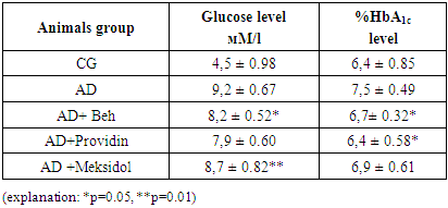
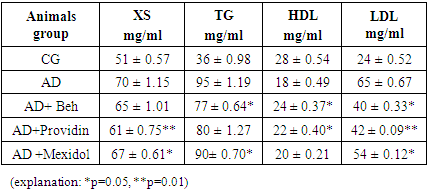
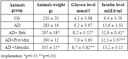
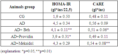
4. Result

The analysis of the obtained results is presented below. The results of blood tests of the animals after inducing the NDHE model showed the following (see Table 1).
Table 1. The amount of individual biochemical indicators in the blood of animals with NDHE (n=5)
     
In experimental NDH, after the glucose level in rat blood reaches 7-8 mmol, glycated hemoglobin exceeds 7%, and cholesterol (CS), triglycerides (TG), and lipoproteins (LP) increase by 20-30% above normal levels. Additionally, cognitive changes in the animals' behavior are observed. Following decapitation, in compliance with ethical guidelines for humane treatment of animals, the blood, brain sections, liver, heart, and other organs of the model animals were collected for histological and biochemical studies. The results of the blood tests after inducing the NDHE model in the animals are shown below (see Table 2).
Table 2. The content of individual biochemical indicators in the blood of animals with NDHE (n=5)
     
As seen from the table, in the context of the animals' weight gain, especially in the intact control group where streptozotocin was not applied, an increase in glucose and glycated hemoglobin levels characteristic of type 2 diabetes is observed.
The blood indicators, insulin levels, and the calculation of insulin sensitivity criteria, such as HOMA-IR and CARE, showed the following results.
Table 3. The effect of an atherogenic diet on insulin resistance
     
The composition of lipid fractions also increases, which is observed not only in diabetes but also in lipid metabolism disorders, indicating potential general atherosclerotic vascular damage.
Table 4. The effect of an atherogenic diet on insulin resistance
     
The table shows that long-term feeding of animals with an atherogenic high-calorie diet leads not only to an increase in body weight, blood glucose, and insulin levels, but also to general insulin resistance, which is confirmed by an increase in HOMA-IR. In the group of Alzheimer's model animals, it was more than twice as high as in the intact group of animals, and experiments showed that after CARE administration, the blood levels of the NDHE group animals increased by 17% and 18%, respectively.

5. Conclusions

In the hippocampus of experimental animals, insulin stimulates neurogenesis by lowering the activation threshold for long-term potentiation and plays a key role in maintaining nerve cells by binding to insulin receptors (InsRs). It was found that insulin plays an important role in glucose uptake by neurons, in glucose uptake by the hypothalamus, and in the formation of spatial memory in the hippocampus.

References

[1]  Ishankhodjaev T. et al. Study on Effects of Liposomal Quercetin on Biochemical Parameters of the Nigrostriatal System of Rats with Experimentally Induced Neurodegenerative Disease // Annals of the Romanian Society for Cell Biology. – 2021. – С. 6128-6143.
[2]  Mukhammadjon M. et al. The effect of ngf on indicators of the antioxidant system in rat brain tissue // Universum: химия и биология. – 2021. – №. 9 (87). – С. 82-86.
[3]  Saatov T. et al. Antioxidant and hypoglycemic effects of gossitan // Endocrine Abstracts. – Bioscientifica, 2019. – Т. 63.
[4]  Saatov T. et al. Study on hypoglycemic effect of polyphenolic compounds isolated from the Euphorbia L. plants growing in uzbekistan // Endocrine Abstracts. – Bioscientifica, 2020. – Т. 70.
[5]  Saatov T. et al. Correction of oxidative stress in experimental diabetes mellitus by means of natural antioxidants // Endocrine Abstracts. – Bioscientifica, 2021. – Т. 73.
[6]  Irgasheva S. et al. Study on compositions of lipids in tissues of rats with alimentary obesity //Endocrine Abstracts. – Bioscientifica, 2019. – Т. 63.
[7]  Mamadalieva N. I., Mustafakulov M. A., Saatov T. S. The effect of nerve growth factor on indicators of the antioxidant system in rat brain tissue // eurasian union of scientists. series: medical, biological and chemical sciences Учредители: ООО" Логика+". – 2021. – №. 11. – С. 36-40.
[8]  Saatov T. et al. Study on antioxidant and hypoglycemic effects of natural polyphenols in the experimental diabetes model //Endocrine Abstracts. – Bioscientifica, 2018. – Т. 56.
[9]  Mustafakulov M. et al. Determination of antioxidant properties of l-cysteine in the liver of alloxan diabetes model rats // International Journal of Contemporary Scientific and Technical Research. – 2023. – №. Special Issue. – С. 47-54.
[10]  Andrieu-Abadie N., Gouaze V., Salvayre R., Levade T. // Free Radic. Biol. Med.- 2001. - N 31. – P. 2041-2048.
[11]  Ayasolla K., Khan M., Singh A.K., Singh I. Inflammatory mediator and beta-amyloid (25-35)-induced ceramide generation and iNOS expression are inhibited by vitamin E // Free Radic. Biol. Med. -2004. - N 37. – P. 325-338.
[12]  Vincent I., Pae C.I., Hallows J.L. The cell cycle and human neurodegenerative disease // Progr. CellCycle Res. – 2003. - N 5. – P. 31-41.
[13]  Currais A., Hortobagyi T., Soriano S. The neuronal cell cycle as a mechanism of pathogenesis in Alzheimer's disease // Aging. -2009. - N 1. – P. 363-369.
[14]  Ogawa O., Lee H.G., Zhu X., Raina A., Harris P.L., Castellani R.J., Perry G., Smith M.A. Increased p27, an essential component of cell cycle control, in Alzheimer's disease // Aging Cell. – 2003. - N 2. – P. 105-110.
[15]  Yang Y., Geldmacher D.S., Herrap K. DNA replication precedes neuronal cell death in Alzheimer's disease // J. Neurosci. – 2001. - N 21. – P. 2661-2668.
[16]  Vincent I., Bu B., Hudson K., Husseman J., Nochlin D., Jin L.-W. Constitutive Cdc25B tyrosine phosphatase activity in adult brain neurons with M phase-type alterations in Alzheimer's disease // Neurosci. -2001. - N 105. – P. 639-650.
[17]  Barrera G., Pizzimenti S., Laurora S., Mononi E., Giglioni B., Dianzan M.U. 4-Hydroxynonenal affects pRb/E2F pathway in HL-60 human leukemic cells// Biochem. Biophys.Res. Commun. - 2002. - N 295. – P. 267–275.
[18]  Ranganathan S., Scudiere S., Bowser R. Hyperphosphorylation of the retinoblastoma gene product and altered subcellular distribution of E2F-1 during Alzheimer's disease and amyotrophic lateral sclerosis // J. Alzheimers Dis.- 2001. - N 3. – P. 377-385.
[19]  Barrera G., Pizzimenti S., Muraca R., Barbiero G., Bonelli G., Baccino F., Fazio V.M., Dianzani M.U. (1996) Free Radic. Biol. Med., 20, 455-462.
[20]  Дубинина Е.Е., Дадали В.А. 4-гидрокси-транс-2- ноненаль в функциональной активности клеток. Обзор // Биохимия. – 2010. - N 75. – P. 1189-1212.
[21]  Tomashevski A., Husseman J., Jin L.W., Nochlin D.,Vincent L. Constitutive Wee1 activity in adult brain neurons with M phase-type alterations in Alzheimer neurodegeneration // Alzheimers Dis.- 2001. - N 3. – P. 195-207.
[22]  Kesavapany S., Patel V., Zheng H., Pareek T.K., Bjelogrlic M., Albers W., Amin N., Jaffe H., Gutkind J.S., Strong M.J., Grant P., Pant H.C. Inhibition of Pin1 reduces glutamate-induced perikaryal accumulation of phosphorylated neurofilament-H in neurons // Mol. Biol. Cell. -2007. -N18. – P. 3645-3655.
[23]  Ma S.L., Pastorino L., Zhou X.Z., Lu K.P. Prolyl isomerase Pin1 promotes amyloid precursor protein (APP) turnover by inhibiting glycogen synthase kinase-3β (GSK3β) activity: novel mechanism for Pin1 to protect against Alzheimer disease. -2012. - N 287. – P. 6969-6973.
[24]  Ryo A., Nakamura M., Wulf G., Liou Y.C., Lu K.P. Pin1 regulates turnover and subcellular localization of beta-catenin by inhibiting its interaction with APC // Nat. Cell Biol. -2001. - N 3. – P. 793-801.
[25]  Thorpe J.R., Morley S.J., Rulten S.L. Utilizing the peptidyl-prolyl cis-trans isomerase pin1 as a probe of its phosphorylated target proteins. Examples of binding to nuclear proteins in a human kidney cell line and to tau in Alzheimer's diseased brain // J. Histochem. Cytochem. – 2001. - N 49. – P. 97-108.
[26]  Zheng H., You H., Zhou Y.Z. et.al. The prolyl isomerase Pin1 is a regulator of p53 in genotoxic response // Nature. – 2002. - N 419. – P. 849-853.
[27]  Bayer E., Goettsch S., Mueller J.W., Griewel B., Guiberman E., Mayr L.M., Bayer P. Structural analysis of the mitotic regulator hPin1 in solution: insights into domain architecture and substrate binding // J. Biol. Chem. -2003. -N 278. – P. 26183-26193.
[28]  Joseph J.D., Yeh E.S., Swenson K.I., Means A.R. The peptidyl-prolyl isomerase Pin1 // Progr. Cell Cycle Res. -2003. - N 5. – P. 477-487.
[29]  Schmetsdorf S., Gartner U., Arendt T. Constitutive expression of functionally active cyclin-dependent kinases and their binding partners suggests noncanonical functions of cell cycle regulators in differentiated neurons // Cereb. Cortex. -2007. - N 17. – P. 1821-1829.
[30]  Ogawa O., Zhu X., Lee H.-G., Raina A., Obrenovich M.E., Bowser R., Ghanbari H.A., Castellani R.J., Perry G., Smith M.A. Ectopic localization of phosphorylated histone H3 in Alzheimer's disease: a mitotic catastrophe? // ActaNeuropathol. – 2003. - N 105. – P. 524-528.