-
Paper Information
- Previous Paper
- Paper Submission
-
Journal Information
- About This Journal
- Editorial Board
- Current Issue
- Archive
- Author Guidelines
- Contact Us
International Journal of Genetic Engineering
p-ISSN: 2167-7239 e-ISSN: 2167-7220
2025; 13(3): 40-45
doi:10.5923/j.ijge.20251303.02
Received: Mar. 9, 2025; Accepted: Mar. 26, 2025; Published: Apr. 6, 2025

Introduced in Uzbekistan Conditions Lagerstroemia Indica L. Taxonomic Information about the Anatomical Features of Vegetative Organs
 Abstract
 Reference
 Full-Text PDF
 Full-text HTML
Sharapova Mokhidil Amanovna
Department of Microbiology and Biotechnology, Karshi State University, Karshi, Uzbekistan
Correspondence to: Sharapova Mokhidil Amanovna, Department of Microbiology and Biotechnology, Karshi State University, Karshi, Uzbekistan.
| Email: |  |
Copyright © 2025 The Author(s). Published by Scientific & Academic Publishing.
This work is licensed under the Creative Commons Attribution International License (CC BY).
http://creativecommons.org/licenses/by/4.0/

This article incorporates information from the region of Kashkadarya from the introduction of Lagerstroemia indica L. the results of data that were first analyzed about the worldwide distribution of the species as well as the anotomic characteristics of the species are given. Analysis of anatomical data in the shell region as well as studies of the characteristics of the acclimatized species, analytical data on changes in the anatomical characteristics of the species due to the influence of environmental factors are presented. Methods: Thin-cut cuts were made. A few drops of 99% ethyl alcohol on the incision were kept in washed water. The alcohol solution was added to soften the tissues, and then 2-3 drops of saffron paint were dripped, after the excess paint was washed off with water, a drop of glycerin was rinsed with distilled water, and a 2-5 cm cut of the leaf epidermis was examined after processing. Micrographs were taken with a computer microphoto attachment with a Canon A123 digital camera under a Motic B1-220A-3 microscope.Linear drawings were obtained using a drawing apparatus in the light of the camera. Results: The leaves of Lagerstroemia indica are almost sessile, oblong, elliptical or obovate, almost leathery, fluffy along the veins below. For the first time, the anatomical structure of the vegetative organs of Lagerstroemia indica L. was studied under the conditions of the introduction of Kashkadarya, and the diagnostic signs were determined. Conclusions: Changes in the structural and functional properties of plants under the influence of environmental factors, the emergence of adaptation mechanisms at different levels, aspects of adaptation to environmental factors have been studied.
Keywords: Introduction, Ornamental plants, Vegetative organs, Anatomical structure, Diagnostic signs, Lagerstroemia, Climate
Cite this paper: Sharapova Mokhidil Amanovna, Introduced in Uzbekistan Conditions Lagerstroemia Indica L. Taxonomic Information about the Anatomical Features of Vegetative Organs, International Journal of Genetic Engineering, Vol. 13 No. 3, 2025, pp. 40-45. doi: 10.5923/j.ijge.20251303.02.
1. Introduction
- Indian Lagerstroemia (Lagerstroemia indica L.) is a type species of the genus Lagerstroemia L. belongs to the family (Lythraceae J.St.-Hil.), Whose representatives are distributed throughout the globe, with the greatest diversity in the tropics. This beautiful deciduous tree is native to China and is widely cultivated throughout Southeast Asia. The first acquaintance of Europeans with lagerstremia occurred at the end of the 18th century in India, hence its trivial name "Indian lilac". Lagerstremia india is perhaps one of the best flowering woody plants on the Black Sea coast. The culture deserves the attention of breeders, the breeding of winter-hardy forms will make it possible to use this valuable breed in urban landscaping in more northern regions [1].Its representatives are distributed around the world, with about 50 species widely distributed around the world. Lagerstroemia indica L. is an evergreen shrub and darxt in the form of Indian subcontinent, Southeast Asia, Northern Australia and its southern parts, and is also acclimatized in African states, one of the warm-climate regions worldwide [1], [2]. Lagerstroemia indica L. to give this species a beautiful view of the surroundings of urban gardens and Acholi houses in the form of an ornamental shrub originally in Nigeria. Pers. and Lagerstroemia speciosa L. species begin to be planted. This species is considered economically profitable, and its wood is also used in the preparation of unique furniture, home decor.In folk medicine, decoctions of flowers and seeds are used to eliminate diseases of species tumors, diabetes mellitus, urinary incontinence. It is used in home conditions for fever and digestive disorders, in the form of decoctions to reduce cholesterol levels, lower blood pressure and lose weight. L.indica L helps prevent erosion in soils where drought is present in large quantities when planted and bovine soil warms up [6]. L. there is little data on the anatomical signs of indica L and its structure. In this regard, there is information in the form of reports from various scientists, including Data is provided in [7] work on the location of stomata in the cell of the plant. Insufficient data on the anotomic properties of this species, increasing the relevance of this study, the main goal is L.indica provides an opportunity to tax the anatomical data as well as to study the specifics of the acclimated species in the Chestnut houdis, comparing the changes that environmental factors have affected the anatomy of the species.The cultivation of wild species in conditions that differ from their natural habitat in nature affects the rhythmological, anatomical and biomorphological processes inherent in the adaptation of plants to a new habitat. Plant introduction testing often refers to the study of plant species diversity within a genus. In this case, information is important not only about biomorphological variability, but also about the anatomical features inherent in the adaptation of plants in a new habitat. The structure of the leaf is determined by the internal laws of the organism, which have developed in its phylogenesis, and not every species is plastic, in which the hereditary reason is expressed. It is known that when plants are transferred from one condition to another, quantitative changes in the anatomical structure of the leaf are manifested and parallelism between modification and hereditary adaptations is revealed [1-2].The anatomical structure of the vegetative organs of Lagerstroemia indica under the conditions of the introduction of Kashkadarya has not been studied. This determines the relevance and novelty of our research.The aim of the study is to study the anatomical structure of the vegetative organs of the decorative species Lagerstroemia indica, to identify diagnostic signs and structural features of this species.
2. Materials and Method
- Acclimatized L. in the country for research.the leaf and stem of indica were used. The resulting na'munas are mostly derived from their growing location and The anatomical modifications from the methods of Ajayi and others [8] Kadiri and Ayodele [10] to authenticate the assembled herbarium were done using Wilkinson's terminologies. Thin-cut cuts were made. A few drops of 99% ethyl alcohol on the incision were kept in washed water. The alcohol solution was added to soften the tissues, and then 2-3 drops of saffron paint were dripped, after the excess paint was washed off with water, a drop of glycerin was rinsed with distilled water, and a 2-5 cm cut of the leaf epidermis was examined after processing. Greases from the standard median part of the Leaf lamina near the middle on the leaf epidermis [11], [12], [13] were taxied by methods. Take the sample for 8-24 hours to soften the mesophyll. Tissue breakdown the layers of vesicles and epidermis were separated and water was placed for cleaning. Different levels of ethanol droplets of up to 50% -100% were added in turn to dehydrate cells.The objects of research are the perennial shrub Lagerstroemia indica L. of the genus Lagerstroemia from the family Lythraceae, growing under the conditions of introduction of the Kashkadarya region of Uzbekistan. Along with the morphological description, vegetative organs (leaf, petiole and stem) were recorded in 70° ethanol for anatomical study. For the preparation of sections of vegetative organs, a manual method was used. The epidermis was examined on paradermal and cross sections. The cross-sections of the leaf, stem and root were prepared by hand using a safety razor. Cross-sections of the leaf are made through the middle, and the petiole and stem are made through the base. Sections were stained with methylene blue and safranin, followed by gluing in glycerol-gelatin [4]. Descriptions of the main tissues and cells are given according to K. Esau (1969), N.S. Kiseleva (1971), epidermis - according to S.F. Zakharevich (1954). Micrographs were taken with a computer microphoto attachment with a Canon A123 digital camera under a Motic B1-220A-3 microscope. Linear drawings were obtained using a drawing apparatus in the light of the camera.
3. Results and Discussion
- The leaves of Lagerstroemia indica are almost sessile, oblong, elliptical or obovate, almost leathery, fluffy along the veins below. On the paradermal section, the outlines of epidermal cells on the adaxial side are rectilinear, the projection is polygonal, the abaxial one is slightly tortuous, the projection is polygonal. The cells of the adaxial (upper) epidermis are larger than those of the abaxial (lower) epidermis. On the adaxial structure of the leaf epidermis there are numerous mucus passages than the abaxial one. Adaxial and abaxial leaf epidermis are pubescent with simple, unicellular trichomes (Figure 1).Leaves are hypostomatic - the stomata are located on the abaxial (lower) side of the epidermis of the leaf blade and are located transversely to the longitudinal axis of the leaf. All this leads to a reduction in water loss from the leaf surface. The shape of the stomatal cells (from the surface) is oval; the stomata are of the lenticular-equally thickened type, in which two identical crescent-shaped cells are arranged symmetrically [8]. On the frontal plane, the thickened shells are almost uniform. The slit is fusiform. The stomata are not submerged, of an anocytic type (Figures 1, 2).
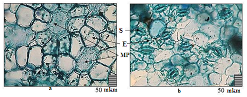 | Figure 1. Anatomical structure of the epidermis of the Lagerstroemia indica leaf in a longitudinal section: a - upper (adaxial) epidermis; b – lower (abaxial) epidermis. Legend: MP - mucus passages, S – stomata, E - epidermis |
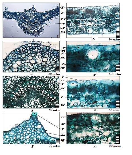 | Figure 2. Anatomical structure of the Lagerstroemia indica leaf in a cross section: a - detail of the main vein of the leaf; b - detail of the leaf mesophyll; c - epidermis and collenchyma; d - mucus passages; e - conducting beams; e - epidermis and palisade parenchyma; g - epidermis and collenchyma; h - mucs passages and spongy parenchyma |
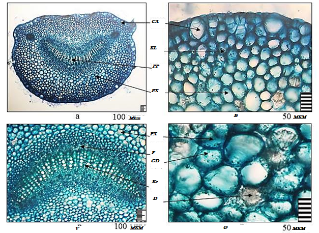 | Figure 3. Anatomical structure of the leaf petiole of Lagerstroemia indica in a cross section: a - general view; b- - epidermis and parenchyma;c - conducting beam; d - parenchymal cells and druses of calcium oxalate. Legend: HD - hydrocytic cells, D - drusen, CL - collenchyme, Kc - xylem, PP - conductive bundle, CX - mucus passages, F - phloem |
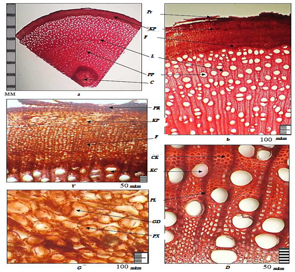 | Figure 4. Anatomical structure of the stem of Lagerstroemia indica in a cross section: a - general view of the stem; b - detail; c - crustal parenchyma; g - core; e - secondary conducting beams. Legend: HD - hydrocytic cells, CP - crustal parenchyma, Kc - xylem, L - libriform, RL - radial rays, PP - conducting bundles, Pr - periderm, C - core, SC - sclerenchyma, F - phloem |
4. Conclusions
- The article presents the results of studying the perennial ornamental shrub Lagerstroemia indica L. introduced in the Kashkadarya region.For the first time, the anatomical structure of the vegetative organs of Lagerstroemia indica L. was studied under the conditions of the introduction of Kashkadarya, and the diagnostic signs were determined. Changes in the structural and functional properties of plants under the influence of environmental factors, the emergence of adaptation mechanisms at different levels, aspects of adaptation to environmental factors have been studied. On the basis of diagnostic features, a high xeromorphicity of this species, its full adaptation to the arid conditions of the Kashkadarya region, was established.