International Journal of Brain and Cognitive Sciences

p-ISSN: 2163-1840    e-ISSN: 2163-1867

2015;  4(2): 33-40

doi:10.5923/j.ijbcs.20150402.03

Impact of AlCl3-Induced Neurotoxicity in Protein-Malnourished Sprague-Dawley Rats

Ebuehi O. A. T., Adeyelu T. T.

Department of Biochemistry, College of Medicine, University of Lagos, Lagos, Nigeria

Correspondence to: Ebuehi O. A. T., Department of Biochemistry, College of Medicine, University of Lagos, Lagos, Nigeria.

Email:

Copyright © 2015 Scientific & Academic Publishing. All Rights Reserved.

Abstract

Aluminium is an ubiquitous and one of the most abundant element on earth. It is known as a toxic agent with its effect predominantly on the central nervous system. Aluminium toxicity and protein deficiency resulted in impairment of the neurobehavioural development, as well as learning. The objective of this study was to ascertain the impact of AlCl3 –neurotoxicity on protein malnutrition in male rats. Thirty two young adult male albino rats (142.73 ± 3.09g) were divided equally into four groups. The control group was fed rat chow and water, while the other three groups were intraperitoneally administered with AlCl3 at a dose of 4.2mg/kg body weight per day for 28 days, and were fed 18%, 5.87% or 29.3% protein diet respectively. The brain and liver tissues were excised and the serum collected for biochemical analysis. The short and long term memories as an index of cognitive function were determined in rats administered aluminium and protein diets using Shuttle box. The activities of antioxidant enzymes and acetyl cholinesterase, levels of brain acetylcholine and glutamate of rats exposed to aluminium and protein diets were assayed. There was a decrease in the body weight and cognitive function of rats administered AlCl3 and low protein diet as compared to the control. The administration of AlCl3 in rats significantly increased serum activities of alanine amino transferase (ALT), aspartate amino transferase (AST) and alkaline phosphatase (ALP). The activities of the antioxidant enzymes were significantly reduced in the protein deficient diet fed rats as compared to the control, while it increased lipid peroxidation in brain and liver tissues. The brain activities of acetyl cholinesterase, and levels of brain acetylcholine and Ý-amino butyric acid (GABA) were significantly reduced in the low protein fed rats, while the brain glutamate level was increased. Therefore, data of the study suggest that adequate dietary protein consumption may alleviate the adverse effect of neurotoxicity associated with aluminium exposure in rats.

Keywords: Aluminium, Dietary protein, Malnutrition, Cognitive function, Antioxidants, Neurotoxicity, Rats

Cite this paper: Ebuehi O. A. T., Adeyelu T. T., Impact of AlCl3-Induced Neurotoxicity in Protein-Malnourished Sprague-Dawley Rats, International Journal of Brain and Cognitive Sciences, Vol. 4 No. 2, 2015, pp. 33-40. doi: 10.5923/j.ijbcs.20150402.03.

1. Introduction

Malnutrition is lack of proper nutrition, caused by not having enough to eat, not eating enough of the right things, or being unable to use the food that one does eat, to ensure optimal growth and function [1]. Over 40,000 children die every day from malnutrition and disease. Therefore, nutrition is an essential tool for development and it is known as the principal non-genetic factor that affects development [2, 3]. In some part of the developing countries, protein malnutrition has been recognised as a major health and nutritional problem having a pronounced effect on the brain [3-5]. There have been reports showing the impact of protein malnutrition on cognitive behaviour and function (6,7). Metal toxicity impact on tissues has however been shown to be worsened as a result of malnutrition [5].
Aluminium is an ubiquitous metal and is the third most abundant element on earth [8]. It has no known physiological role in the body, but had been known to cause adverse physiological responses [9]. Its ubiquity and extensive use in product and processes coupled with its ability to cause neurodegeneration has made aluminium a cause of health concern [10]. Presently, aluminium utensils are widely used in developing countries [11], it is also in food products, drinking water, salts, herbs, cosmetics, toothpaste and also widely used in medicine as antacids, phosphate binder, buffered aspirin, vaccines and injectable allergens [12-14].
Aluminium is toxic to humans and is distributed mainly in the bone, liver, testis, kidney and brain [15]. Its route of absorption includes oral, intranasal, transdermal and parenteral [16]. The entry of aluminium into the brain occurs mainly because it crosses the blood brain barrier and causes alteration of major neurotransmitters and it has been reported that this penetration occurs with complex with transferring by receptor mediated endocytosis [17]. Due to the deleterious effects and ubiquity of aluminium, it is imperative to decipher the impact of aluminium on male Sprague Dawley rats fed with different levels of dietary protein.

2. Materials and Methods

Chemicals: All chemicals and reagents, including AlCl3 used for the study were of analytical grade and purchased from reputable chemical industries.
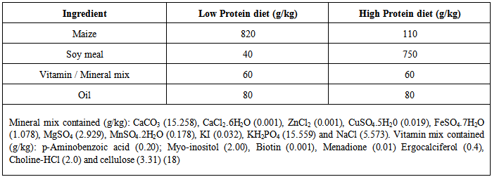
Formulation of Diets: The experimental diets were formulated according to the method of Adelusi and Olowokere [18], with modification. The composition of the diets is shown in Table 1.
Table 1. Formulation of Experimental Diets (g/kg)
     
Experimental Animals: Thirty two male albino Sprague-Dawley rats (135-145g) were used for this study. The rats were purchased from the Laboratory Animal Centre of the College of Medicine, University of Lagos, Lagos, Nigeria. They were acclimatized for 14 days and housed in metallic cages and fed with commercial rat chow and water ad libitum.
Experimental Design: Aluminium chloride (AlCl3.6H20) was administered intraperitoneally at a dose of 4.2 mg/kg body weight and it was given to the rats fed different protein diets as indicated in the different groups for 28 days. The Group 1 rats were fed rat chow (control diet) and water, while Group 2 rats were fed rat chow + 4.2 mg/kg body weight of AlCl3. The Group 3 rats were fed low protein diet (5.87% protein) + 4.2 mg/kg body weight of AlCl3, while Group 4 rats were fed high-protein (29.20% protein) + 4.2 mg/kg body weight of AlCl3.
Collection of Serum: After the rats were fed the respective diets for 28 days, they were subjected to overnight fast and sacrificed by cervical dislocation. The blood samples were taken and centrifuged at 3000g for 10min, to obtain the serum which was used for biochemical analysis.
Preparation of Liver and Brain homogenates: The brain and liver were quickly removed, washed in ice-cold isotonic saline and blotted individually on ash-free filter paper. The tissues were homogenized in ice-cold 0.25M sucrose buffer solution, pH 7.4 and centrifuged at 10,000g for 10min. The supernatant was collected and frozen until use.
Proximate analysis of Experimental Diets: The proximate analysis of the experimental diets was carried out by the method of AOAC [20].
Cognitive Function assessment: The short and long term memories of the rats were assessed through the use of the Shuttle box [21]. The shuttle box consisted of two wooden compartments of identical dimensions (28 by 15cm). The floor consisted of 6mm diameter wire rods (spaced 1.7 cm apart at the centre) through which 1.5mA of scrambled foot shock was administered. The rods were connected to a set down transformer with a regular dimmer which could be switched on and off to deliver an instant scrambled foot shock to either compartment. A miniature lamp was in the ceiling of the first compartment.
The floor was divided into two equal compartments by a wooden door that the mouse crossed to avoid or escape foot shock. The door could be raised to permit entry of the rats into any of the two compartments. The training or learning task commenced after rats were acclimatized. At the beginning of each trial, the light was turned on in the compartment. At the first day of training, all rats were placed in the shuttle box and allowed to have access to both the light and dark compartment for a period of 1h. On the third day of the first week of training, rats from each group were placed in the illuminated compartment and 30 sec later the door was raised. The dark compartment indicated a "safer" compartment in which the rats would not receive foot shock. To avoid foot shock and the light compartment, the rats had to cross into the safer compartment within 5s. Failing to do so resulted in foot shock. The inter-trial intervals were randomized at 30, 45 and 60s [3].
For each rat, the weekly training session consisted of about 2 trials. Such training trials were conducted for about 4weeks. The measure of acquisition was the number of avoidance responses per day and the number of rats making about 70% avoidances in a block of 2 trials. The learning procedure was repeated for about 4 weeks. For short-term testing, 24 h after training (day 2), each rat from the four groups was placed in illuminated chamber and 30 sec later the door was raised, and the time spent in the light compartment before entering the dark compartment was recorded. Learning skills acquired on week 3 and 4 (day 21 and 27) depicted long-term memory [7].
Assay of Liver Enzymes: Aspartate aminotransferase (AST), alanine aminotransferase (ALT) and alkaline phosphatase (ALP) were assayed using commercial kits (Randox Kits), following the manufacturer’s instruction.
Determination of Lipid Peroxidation: This was assayed by measuring the TBA reactive products present in the test sample using the procedure of Vashney and Kale [22] and expressed as micromolar of malondialdehyde (MDA)/g tissue.
Determination of Reduced Glutathione (GSH) Level: The total sulphydryl groups, protein – bound sulphydryl groups and free sulphydryl groups, like GSH in biological samples were determined using Ellman’s reagent, 5,5'-dithio-bis-2-nitrobenzoic acid (DTNB) as described by Jollow et al, [23].
Assay of Catalase Activity: Catalase activity was determined according to the method of Sinha [24]. In this method, dichromate in acetic acid is reduced to chromic acetate when heated in the presence of H2O2. Perchromic acid is formed which is an unstable intermediate. The chromic acid produced was measured colorimetrically at 570nm.
Assay of Superoxide Dismutase Activity: The activity of superoxide dismutase in the tissues was determined by the method of Misra and Fridovich [25]. In this method, 0.1ml of sample was diluted in 0.9ml of distilled water to make a 1 in 10 dilution. An aliquot of 0.2ml of the diluted enzyme preparation was added to 2.5ml of 0.05M carbonate buffer (pH 10.2) to equilibrate in the spectrophotometer, and the reaction was started by adding 0.3ml of fleshly prepared 0.3mM adrenaline to the mixture which was quickly mixed by inversion. The reference cuvette contains 2.5ml of 0.05M carbonate buffer, 0.3ml of adrenaline (substrate) and 0.2ml of distilled water. The increase in absorbance at 480nm was monitored every 30seconds for 150 seconds.
Assay of Brain Acetylcholine Level: This assay was carried out using the Enzylite Acetylcholine Assay Kit according to the manufacturer’s instruction. In this assay, acetylcholinesterase catalyzes the reaction of acetylcholine to choline. Choline gets oxidized to H2O2 by choline oxidase. In the presence of horsedash peroxidase (HRP), the H2O2 reacts with a specialized fluorescent probe in a 1:1 stoichiometry to generate the red fluorescent oxidation product, resorufin. Resorufin has fluorescent excitation and emission maxima of approximately 571nm and 585nm and the absorbance maxima is at 570nm.
Assay of Brain Acetylcholinesterase Activity: The modified method of Ebuehi and Akinwande (7was used to estimate the acetylcholinesterase activity. 0.4ml aliquot of the homogenate was added to a cuvette containing 2.6ml phosphate buffer (0.1M, pH 8) and 100ul of DNTB. The content of the cuvette were mixed thoroughly by bubbling air and the absorbance was measured at 412nm. When the absorbance reaches a stable value, it was recorded as the basal reading. 2.0ul of the substrate acetylthiocholine is added and the change in absorbance is recorded for a period of 10mins at an interval of 2min. The change in absorbance is thereafter determined.
Determination of Brain Glutamate and GABA Levels: The glutamate and GABA levels in the brain homogenates were determined using multiple development paper chromatography; an adapted method from Sadasivudu and Murthy [26]. After the dissection of the rats and excision of the rat brain, it was homogenized in 80% double distilled ethanol (for every 100mg of the brain tissue, 2ml of 80% alcohol was used). The homogenates were transferred to polypropylene tubes and centrifuged at 1200g for 10 min. One ml of the supernatant was transferred into small test tubes and evaporated to dryness at 70oC in an oven. The residue was reconstituted in 100ml distilled water and 10ml were used for spotting on the Whatman No.1 Chromatography paper. The standard solutions of glutamate and GABA at a concentration of 2mM were also spotted using Eppendorf micropipette and the spot was allowed to dry. The chromatogram was then placed in the chromatography chamber containing butanol: acetic acid: water (65:15:25 v/v) as solvent. When the solvent front reached the top, the paper was removed and dried and sprayed with ninhydrin (0.25% in 99ml of acetone with 1% pyridine) and placed in an oven at 100oC for 4min. The portion with glutamate and GABA spot corresponding to the standard were cut and eluted with 0.005% CuSO4 in 75% ethanol. The absorbance was measured against the blank at 515nm and expressed as mMoles/gram of wet weight tissue.
Statistical Analysis: Data were subjected to one way analysis of variance (ANOVA), with SPSS version 20.0, with significant level of p<0.01. Data were expressed as Mean ± SEM.

3. Results

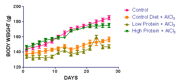
The results of the changes in body weight of rats administered AlCl3 and control diet for 28 days are presented in Fig. 1. Treatment with AlCl3 reduced the body weight of rats, which however was more significantly (p<0.01) pronounced in the low protein diet group.
Figure 1. Changes in body weight of rats administered AlCl3 and subjected to different dietary protein regimens. Results are expressed Mean ± SEM
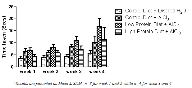
The time spent by the male rats in the light compartment before entering the dark compartment is showed in Fig. 2. The low protein diet rats spent the longest time in the light compartment, followed by the control rats and the high protein diet fed rats exposed to aluminium, while the control rats spent the shortest time in the light compartment.
Figure 2. The time spent (seconds) in crossing from the light compartment to the dark compartment using the Shuttle box1
The activities of serum aspartate aminotransferase (AST), alanine aminotransferase (ALT) and alkaline phosphatase (ALP) of rats administered AlCl3 were significantly increased (p<0.01) as compared to the control as shown in Table 2.
Table 2. The activities of liver enzymes of rats fed protein diets with AlCl3 or rat chow for 28 days
     
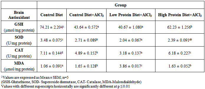
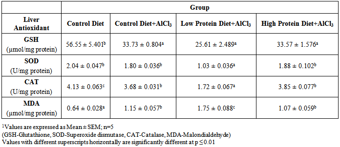
The activities of brain and liver antioxidant enzymes of male rats fed rat chow or protein diets with AlCl3 for 28days are presented in Tables 3 and 4, respectively. The activities of brain catalase, superoxide dismutase and reduced glutathione level were significantly (p<0.01) decreased in rats fed AlCl3 protein diets, while their lipid peroxidation level was elevated when compared to control (Table 3). Similarly, the activities of liver catalase, superoxide dismutase and reduced glutathione level were significantly (p<0.01) decreased in rats fed AlCl3 protein diets, while their lipid peroxidation level was elevated when compared to control (Table 3).
Table 3. Brain antioxidants of male rats fed control or protein diets with AlCl3 for 28 days1
     
Table 4. Liver antioxidants of male rats fed rat chow or protein diets with AlCl3 for 28 days1
     
The brain acetylcholine level was reduced in the AlCl3 treated rats as compared to the control is presented in Table 5. The activity of brain acetylcholinesterase was significantly reduced in rats fed low protein diet as compared to other groups of rats.
Table 5. The brain acetylcholine level and acetyl cholinesterase activity of male rats fed control or protein diets with AlCl3 or rat chow for 28 days1
     
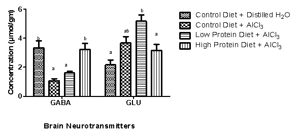
The brain glutamate level was significantly increased as compared to the control in the low protein diet fed rats, while those fed high protein diet, showed no significant( p>0.01) difference as compared to control. However, the brain GABA level was significantly reduced in low protein diet treated rats as compared to the high protein diet fed rats. However, there was no significant difference in GABA level of low or high protein diet fed rats as compared to the aluminium administered rats as presented in Fig. 3.
Figure 3. The brain glutamate and GABA concentrations of male rats fed protein diets with AlCl3 or rat chow for 28 days. (Values are expressed Mean ± SEM; GABA: Ɣ-amino butyric acid, GLU-Glutamate)

4. Discussion

Several studies have shown that aluminium has no known physiological role in the body, but causes deleterious effect by altering various metabolic reactions [9]. This present study reports the effects of dietary protein deficiency and aluminium exposure on oxidative stress, liver enzymes and cognitive and cholinergic function in rats. From the study, dietary protein deficiency and aluminium exposure significantly curtailed growth, as evident in the reduction of body weights of rats. These findings concur with previous reports by Nayak and Chatterjee [5], and Julka et al. [26]. This may suggest that aluminium interferes with the bioavailability of some nutrients which are needed for protein synthesis in rats.
The short and long term memory or learning task were markedly reduced by dietary protein deficiency and aluminium exposure in male rats subjected to 28days. Ebuehi and Akande [7] previously reported a reduction in learning response by zinc deficient rats using Shuttle box model. In the present study, rats subjected to protein deficiency and aluminium exposure spent longer time to cross from the light compartment to dark compartment of the Shuttle box, as compared to the control rats. The results are in agreement with other reports which indicated a significant reduction in learning and cognitive response of rats exposed to aluminium toxicity [27].
The activities of liver function enzymes, namely AST, ALT and ALP significantly increased in rats treated with aluminium as compared to control. This is in agreement with the work of Yousef [28], who reported increased activities of liver enzymes as a result of aluminium toxicity. The impact of the various dietary regimens is also important in consideration as it was observed that there was a significant increase in the activities of liver enzymes of rats fed low protein diet. This shows that the impact of protein malnutrition caused more damage to hepatocytes and thus indicates a combinatorial damage from both protein deficiency as well as aluminium toxicity on the liver.
It is known that the swelling and necrosis of hepatocytes results in the release of liver enzymes into the circulating blood [29]. This is associated with massive centrilobular necrosis, ballooning degeneration and cellular infiltration of the liver [30]. Aluminium has been shown to induce liver damage [31, 32] and it was observed from the present study that there was an overall perturbation in the metabolic profile of serum as demonstrated by an impairment in the metabolism of liver. It is however relevant to quickly mention at this point, the importance of enzymatic changes as manifestation of tissue toxicity. The elevation of AST, ALT and ALP activities are specific and are used as predominant markers of liver damage [33]. Rahman et al., [34] had earlier reported that increase in activities of these enzymes in the blood might be due to liver necrosis, which corroborate enhanced oxidative stress of rats exposed to aluminium. This study therefore support that liver damage occurs as a result of aluminium toxicity. It was also observed that protein malnutrition also contributed to liver damage.
It has been suggested that the toxic effects associated with aluminium are due to generation of free radicals or reactive oxygen species (ROS), which results in oxidative deterioration of cellular lipids, protein and DNA [35, 36]. The result of this study however is in agreement with previous findings that aluminium toxicity in rat brain and liver created an imbalance in the oxidative stress parameters [37, 38].
The brain is highly vulnerable to aluminium induced oxidative stress as compared to the liver [5]. There was a significant lowering activities of the glutathione (GSH), superoxide dismutase (SOD) and catalase (CAT) in the brain of the rats exposed to aluminium when compared with the control. These results are in agreement with other findings that had shown that aluminium inhibits the activities of several antioxidant enzymes in different part of the brain [37-40]. Aluminium is a strong Lewis acid and it may react with superoxide anion, and forms an aluminium superoxide anion, which is a more potent oxidant than superoxide on its own [6].
However, there was a significant (p<0.05) increase in lipid peroxidation and brain MDA increases significantly in the aluminium exposed rats as compared to the control. The impact of oxidative stress induced by aluminium does not only affect the brain but also the liver. The liver which is known for its high metabolic activity is also subject to various toxic invasions by metals. It was seen in this study that the aluminium treated rats had a significantly reduced antioxidants, such as GSH, SOD and CAT, whereas lipid peroxidation levels were increased as evident in the increased MDA concentration in the hepatocytes. This study showed that the rats fed with low protein diet exhibited a significantly higher peroxidation as compared to high protein fed rats.
The impact of aluminium toxicity on the brain neurotransmitters (acetylcholine and GABA) resulted in a decrease in brain acetyl cholinesterase activity, with increase in brain glutamate. Data of the study indicate that aluminium altered the cholinergic neurotransmission. This may occur because aluminium which has a molecular weight of less than 2000 and therefore could easily cross the blood brain barrier and alter the cholinergic pathway (Harris et al., 2003). In addition, adequate protein consumption did not adversely affect the cholinergic system in rats. This study however showed a significant decrease in brain GABA as a result of treatment with AlCl3 under different dietary protein regimens. The alteration of the Glutamate-GABA system alongside the cholinergic pathway could explain the reason for the reduction in cognitive function observed relatively in the rats exposed to aluminium toxicity.

5. Conclusions

Data of the study indicate that aluminium exposure impaired antioxidant system and cholinergic neurotransmission, but adequate supplementation of dietary protein may ameliorate the adverse effects of its neurotoxicity in rats.

ACKNOWLEDGEMENTS

The authors are grateful to Mr. C. Duncan and Mr. S. Adenekan, both technologists in the Departments of Physiology and Biochemistry, respectively, College of Medicine, University of Lagos, Lagos, for providing technical assistance during the study.

References

[1]  De Onis, M. and Blössner, M. (1997). WHO Global Database on Child Growth and Malnutrition; World Health Organization: Geneva, Switzerland; p. 74.
[2]  Galler, J. R. (2001). Children and famine: long term effects on behavioural development. Ambulatory Child Health; 7:85–95.
[3]  Ebuehi, O.A.T. (2012). Nutrients and neurotransmitters: The chemicals of life in the brain. 10th Inaugural Lecture series, delivered at Main Auditorium, University of Lagos, University of Lagos Press, Lagos, Nigeria, March 21, 96pp.
[4]  Ebuehi, O.A.T., Salawu, O.M. and Akinwande, A.I. (1999). Effect of post-weaning protein malnutrition on organ growth, protein malnutrition and lipid composition in rats. Nig. Journal of Medical Research 4: 84-88.
[5]  Nayak, P. and Chatterjee, A. K. (2003). Dietary protein restriction causes modification in aluminum-induced alteration in glutamate and GABA system of rat brain. BMC Neurosci. 4, 4–11.
[6]  Winick, M. and Rosso, P. (1969). The effect of severe early malnutrition on cellular growth of the human brain. Pediatr. Res.; 3:181– 4.
[7]  Ebuehi, O.A.T and Akande, G.A. (2009). Effect of zinc deficiency on memory, oxidative stress and blood chemistry in rats. Int. J. Biol. Chem. Sci. 3(3): 513-523.
[8]  Becaria, A., Campbell, A. and Bondy, S. C. (2002). Aluminum as a toxicant. Toxicol. Ind. Health. 18, 309–320.
[9]  Turgut, G., Kaptanoglu, B., Enli, Y. and Genc, O. (2004). Effect of chronic aluminium administration on blood and liver iron related parameters in mice .Yonsei Med. J. 45(1), 135-149.
[10]  Zatta, P., Lucchini, R., van Rensburg, S. J. and Taylor. A. (2003). The role of metals in neurodegenerative processes: aluminum, manganese, and zinc. Brain Res. Bull. 62, 15–28.
[11]  Yokel, R. A. (2000). The toxicology of aluminum in the brain: a review. Neurotoxicology 21, 813–828.
[12]  Cannata, J.B. Serrano, M. and Fernandez, S.I. (1998). Minimizing the risk of oral aluminium exposure in chronic renal failure. Contrib Nephrol. 71, 81-9.
[13]  Yokel, R. A. and McNamara, P. J. (2001). Aluminium toxicokinetics: an updated minireview. Pharmacol. Toxicol. 88, 159–167.
[14]  Abbasali, K.M., Zhila, T. and Farshad, N. (2005). Developmental toxicity of Aluminum from high doses of AlCl3 in mice. J. Applied Res., 5: 575-579.
[15]  ATSDR (Agency for Toxic Substances and Disease Registry) (1999). Toxicological profile for aluminium (update). Atlanta, GA, U. S. Department of Health and Human Services, Public Health Service (TP-91/01).
[16]  Exley, C. (2004). The pro-oxidant activity of aluminum. Free Radic. Biol. Med. 36, 380–387.
[17]  Roskams, AJ and Connor, JR (1990). Aluminium access to the brain: a role or transferring and its receptor. Proc. Natl. Sci USA, 87,9024-9027
[18]  Adelusi, S.A. and Olowookere, J.O. (1985). A rapid method of induction of post-natal protein-energy malnutrition (PEM) in laboratory animals Nig. J. Appl. Sci., 3: 171-174.
[19]  Gowri Shankar, N. L, Manavalan, R., Venkappayya, D. et al. (2008). Hepatoprotective and antioxidant effects of Commiphora berryi (Arn) Engl bark extract against CCl(4)-induced oxidative damage in rats. Food Chem Toxicol 46: 3182-185.
[20]  AOAC, (2005). Official Methods of Analysis of AOAC International. p. 42-1-42-2. In T.R.Mulvaney (ed.) AOAC International, Arlington, VA.
[21]  Halas, E.S., Eberhardt, M.J., Diers, M.A. and Sandstead, H.H. (1983). Learning and memory impairment in adult rats due to severe zinc deficiency during lactation. Physiol. Behav., 30: 371-381.
[22]  Varshney, R. and Kale, R.K. (1990). Effect of calmodulin antagonists on radiation induced lipid peroxidation in microsomes. Int. J. Radiat. Biol., 58:733–743
[23]  Jollow, D.J., Mitchell, J.R., Zampaglione, N. and Gillete, J.R. (1974). Bromobenzene induced liver necrosis: protective role of glutathione and evidence for 3,4 bromobenzene oxide as the hepatotoxic metabolite. Pharmacology, 11, 151-169.
[24]  Sinha, K.A. (1971). Colorimetric assay of catalase. Anal Biochem., 47: 389-394.
[25]  Misra, A. and Fridovich, H. (1972). Determination of the level of superoxide dismutase in whole blood. Yale Univ. Press New Haven, pp: 101-109.
[26]  Julka, D., Sandhir, R. and Gill, K. D. (1996). Altered cholinergic metabolism in rat CNS following aluminum exposure: implications on learning performance. J. Neurochem. 65, 2157–2164.
[27]  Sadasivudu, B. and Murthy, C.R.K (1978). Effect of ammonia on monoamine oxidase and enzymes of GABA metabolism in mouse brain. Arch. Int. Physiol. Biochem. 86, 67-82
[28]  Abu-Taweel, G.M., Ajarem, J.S. and Ahmad, M. (2012). Neurobehavioural toxic effect of perinatal oral exposure of aluminium on the development motor reflexes, learning, memory and brain neurotransmitters of mice offspring. Pharmacol. Biochem Behav. 101 (1): 49-56.
[29]  Yousef, M. I. (2004). Aluminum-induced changes in hemato-biochemical parameters, lipid peroxidation and enzyme activities of male rabbits: protective role of ascorbic acid. Toxicol. 199, 47-57.
[30]  Nikolova, P., Softova, E., Kavaldzhieva, B. et al. (1994). The functional and morphological changes in the liver and kidneys of white rats treated with aluminum. Eksp Med Morfol 32: 52-61.
[31]  Gowri Shankar, N. L, Manavalan, R., Venkappayya, D. et al. (2008). Hepatoprotective and antioxidant effects of Commiphora berryi (Arn) Engl bark extract against CCl(4)-induced oxidative damage in rats. Food Chem Toxicol 46: 3182-185.
[32]  Bogdanovic, M., Janeva, A.B. and Bulat, P. (2008). Histopathological changes in rat liver after a single high dose of aluminium. Arh Hig Rada Toksikol 59: 97-101.
[33]  Sinha, U. S, Kapoor, A. K. and Singh, A.K. (2005). Histopathological changes in cases of aluminium phosphide poisoning. Indian J Pathol Microbiol 48: 177-80.
[34]  Singhal, R. L. and Merali, Z. (1977). Biochemical toxicity of cadmium. In: Mennear JH. ed. Cadmium toxicity. Marcel Dekker; pp. 61-112.
[35]  Rahman, M. F., Siddiqui, M. K. and Jamil, K. (2000). Acid and alkaline phosphatase activities in a novel phosphorothionate (RPR-11) treated male and female rats. Evidence of dose and time dependent response. Drug Chem Toxicol 23: 497-509.
[36]  El-Demerdash, F.M., Yousef, M.I., Kedwany, F.S. and Baghdadi, H.H. (2004). Role of α-tocopherol and β-carotene in ameliorating the fenvalerate-induced changes in oxidative stress, hematobiochemical parameters and semen quality of male rats. J. Environ. Sci. Health Bull., 39: 443-459.
[37]  Mansour, S., Alan, S. and Norman, B.R. (2006). Aluminum-induced injury to kidney proximal effects on markers of oxidative damage. J. Trace Elem. Med. Biol., 19: 267-273.
[38]  Moumen, R., Ait-Oukhatar, N., Bureau, F., Fleury, C., Boublé, D., Arhan, P., Neuville, D. and Viader, F. (2001). Aluminium increases xanthine oxidase activity and disturbs antioxidant status in the rat. J. Trace Elem. Med. Biol. 15, 89–93.
[39]  Anane, R. and Creppy, E.E. (2001). Lipid peroxidation as a pathway to aluminum cytotoxicity in human skin fibroblast cultures: Prevention by superoxide dismutase plus catalase and vitamins E and C. Hum. Exp. Toxicol., 20: 477-481.
[40]  Nehru, B. and Anand, P. (2005). Oxidative damage following chronic aluminium exposure in adult and pup rat brains. J. Trace Elem. Med. Biol. 19, 203–208.
[41]  Sánchez-Iglesias, S., Méndez-Alvarez, E., Iglesias-González, J., Muñoz-Patiño, A., Sánchez-Sellero, I., Labandeira-García, J. L. and Soto-Otero R. (2009). Brain oxidative stress and selective behaviour of aluminium in specific areas of rat brain: potential effects in a 6-OHDAinduced model of Parkinson's disease. J. Neurochem. 109, 879–88.
[42]  Harris, W. R., Wang, Z. and Hamada Y. Z. (2003). Competition between transferrin and the serum ligands citrate and phosphate for the binding of aluminum. Inorg. Chem. 42, 3262–3273.