International Journal of Brain and Cognitive Sciences

p-ISSN: 2163-1840    e-ISSN: 2163-1867

2015;  4(2): 15-27

doi:10.5923/j.ijbcs.20150402.01

Candidate Biomarkers and CSF Profiles for Alzheimer’s Disease and CADASIL

Bowirrat Abdalla1, Bisharat Bishara2, Nseir William3, Omary Muhamad3, Yassin Mustafa4

1Prof. Dr. of Clinical Neuroscience and Population Genetics – Director of Research Center, EMMS Nazareth Hospital, Faculty of Medicine in the Galilee, Bar Ilan University, Israel

2Senior Physician Specialists in Family Medicine, Director of EMMS Nazareth Hospital, Faculty of Medicine in the Galilee, Bar Ilan University, Israel

3EMMS Nazareth Hospital, Faculty of Medicine in the Galilee, Bar Ilan University, Israel

4Rabin Medical Center, Campus Hasharon, Petah Tikva, Israel

Correspondence to: Bowirrat Abdalla, Prof. Dr. of Clinical Neuroscience and Population Genetics – Director of Research Center, EMMS Nazareth Hospital, Faculty of Medicine in the Galilee, Bar Ilan University, Israel.

Email:

Copyright © 2015 Scientific & Academic Publishing. All Rights Reserved.

Abstract

The differential diagnosis between Alzheimer’s disease (AD) and vascular dementia (VaD) are still roughly problematic in clinical practice, despite the widely used diagnostic criteria to differentiate between the two disorders. There is an increasing evidence that cerebrovascular dysfunction plays a role not only in vascular causes of cognitive alterations but also in AD. Cognitively patients, with AD, show sometimes mixed degrees of associated vascular lesions in 30-60% of AD cases. In opposition, AD pathology may be present in 40%-80% of VaD patients, thus impeding diagnosis accuracy. Therefore, to eliminate this bewilderment and discrepancies in the diagnosis between the AD and VaD, it is worthy to shed light firstly on a disease that is a microangiopathy and represents VaD with clear milestones and features as is the case of cerebral autosomal dominant arteriopathy with subcortical infarcts and leukoencephalopathy (CADASIL). Studying CADASIL CSF biomarkers profile, will help in the differential diagnosis between both diseases sharing the coexisting neurodegeneration, furthermore, CADASIL is a dominantly inherited mid-adult life disorder causing ischemic strokes, which belongs to vasculopathies and symbolizes a genuine prototype of VaD that provides a valuable opportunity for studying its CSF biomarkers. Secondly, examining and evaluating the CSF biomarkers of AD compared to that of CADASIL. The pathogenesis similarities between CADASIL and early onset AD affecting the small vessels of the brain have suggested plausible molecular mechanisms involved in vascular damage and their impact on brain function and also come from the fact that in both diseases genetic mutations occur. CADASIL mutations in NOTCH3 gene generate toxic protein aggregates (Granular Osmiophilic Material- GOM) in the vicinity of vascular smooth muscle cells (VSMCs) causing degeneration and loss of VSMCs in small arteries and arterioles of white matter regions of the brain that lead to dementia, similar to those attributed to mutant forms of the Amyloid Precursor proteins (APP) and presenilins genes who cause overproduction and accumulations of the toxic Aβ42 protein in the brain and collapse of Aβ42 clearance mechanisms in AD. Despite the presumed pathological similarities, substantial differences between the two phenomena may exist especially in the CSF neurochemical phenotypes. To examine this aspect, which may help in the differential diagnosis, we carried out this review.

Keywords: Biomarkers, CSF, Aß, Tau, Vascular dementia, Alzheimer’s diseases, CADASIL

Cite this paper: Bowirrat Abdalla, Bisharat Bishara, Nseir William, Omary Muhamad, Yassin Mustafa, Candidate Biomarkers and CSF Profiles for Alzheimer’s Disease and CADASIL, International Journal of Brain and Cognitive Sciences, Vol. 4 No. 2, 2015, pp. 15-27. doi: 10.5923/j.ijbcs.20150402.01.

1. Introduction

Alzheimer's disease (AD) is an insidious neurodegenerative disease and a genetically heterogeneous disorder causing dementia in elderly and leading to a massive burden on AD individuals, their families, and on social and health care systems [1].
Its diagnosis is subjective, definite AD can only be diagnosed after pathological brain specimens are examined by either biopsy or autopsy, and it covers 50-60% of all dementia cases. It is estimated that, by 2050, the number of people aged 80 years or older will approach 370 million worldwide and that 50 percent of those aged 85 years or older will be affected with AD [2].
Causes of the disease are multifactorial; where genetics and environmental risk factors work in harmony to cause the disease [3, 4]. Neuropathological features of AD depends on finding extracellular deposits of β-amyloid peptides (Aβ) that lead to senile plaque formation and intracellular neurofibrillary tangles of hyperphosphorylated tau (p-tau), and total tau protein (t-tau). Which all together represent well accepted biomarkers of AD [5, 6, 7]. However, increasing evidence has suggested that vascular pathology plays a critical role in AD pathogenesis as well [8].
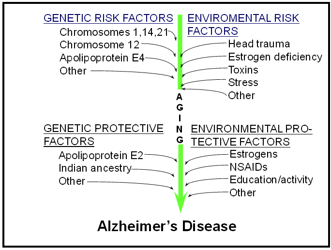
Risk factors and protective factors for Alzheimer’s disease
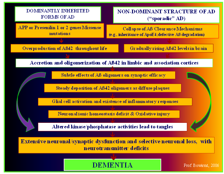
The mechanism of Alzheimer’s pathology
Importantly, the manifestation of ischemic and neurodegenerative pathology was found to have a profound influence on the appearance of dementia, suggesting reciprocal interactions between ischemia and neurodegeneration. Indeed, another entity, called vascular dementia (VaD), comprises a less defined group of dementia patients having various vascular diseases that especially emerge in the elderly population and require valid options for examination and differential diagnosis [9].
The differential diagnosis between AD (the most common and perhaps best known cause of Dementia) and VaD (The second common cause of dementia after Alzheimer’s dementia) are still roughly problematic in clinical practice, despite the widely used diagnostic criteria to differentiate between the two disorders [10].
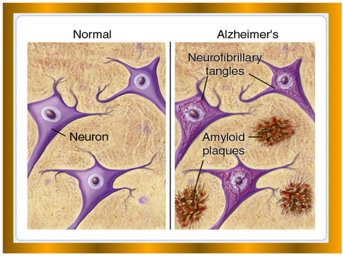
Comparison between Normal and abnormal neuron cells in AD.
There is an increasing evidence that cerebrovascular dysfunction plays a role not only in vascular causes of cognitive alterations but also in AD [11, 12, 13, 14]. Cognitively patients, with AD, show sometimes mixed degrees of associated vascular lesions in 30-60% of AD cases. In opposition, AD pathology may be present in 40%-80% of VaD patients, thus impeding diagnosis accuracy [15, 16, 17]. Therefore, to eliminate this bewilderment and discrepancies in the diagnosis between the AD and VaD, it is worthy to shed light firstly on a disease that is a microangiopathy and represents VaD with clear milestones and features as is the case of cerebral autosomal dominant arteriopathy with subcortical infarcts and leukoencephalopathy (CADASIL).
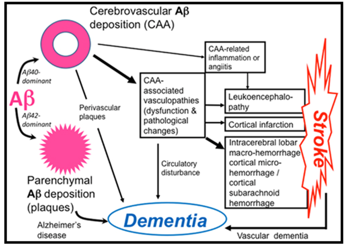
Pathophysiology of cerebral amyloid angiopathy (CAA)-related disorders. Aβ shows parenchymal or vascular deposition, depending on dominance of Aβ42 or Aβ40, respectively, in elderly individuals and patients with Alzheimer’s disease (AD). Cerebrovascular amyloid deposition, CAA, is related to stroke and dementia.
Studying CADASIL CSF biomarkers profile, will help in the differential diagnosis between both diseases sharing the coexisting neurodegeneration, furthermore, CADASIL is a dominantly inherited mid-adult life neurodegenerative disease, which belongs to vasculopathies and symbolizes a genuine prototype of VaD that provides a valuable opportunity for studying its CSF biomarkers. Secondly, examining and evaluating the CSF biomarkers of AD compared to that of CADASIL [18, 19, 20]. The pathogenesis similarities between CADASIL and early onset AD come from the fact that in both diseases genetic mutations occur in early adulthood [21, 22, 23]. CADASIL mutations in NOTCH3 gene generate toxic protein aggregates (Granular Osmiophilic Material-GOM) in the vicinity of vascular smooth muscle cells (VSMCs) causing degeneration and loss of VSMCs in small arteries and arterioles of white matter regions of the brain [24, 25, 26]. The deposition and discrepancy of clearance of the neurotoxic GOM lead to neurodegenerative subcortical dementia, similar to those attributed to mutant forms of the Amyloid Precursor Proteins (APP) and presenilins genes which cause overproduction and accumulations of the toxic Aβ42 protein in the brain and collapse of Aβ42 clearance mechanisms which lead to neurodegenerative cortical dementia (AD). Despite the presumed pathological similarities, substantial differences between the two phenomena may exist especially in the CSF neurochemical phenotypes [27]. Our advance knowledge of the underlying molecular pathology has set the stage for clinically meaningful advances in the development of biomarkers. The discovery of novel diagnostic methods based on biochemical and imaging biomarkers of disease specific pathology increase the capability to supply efficient measures of natural history, biological activity and markers of surrogate endpoints. Markers of biological activity should be also evaluated regarding their value to reflect disease progression, heterogeneity of the clinical population, for early decision making and characterization of new treatments [28]. On this basis we reviewed the body of literature that has examined CSF total tau (t-tau), β-amyloid protein 1-42, and phosphorylated tau (p-tau) as diagnostic tests for AD and, for a genuine prototype of VaD (CADASIL) that provide a unique opportunity for studying their CSF biomarkers.

2. Overview of Biomarkers Properties

Access to molecular and biochemical markers of neurodegenerative diseases would complement clinical approaches, and further the goals of early and accurate diagnosis. Hence, the importance of the biological markers studies which are quantitative measurements that provide information about: intrinsic biological processes, a disease circumstances and risk of developing an illness (antecedent biomarkers); assist in diagnosing disease (diagnostic biomarkers); response to treatment (prognostic biomarkers), providing much-needed insight into preclinical and clinical data, all of these are still valid procedures for early detection of diseases [29].
Detection the subject’s susceptibility to the disease prior to appearance of prodromal signs, and detection of neural dysfunction before irreversible cellular damage, will be tremendously valuable for developing; prevention and intervention strategies and early treatments. From here stems the truthfulness and reliability of biomarker to distinguish between normal and interested disease [30].
In fact, biomarkers are set of factors used to measure anatomic, physiologic, biochemical, pharmacological, or molecular parameters associated with the presence and severity of particular disease states or processes in humans and animals [31, 32].
These characteristics are objectively measured and carefully evaluated as indicators of normal biologic or pathogenic processes or pharmacological responses to a therapeutic intervention. Actually, define biomarker panels comprehensively to quantify risk, assess prognosis, and determine response to therapy [33]. Years ago, biomarkers were primarily physiological indicators such as blood pressure or heart rate. More recently, biomarker is becoming an important tool in different disciplines such as in field of oncology, immunology, cardiovascular diseases and metabolic diseases [34]. Synonym for molecular biomarker, such as elevated prostate specific antigen (PSA) as a molecular biomarker for prostate cancer, or using enzyme assays as liver function tests [35].
Biomarkers also cover the use of molecular indicators of environmental exposure in epidemiologic studies such as human papilloma virus (HPV) or certain markers of tobacco exposure such as 4 - (methylnitrosamino) - 1- (3-pyridyl)-1-butanone (NNK) which is a nitrosamine present in tobacco that is a potent procarcinogen. It is activated by CYP2A6, and plays a role as a biomarker of exposure to cigarette smoke, produced upon the curing of tobacco [36, 37]. Also, Genomic biomarkers have principal role in investigation diseases; such as Apolipoproteine Epsilon-4 allele (APOE-ε4) for AD, and HLA for looking for narcolepsy, CD19, Sialophorin, CD11 integrin cluster, and IL-4 receptor – for Crohn’s disease. Other methods and assays are used like; Serum or Spinal fluid substance, Neuroimaging and Physiologic parameters [38, 39, 40, 41].
A. Overview of the CSF Biomarkers for AD and CADASIL.
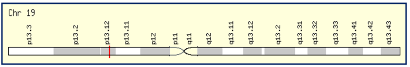
A. CADASIL Gene located at Chromosome 19p13.2
Plethora of biomarkers has been evaluated for AD. According to the literature more than 25 potential biomarkers for AD had previously been identified and new ones are still under investigation [42].
The CSF is the direct target because of its straight contact with the extracellular space of the brain where many biochemical processes in the brain take place and are reflected in the CSF. Since AD pathology is mainly restricted to the brain, CSF is an obvious source and justified biomarkers for AD. Indeed, early biomarker discovery efforts for AD is based on immunoassays to detect and measure pathophysiological molecules of AD, such as Cerebrospinal fluid–derived β-amyloid protein 1-42, total tau protein, and phosphorylated tau (181,199,231) protein [43]. These CSF classical biomarkers reflect the neuropathological changes taking place in AD brains, thus disclosing the disease in its prodromal phase. However, it has been demonstrated that variations in the levels of these biomarkers in the CSF also appear in different degrees in other neurological diseases such as stroke, VaD and CADASIL. With the aim to investigate the usefulness of these biomarkers in the differential diagnosis between the various pathologies mentioned above and to evaluate the power of each biomarker and/or their combination in predicting different diseases progression, we have compared the levels of these CSF biomarkers in AD, VaD and CADASIL.
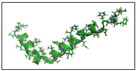
B. Amyloid βeta (Aβ)
Although the production pathways and the absolute function of Aβ in the brain still vague, its presence as a main component of senile plaques is unambiguous. Aβ is created by a cluster of peptides formed by proteolytic cleavage of the type I transmembrane straddling glucoprotein amyloid precursor protein (APP, OMIM 104760, chromosome 21q21) through sequential cleavages by BACE1 (The major β-secretase in the brain) and γ-secretase complex. The γ secretase, which produces the C-terminal end of the Aβ peptide, cleaves within the transmembrane region of APP and can generate a number of isoforms of 36-43 amino acid residues in length. The most common isoforms are Aβ40 and Aβ42; the longer form is typically produced by cleavage that occurs in the endoplasmic reticulum, while the shorter form is produced by cleavage in the trans-Golgi network- “amyloidogenic pathway” [44]. Missense mutations in the genes of APP and presenilins (PSEN1, OMIM 104311, chromosome 14q24.3) and presenilin 2 (PSEN2, OMIM 600759, chromosome 1q31-q42) genes that are located on chromosomes 21, 14, and 1, respectively share the common feature of altering the γ-secretase cleavage of APP to increase the production of the amyloidogenic Aβ42, the primary component of amyloid plaques in both familial and sporadic AD [45, 46]. Paradoxically, “non-amyloidogenic pathway”, APP is first cleaved by the α-secretase, members of the ADAM (a disintegrin and metalloprotease) family of zinc metalloproteases, within the Aβ sequence thus precluding production of intact Aβ peptides [47].
The peptides, particularly Aβ1–42, are aggregation prone, self-assembling to form a heterogeneous mixture of soluble oligomers, protofibrils and fibrils. Only levels of the soluble, fibrillar oligomers were found to be elevated significantly in AD brains, where their levels correlate strongly with AD onset-severity, and are therefore proposed to be the major neurotoxic species in AD [48].
Consequent deleterious effects include neurotoxicity, memory impairments, inhibition of long-term potentiation (LTP), loss of dendritic spines and synaptic dysfunction [49].
Although the function of APP remains to be fully elucidated, understanding APP trafficking and processing would also provide new insights into the regulatory mechanism of the amyloidogenic pathway. The processing of APP involves numerous steps, including APP sorting, transport, internalization and sequential proteolysis. Altered routing of APP trafficking and distribution in neurons might lead to the amyloidogenic pathway, which is implicated in the pathology of AD. Hence, the intracellular distribution and transport of APP are critical for Aβ production [50]. Indeed, increased Aß42 production throughout life and faulty clearance of Aß mechanisms lead to accumulation of oligomerization of Aß42 in limbic and association cortices, due to the altered in the numerous proteases in the brain that participate in Aβ degradation and clearance including cathepsins, gelatinases, endopeptidases, aminopeptidase, neprilysin, serine protease, and insulin-degrading enzyme [51, 52].
Aß42 makes up less than 10% of total Aß and it is the initial and major component of amyloid plaque deposits in AD [53].
The detection that Aβ42 peptide forms the essential component of AD plaques and that is secreted by cells led to examinations of Aβ42 in the CSF. Previous studies showed a decrease in CSF-Aβ42 to about 40–50% of control levels has been found in AD in several papers [54].
It is not clear why Aβ42 is reduced in AD patients, but it is thought that its decrease reflects trapping of Aß42 in the amyloid plaques in the brain. Indeed, studies suggest that decreased CSF Aß42 correlates well with the levels of amyloid plaques in the AD brain as determined by amyloid imaging [55].
According to the amyloid cascade hypothesis, the pathogenesis of AD states that Aβ accumulation in the brain begins in the early prodromal stage of AD, and it is a key factor that initiates the neurodegenerative process. Accumulation of Aβ in the brain of presymptomatic AD patients results consequently in decreasing the level of CSF Aß42. Therefore, CSF biomarkers (Aß42) are altered very early giving the possibility to detect AD patients at risk or to be used as predictors of disease progression in persons with mild cognitive impairment (MCI). More precisely, amyloid-peptide burden and changes in APP metabolism are altered at first up to 10 years before clinical symptoms. Considering the diagnostic sensitivity and specificity levels of Aβ42 in CSF – AD patients that ranged between 80% and 90%, may enhance its use as potential test in combinations with other tests [56, 57, 58].
More than two decade ago a new broader term called vascular dementia (VaD) appeared on the screen as a wide spectrum of cognitive impairments pathology associated with cerebrovascular pathologies that do not fulfill AD criteria [59]. New criteria and diagnostic standards for this new disorder were recognized and have gained the attention of the clinical neuroscientists [20, 60, 61, 62].
Intensive studies in this topic indicate that cerebrovascular diseases and AD share the same risk factors [63] and this notion enhanced the thought that simultaneous ischemic events and neurodegenerative pathology have a profound impact on the expression of the dementia, suggesting mutual interactions between ischemia and neurodegeneration and point out that vascular factors may play a role in the pathogenesis of AD [64, 65, 66, 67].
Indeed, Aβ has potent cerebrovascular effects, and hypoxia–ischemia is a powerful modulator of cerebral amyloidogenesis [68]. Conversely, the cerebrovascular dysregulation induced by Aβ could hazard perfusion; decrease vascular reserves, and enhance the propensity to ischemic damage.
In addition, hypoxia and/or ischemia encourage the cleavage of Aβ from the amyloid precursor protein (APP) by upregulating β-secretase expression and activity [69, 70, 71].
On the light of these findings we can conclude that ischemia events exacerbates Aβ accumulation in the brain by decreasing the major elimination pathway of this neurotoxic peptide and in the end its brain clearance [72, 73].
The increasing evidence that cerebrovascular dysfunction plays a role not only in VaD but also in AD, comes from the fact that both impair the neurovascular unit, boost the view that a remarkable overlapping is exist between VaD and AD not only in the underlying risk factors mentioned above, but may also in sharing some CSF neurochemical phenotypes abnormalities.
For example, the CSF profile of Aβ proteins in patients with AD is highly decreased (A pathological decreased amyloid β1–42 assay was considered at <450 pg/ml according to manufacturer’s instruction) but Aβ CSF levels in VaD have been reported to be moderately decreased or significantly overlapping with AD. Since previous studies showed conflicting results in VaD, we investigated CSF biomarkers in CADASIL, since CADASIL represents a model of pure subcortical vascular dementia occurring in young adults, and unlikely to share associated onset age with that of AD and it is the most common single gene disorder leading to ischaemic stroke, characterized by a systemic arteriopathy, which specifically affects the cerebral small vessels causing diffuse changes which result in hypo-perfusion, ischemia and inability of the cerebral vessels to autoregulate [74], we can expect that CSF biomarkers, especially Aβ, t-tau and p-tau levels, sufficiently discriminate between AD on one hand and CADASIL on other hand.
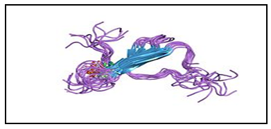
C. Total tau protein (Microtubule-associated protein tau)
While tau has long been implicated in neurodegenerative conditions, its functions in the adult brain and the precise mechanisms by which it contributes to neuronal dysfunction and degeneration in these disorders remain to be elucidated. Physiologically, tau proteins are an intracelluar microtubule-associated protein acting as stabilizers microtubules in the cell cytoskeleton, and pathologically, tau proteins characterize the main component relating to intraneuronal changes in AD patients.
A flurry of recent studies has challenged major dogmas in this field, including the vision that filamentous tau aggregates are the most pernicious forms of tau, that failure of tau function plays a major role in the pathogenesis of tauopathies. Provocative discoveries suggest that tau regulates neuronal excitability and that it is required for Aβ and other neurotoxins to cause neuronal deficits, aberrant network activity and cognitive decline [75].
T-Protein is a microtubule-associated protein located in the neuronal axons. Because of alternative splicing of T-mRNA, there are six isoforms ranging in size from 352 to 441 amino acids, with molecular weights of approximately 50–65 kDa [76].
In normal situations, tau is coordinated by phosphorylation. In abnormal conditions, tau becomes hyperphosphorylated (phospho-tau) and accumulates as paired helical filaments that aggregate into masses inside the neurons as neurofibrillary tangles (NFT), which represent one of the hallmarks of AD [77].
The logic for considering tau as biomarker is the presence of abnormal intraneuronal aggregates of phospho-tau observed in many tauopathies, including AD, Tau aggregates can be examined in the Brain and peripheral fluids. Biochemical and Immunohistochemical properties of Tau cumulative in brain permit postmortem categorization and differential diagnosis of tauopathies [78].
The first report on CSF T-T as a biomarker for AD was published in 1993. In that paper, an enzyme-linked immunosorbent assay (ELISA) with a polyclonal reporter antibody was used [79].
Total tau concentrations especially phosphorylated tau (181,199,231) proteins can be measured in the CSF as Aß, and show a good correlation with the diagnosis of AD [80].
Previous studies have established 300% increase in the concentration of total-Tau in CSF Alzheimer’s patients 70 years and older versus control subjects younger than 50 years [(>600 pg/mL vs <200 pg/mL), respectively] [81].
A strong correlation between age and t-tau in healthy individuals has been determined with a cut off value of > 500 pg/mL (> 70 years) versus 450 pg/mL (< 70 years) [82].
CSF t-tau levels in AD patients have a sensitivity of 90% and specificity of 81% compared to healthy Control [83], compared to other dementias, the sensitivity and specificity drops to 50%-60% [84].
Notwithstanding, the relative high sensitivity and specificity that CSF T-tau level plays as discriminator between AD patients and control, its presence in other neurological diseases for instance, VaD, progressive supranuclear palsy, corticobasal degeneration, CADASIL, and its notable high concentration in Spongiform Encephalopathies (3000 pg/mL), decreases at some level its validity as AD specific biomarker [85, 86].
However, the absence of specific novel biomarker for AD and related disorders require a combination of different candidate’s biomarkers. The decreased CSF levels of Aβ42 in AD, VaD and CADASIL enhance the effort to find additional biomarkers that may be utilized to differentiate between them. Thus, we studied CSF changes in CSF biomarker profiles of commonly used in patients with AD, VaD and CADASIL. With special respect to total tau, which is known as an indicator of neuronal damage and considered to be altered in CSF of AD and VaD patients. The CSF profile of total tau in patients with AD is characterized by increased levels. On the contrary in VaD, studies on this CSF biomarker showed conflicting results: t-tau levels have been reported to be increased, normal or intermediate or significantly overlapping with AD whereas in CADASIL t-tau levels were normal and significantly different with respect to AD.
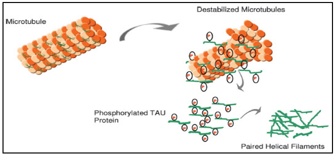
D. Human Phosphorylated tau (P-tau: phosphorylated at Thr181)
Tau proteins belongs to a group of proteins referred to as microtubule-associated phosphoproteins that are abundant in neurons in the central nervous system and are less common elsewhere. It has been almost 38 years since tau was discovered as a heat resistant and limited affected by acid treatment without loss their function [87].
Remarkably, soluble neuronal microtubule-associated protein that normally functions to support the assembly and stabilization of the microtubule cytoskeleton [88].
There is significant evidence that a deviations from normal phosphorylation process (Hyperphosphorylation) results in tau dysfunction and modification of the conformation of tau and decreasing its affinity to microtubules [89].
Discussions with regulatory authorities gain momentum defining the role of tau biomarkers for trial designs and how they may be further qualified for surrogate marker status [90].
Numerous studies have evaluated the diagnostic value of CSF markers for AD cases, finding high CSF-T-tau and P-tau, followed by MCI, Stroke and VaD (the pathological elevated phosphorylated tau level in AD was considered at >61 pg/ml according to manufacturer’s instruction). It is worthy to mention that paradoxical results on CSF biomarkers were found conflicting in VaD (the prototype model of CADASIL) which can be due to possible presence of other underlying pathological alterations [91].
These findings encourage the investigation of P-tau as well as T-tau levels in CADASIL rather than in VaD, since CADASIL reflects a comprehensible pathological model almost exclusively due to cerebrovascular features, making it a clearer model than VaD. Since the levels of P-tau and total tau (T-tau) in the CSF of CADASIL patients were normal, in contrast with elevated values in AD, it’s legitimate to consider CADASIL in the differential diagnosis between various neurological diseases.
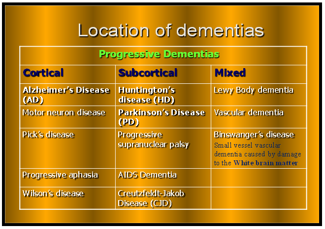
Different anatomical location of dementias within the brain (Cortical area, Subcortical and Mixed form)

3. Conclusions

The differential diagnosis between AD and VaD are still roughly problematic in clinical practice. There is a growing evidence that cerebrovascular dysfunction plays a role not only in vascular causes of cognitive impairments but also in AD. Therefore, to eliminate this bewilderment and discrepancies in the diagnosis between the AD and VaD, it is worthy to examine a disease that is a microangiopathy and represents VaD with clear milestones and features as is the case of cerebral autosomal dominant arteriopathy with subcortical infarcts and leukoencephalopathy (CADASIL). CADASIL is a dominantly inherited mid-adult life disorder causing ischemic strokes, which belongs to vasculopathies and symbolizes a prototype of VaD that provides a unique opportunity for studying its CSF biomarkers and comparing it with CSF biomarkers of AD.
The pathogenesis similarities between CADASIL (a relatively mid-adult disease) with early onset of AD affect the small vessels of the brain suggest plausible molecular mechanisms that are involved in vascular damage and come from the fact that in both diseases genetic mutations occur. CADASIL mutations in NOTCH3 gene generate toxic protein aggregates (Granular Osmiophilic Material- GOM) in the vicinity of vascular smooth muscle cells (VSMCs) causing degeneration and loss of VSMCs in small arteries and arterioles of white matter regions of the brain that lead to dementia, similar to those attributed to mutant forms of the Amyloid Precursor proteins (APP) and presenilins genes who cause overproduction and accumulations of the toxic Aβ42 protein in the brain and collapse of Aβ42 clearance mechanisms in AD. Despite the presumed pathological similarities, substantial differences between the two phenomena may exist especially in the CSF neurochemical phenotypes.
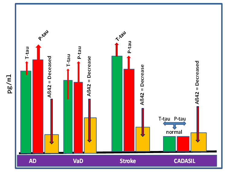
The figure illustrates the variations in levels of the classical biomarkers Aß42, t-tau and P-tau), altered generally in the AD, VaD, Stroke and CADASIL.
The figure below comes to illustrate the dissimilarity of commonly used biomarkers (Aß42, t-tau and P-tau) that are found in the CSF of different neurological diseases such as: AD, VaD, Stroke and CADASIL. Indeed, these divergences are important for the differential diagnosis between the mentioned neurological diseases above. In fact, there is a clear advantage when highlighting the difference in CSF biomarkers between AD and CADASIL that serves to increase the diagnostic accuracy when they used with other biomarkers such as imaging markers.
This figure was made according to data from previous studies and from the literature to illustrate the variations in levels of the classical biomarkers (Aß42, t-tau and P-tau), altered generally in the AD, VaD, Stroke and CADASIL. Levels measured by (pg/ml) for all the biomarkers, showed Aß42 with lowest level in AD followed by stroke and moderately decreased in VaD. Total-tau levels are highly increased in stroke followed by AD. On the contrary in VaD, studies on these CSF biomarkers showed conflicting results: t-tau levels have been reported to be increased, normal or intermediate, but in any case much lower than in AD. Phosphorylated-tau (P-tau) levels are highly increased in AD followed by stroke but levels of (P-tau) have been reported to be either normal or increased in VaD. In CADASIL total-tau levels and P-tau levels are normal, and Aß42 are markedly deceased and considerably overlapped with AD [19, 92].

ACKNOWLEDGMENTS

We thank Miss Aia Bowirrat for her contribution in revising and editing the manuscript.

References

[1]  Koffie RM, Hyman BT and Spires-Jones TL (2011) Alzheimer’s diseases: synapses gone cold. Molecular Neurodegeneration 6:63. Doi:10.1186/1750-1326-6-63
[2]  Yassin M, Armaly Z, Bisharat B, Bowirrat A (20-13) Biomarkers for Alzheimer’s Disease: Imagination or Reality-View and Review! Journal of Behavioral and Brain Science 3: 393-402.
[3]  Guerreiro RJ, Gustafson DR, Hardy J (2012) The genetic architecture of Alzheimer’s disease: Beyond APP, PSENs AND APOE. Neurobiol Aging 33(3): 437–456.
[4]  Bowirrat A., Friedland RP., Farrer L., Baldwin C., Korczyn A (2002) Genetic and Environmental Risk factors for Alzheimer’s disease in Israeli Arabs. Journal of Molecular Neuroscience 19(1-2):239-245.
[5]  Waldemar G, Dubois B, Emre M (2007) Recommendations for the diagnosis and management of Alzheimer’s disease and other disorders associated with dementia: EFNS guideline. European Journal of Neurology 14(1):e1–e26.
[6]  Clifford RJ, Knopman DS, Jagust WJ, Shaw LM, Aisen PS, Weiner MW, Petersen RC, Trojanowski JQ (2010) Hypothetical model of dynamic biomarkers of the Alzheimer’s pathological cascade. Lancet Neurol 9(1): 119. doi: 10.1016/S1474-4422(09)70299-6.
[7]  Jack CR, Holtzman DM (2013) Biomarker modeling of Alzheimer's disease. Neuron 80(6):1347-58.
[8]  Leszek J, Sochocka M, Gąsiorowski K. Vascular factors and epigenetic modifications in the pathogenesis of Alzheimer's disease (2012) J Neurol Sci 15;323(1-2):25-32. doi: 10.1016/j.jns.2012.09.010. Epub 2012 Sep 29. Review.
[9]  Iadecola C (2010) The overlap between neurodegenerative and vascular factors in the pathogenesis of dementia. Acta Neuropathol 120(3): 287–296. doi:10.1007/s00401-010-0718-.
[10]  Grand Jacob HG, Caspar S, MacDonald Stuart WS (2011) Clinical features and multidisciplinary approaches to dementia care. Journal of Multidisciplinary Healthcare 4 125–147.
[11]  Iadecola C, Park L, Capone C (2009) Threats to the Mind Aging, Amyloid, and Hypertension. Stroke 40(3 Suppl): S40–S44.
[12]  Esiri MM, Nagy Z, Smith MZ, Barnetson L, Smith AD (1999) Cerebrovascular disease and threshold for dementia in the early stages of Alzheimer’s disease. Lancet 354:919–920.
[13]  Meng Y, Baldwin CT, Bowirrat A, Inzelberg R, Waraska K, Friedland RP, Farrer LA (2006) Association of Polymorphism in the Angiotensin-converting Enzyme Gene With Alzheimer's Disease in an Israeli-Arab Community. Am J Hum Genet 78:871-877.
[14]  Schneider JA, Wilson RS, Bienias JL, Evans DA, Bennett DA (2004) Cerebral infarctions and the likelihood of dementia from Alzheimer disease pathology. Neurology 62:.1148–1155.
[15]  Jellinger KA (2013) Pathology and pathogenesis of vascular cognitive impairment—a critical update. Front Aging Neurosci 5: 17.
[16]  Jellinger KA (2007) The enigma of vascular cognitive disorder and vascular dementia. Acta Neuropathol 113(4): 349–388.
[17]  Kalaria RN, Ballard C (1999) Overlap between pathology of Alzheimer disease and vascular dementia. Alzheimer Disease and Associated Disorders 13(3):S115–S123.
[18]  Jellinger KA, Attems J (2010) Prevalence of dementia disorders in the oldest-old: an autopsy study. Acta Neuropathol 119:421–433. [PubMed]
[19]  Formichi P, Parnetti L, Radi E, Cevenini G, Dotti MT, and Federico A (2010) “CSF Biomarkers Profile in CADASIL—A Model of Pure Vascular Dementia: Usefulness in Differential Diagnosis in the Dementia Disorder,” International Journal of Alzheimer’s Disease Article Volume 2010, ID 959257, 6 pages. doi:10.4061/2010/959257.
[20]  Roman GC, Tatemichi TK, Erkinjuntti T, Cummings JL, Masdeu JC, Garcia JH, Amaducci L, Orgogozo JM, Brun A, Hofman A, Moody DM, O’Brien MD, Yamaguchi T, Grafman J, Drayer BP, Bennet DP, Fisher M, Ogata J, Kokmen E, Bermejo F, Wolf PA, Gorelick PB, Bick KL, Pajeau AK, Bell MA, DeCarli C, Culebras A, Korczyn AD, Bogousslavsky J, Hartman A, Scheinberg P (1993) Vascular dementia: diagnostic criteria for research studies. Report of the NINDS-AIREN International Workshop. Neurology 43: 250 – 260.
[21]  Marchesi VT (2014) Alzheimer's disease and CADASIL are heritable, adult-onset dementias that both involve damaged small blood vessels. Cell Mol Life Sci 71(6):949-55. doi: 10.1007/s00018-013-1542-7. Epub 2013 Dec 31.
[22]  Xinzhen Yin, Dingwen Wu, Jinping Wan, Shenqiang Yan, Min Lou, Guohua Zhao, Baorong Z (2014) Cerebral autosomal dominant arteriopathy with subcortical infarcts and leukoencephalopathy: Phenotypic and mutational spectrum in patients from mainland China. International Journal of Neuroscience 8:1-22.
[23]  Joutel A, Corpechot C, Ducros A, Vahedi K, Chabriat H, Mouton P, Alamowitch S, Domenga V, Cecillion M, Marechal E, Maciazek J, Vayssiere C, Cruaud C, Cabanis EA, Ruchoux MM, Weissenbach J, Bach JF, Bousser MG, Tournier-Lasserve E (1996) Notch3 mutations in CADASIL, a hereditary adult-onset condition causing stroke and dementia. Nature 24:383:707-10.
[24]  Goebel HH, Meyermann R, Rosin R, Schlote W (1997) Characteristic morphologic manifestation of CADASIL, cerebral autosomal- dominant arteriopathy with subcortical infarcts and leukoencephalopathy, in skeletal muscle and skin. Muscle Nerve 20:625–7.
[25]  LaPoint SF, Patel U, Rubio A (2000) Cerebral autosomal dominant arteriopathy with subcortical infarcts and leukoencephalopathy (CADASIL). Adv Anat Pathol 7(5): 307-21.
[26]  Malandrini A, Gaudiano C, Gambelli S (2007) Diagnostic value of ultrastructural skin biopsy studies in CADASIL. Neurology 68(17):1430-1432.
[27]  Bowirrat A (2014) The Pathogenesis of Cerebral Autosomal Dominant Arteriopathy with Subcortical Infarcts and Leukoencephalopathy (CADASIL, OMIM #125310). Ann Clin Cytol Pathol 1(1): 1002.
[28]  Hampel H, Mitchell A, Blennow K, Frank RA, Brettschneider S, Weller L, Möller HJ (2004) Core biological marker candidates of Alzheimer's disease - perspectives for diagnosis, prediction of outcome and reflection of biological activity. J Neural Transm 111(3): 247–272. doi:10.1007/s00702-003-0065-z.
[29]  Frangogiannis NG (2012) “Biomarkers: Hopes and Challenges in the Path from Discovery to Clinical Practice,” Transla-tional Research 159(4): 197-204. doi:10.1016/j.trsl. 2012.01.023.
[30]  Shaw M, Korecka M, Clark CM, Lee VMY and Trojanowski JQ (2007) “Biomarkers of Neurodegeneration for Diagnosis and Monitoring Therapeutics,” Nature Reviews Drug Discovery 6(4): 295-303. doi:10.1038/nrd2176.
[31]  Downing G (2001) “Biomarkers Definitions Working Group. Biomarkers and Surrogate Endpoints,” Clinical Pharma- cology & Therapeutics 69; 89-95.
[32]  Lee J, Devanarayan V, Barrett U, Weiner R, Allinson J, Fountain S, Keller S, Weinryb I, Green M and Duan L (2006) “Fit-for-Purpose Method Development and Validation for Successful Biomarker Measurement,” Pharmaceutical Research 23(2): 312-328. doi:10.1007/s11095-005-9045-3
[33]  Frank R and Hargreaves R (2003) “Clinical Biomarkers in Drug Discovery and Development,” Nature Reviews Drug Dis- covery 2(7): 566-580. doi:10.1038/nrd1130
[34]  Harald H, Simone L and Khachaturian ZS (2012) “Develop- ment of Biomarkers to Chart All Alzheimer’s Disease Stages: The Royal Road to Cutting the Therapeutic Gor- dian Knot,” Alzheimer’s & Dementia 8(4): 312-336.doi:10.1016/j.jalz.2012.05.2116.
[35]  Dhanasekaran SM (2001) “Delineation of Prognostic Biomar- kers in Prostate Cancer,” Nature 412(6849): 822-826. doi:10.1038/35090585
[36]  Wagner PD, Maruvada P and Srivastava S (2004) “Molecular Diagnostics: A New Frontier in Cancer Prevention,” Ex- pert Review of Molecular Diagnostics 4(4): 503-511. doi:10.1586/14737159.4.4.503.
[37]  Gerde P, Muggenburg BA, Stephens T, Lewis JL, Pyon KH and Dahl AR (1998) “A Relevant Dose of 4-(Me- thylnitrosamino)-1-(3-pyridyl)-1-butanone Is Extensively Metabolized and Rapidly Absorbed in the Canine Tra- cheal Mucosa,” Cancer Research 58(7): 1417-1422.
[38]  Prashanthi V (2010) “Effect of Apolipoprotein E on Biomarkers of Amyloid Load and Neuronal Pathology in Alzheimer Disease,” Annals of Neurology 67(3): 308-316.
[39]  Johnston JA and Hill M (2011) “The Risk and Gamble We Take with Our Patients on Dopamine Agonists,” Journal of Neurology, Neurosurgery & Psychiatry 83(3): e1-e1. doi:10.1136/jnnp-2011-301993.155.
[40]  Golias CH (2005) “Adhesion Molecules in Cancer Invasion and Metastasis,” Hippokratia 9(1): 106-114.
[41]  Kader HA, Tchernev VT, Satyaraj E, Lejnine S, Kotler G, Kingsmore SF and Patel D (2005) “Protein Microar- ray Analysis of Disease Activity in Pediatric Inflamma- tory Bowel Disease Demonstrates Elevated Serum PLGF, IL-7, TGF-β1, and IL-12p40 Levels in Crohn’s Disease and Ulcerative Colitis Patients in Remission versus Ac- tive Disease,” The American Journal of Gastroenterology 100(2): 414-423. doi:10.1111/j.1572-0241.2005.40819.x.
[42]  Carrette O, Demalte I, Scherl A, Yalkinoglu O, Corthals O, Burkhard G and Sanchez JC (2003) “A Panel of Cerebrospinal Fluid Potential Biomarkers for the Diagno- sis of Alzheimer’s Disease,” Proteomics 3(8): 1486-1494. doi:10.1002/pmic.200300470
[43]  Ibach B, Binder H, Dragon M, Poljansky S, Haen E, Schmitz E and Hajak G (2005) “Cerebrospinal Fluid tau and β- Amyloid in Alzheimer Patients, Disease Controls and an Age-Matched Random Sample,” Neurobiology of Aging 27(9): 1202-1211. doi:10.1016/j.neurobiolaging.2005.06.005
[44]  Hartmann T, Bieger SC, Brühl B, Tienari PJ, Ida N, Allsop D, Roberts GW, Masters CL, Dotti CG, Unsicker K, Beyreuther K (1997) "Distinct sites of intracellular production for Alzheimer's disease A beta40/42 amyloid peptides". Nat Med 3(9): 1016–20. doi:10.1038/nm0997-1016. PMID
[45]  Allam AR, Kiran KR, Hanuman T (2008) Bioinformatic analysis of Alzheimer's. Disease using functional protein sequences. J Proteomics Bioinform 1: 036–042.
[46]  Wang R, Wang B, He W, Zheng H (2006) Wild-type presenilin 1 protects against Alzheimer disease mutation-induced amyloid pathology. J Biol Chem 281:15330–15336.
[47]  Kessels HW, Nguyen LN, Nabavi S, Malinow R (2010) The prion protein as a receptor for amyloid- beta. Nature 466: E3–E4.
[48]  Tomic JL, Pensalfini A, Head E, Glabe CG (2009) Soluble fibrillar oligomer levels are elevated in Alzheimer's disease brain and correlate with cognitive dysfunction. Neurobiol Dis 35:352–358.
[49]  Gimbel DA, Nygaard HB, Coffey EE, Gunther EC, Laurén J, Gimbel ZA (2010) Memory impairment in transgenic Alzheimer mice requires cellular prion protein J Neurosci 30: 6367–6374.
[50]  Yang M, Virassamy B, Vijayaraj SL, Lim Y, Saadipour K, Wang YJ (2013) The intracellular domain of sortilin interacts with amyloid precursor protein and regulates its lysosomal and lipid raft trafficking. PLoS ONE 8(5): e63049
[51]  Butler D and Bahr BA (2006) “Oxidative Stress and Lyso- somes: CNS-Related Consequences and Implications for Lysosomal Enhancement Strategies and Induction of Autophagy,” Antioxidants & Redox Signaling 8(1-2): 185-196.
[52]  Avila J, Lucas JJ, Perez M, Hernandez F (2004) Role of tau protein in both physiological and pathological conditions. Physiol Rev 84: 361–384.
[53]  Graff-Radford NR, Crook JE, Lucas J, Boeve BF, Knopman DS, Ivnik RJ and Younkin SG (2007) “Associa- tion of Low Plasma Abeta42/Abeta40 Ratios with In- creased Imminent Risk for Mild Cognitive Impairment and Alzheimer Disease,” Archives of Neurology 64(3): 354.doi:10.1001/archneur.64.3.354.
[54]  Blennow K, Vanmechelen E and Hampel H (2001) “CSF To- tal tau, Ab42 and Phosphorylated tau Protein as Bio- markers for Alzheimer’s Disease” Molecular Neurobiol- ogy 24(1-3): 87-97.
[55]  Fagan AM, Mintun MA, Mach RH, Lee SY, Dence CS, Shah AR, LaRossa GN, Spinner ML, Klunk WE, Mathis CA, DeKosky ST, Morris JC Holtzman DM (2006) “Inverse Relation between in Vivo Amy- loid Imaging Load and Cerebrospinal Fluid Abeta42 in Humans,” Annals of Neurology 59(3): 512-519. doi:10.1002/ana.20730
[56]  Fagan AM, Roe CM, Xiong C, Mintun MA, Morris JC and Holtzman DM (2007) “Cerebrospinal Fluid tau/ beta-Amyloid(42) Ratio as a Prediction of Cognitive De- cline in Nondemented Older Adults,” Archives of Neu- rology 64(3): 343-349. doi:10.1001/archneur.64.3.noc60123
[57]  Sjögren M, Davidsson P and Aallin A (2002) “Decreased CSF β-Amyloid42 in Alzheimer’s Disease and Amyotrophic Lateral Sclerosis May Reflect Mismetabolism of β- Amyloid Induced by separate mechanisms,” Dementia and Geriatric Cognitive Disorders 13: 112-118. doi:10.1159/000048642.
[58]  Strozyk D, Blennow K, White LR and Launer LJ (2003) “CSF Aβ42 Levels Correlate with Amyloid-Neuropatho- logy in a Population-Based Autopsy Study,” Neurology 60(4): 652-656. doi:10.1212/01.WNL.0000046581.81650.D0
[59]  Hachinski VC, Bowler JV (1993) Vascular dementia. Neurology 43:2159–2160.
[60]  Chui HC, Victoroff JI, Margolin D (1992) Criteria for the diagnosis of ischemic vascular dementia proposed by the State of California Alzheimer’s Disease Diagnostic and Treatment Centers. Neurology 42:473–480.
[61]  Jellinger KA (2008) Morphologic diagnosis of “vascular dementia”—a critical update. J Neurol Sci 270:1–12.
[62]  Erkinjuntti T, Gauthier S (2009) The concept of vascular cognitive impairment. Front Neurol Neurosci 24:79–85.
[63]  Breteler MM (2000) Vascular risk factors for Alzheimer’s disease: an epidemiologic perspective. Neurobiol Aging 21:153–160.
[64]  Iadecola C, Gorelick PB (2003) Converging pathogenic mechanisms in vascular and neurodegenerative dementia. Stroke 34:335–337.
[65]  Kalaria RN (2000) The role of cerebral ischemia in Alzheimer’s disease. Neurobiol Aging 21:321–330.
[66]  Nagy Z, Esiri MM, Jobst KA (1997) The effects of additional pathology on the cognitive deficit in Alzheimer disease. J Neuropathol Exp Neurol 56:165–170.
[67]  Snowdon DA, Greiner LH, Mortimer JA (1997) Brain infarction and the clinical expression of Alzheimer disease. The Nun Study. JAMA 277:813–817.
[68]  Iadecola C (2004) Neurovascular regulation in the normal brain and in Alzheimer’s disease. Nat Rev Neurosci 5:347–360.
[69]  Koike MA, Green KN, Blurton-Jones M, LaFerla FM (2010) Oligemic hypoperfusion differentially affects tau and amyloid-{beta} Am J Pathol 177(1): 300-310. doi: 10.2353/ajpath.2010.090750. [PMC free article] [PubMed]
[70]  Li L, Zhang X, Yang D (2009) Hypoxia increases Abeta generation by altering beta- and gamma-cleavage of APP. Neurobiol Aging 30:1091–1098. [PubMed]
[71]  Tesco G, Koh YH, Kang EL (2007) Depletion of GGA3 stabilizes BACE and enhances beta-secretase activity. Neuron 54:721–737. [PMC free article] [PubMed]
[72]  Cirrito JR, Yamada KA, Finn MB (2005) Synaptic activity regulates interstitial fluid amyloid-beta levels in vivo. Neuron 48:913–922. [PubMed]
[73]  Deane R, Wu Z, Sagare A (2004) LRP/amyloid beta-peptide interaction mediates differential brain efflux of Abeta isoforms. Neuron 43:333–344. [PubMed]
[74]  Pfefferkorn T, von Stuckrad-Barre S, Herzog J, Gasser T, Hamann GF, Dichgans M. Reduced cerebrovascular CO(2) reactivity in CADASIL: a transcranial Doppler sonography study. Stroke 32: 17–21.
[75]  Morris M, Maeda S, Vossel K and Mucke L (2011) “The Many Faces of Tau,” Neuron 70(3): 410-426. doi:10.1016/j.neuron.2011.04.009.
[76]  Buée L, Bussiere T, Buee-Scherrer V, Delacourte A and Hof PR (2000) “Tau Protein Isoforms, Phosphorylation and Role in Neurodegenerative Disorders,” Brain Re- search Reviews 33(1): 95-130. doi:10.1016/S0165-0173(00)00019-9.
[77]  Feijoo C, Campbell DG, Jakes R, Goedert M and Cuenda A (2005) “Evidence That Phosphorylation of the Micro- tubule-Associated Protein Tau by SAPK4/p38δ at Thr50 Promotes Microtubule Assembly,” Journal of Cell Sci- ence 118(2): 397-408. doi:10.1242/jcs.01655.
[78]  Boekhoorn K, Terwel D, Biemans B, Borghgraef P, Wiegert O, Ramakers GJ and Lucassen PL (2006) “Improved Long-Term Potentiation and Memory in Young tau- P301L Transgenic Mice before Onset of Hyperphos- phorylation and Tauopathy. J Neurosci 26(13): 3514-3523.
[79]  Vandermeeren M, Mercken M and Vanmechelen E (1993) “Detection of τ Proteins in Normal and Alzheimer’s Dis- ease Cerebrospinal Fluid with a Sensitive Sandwich En- zyme-Linked Immunosorbent Assay. Journal of Neuro- chemistry 61: 1828-1834.doi:10.1111/j.1471-4159.1993.tb09823.x.
[80]  Hansson O, Zetterberg H, Buchhave P, Londos E, Blennow K and Minthon L (2006) “Association between CSF Biomarkers and Incipient Alzheimer’s Disease in Patients with Mild Cognitive Impairment: A Follow-Up Study. The Lancet Neurology 5(3): 228-234.doi:10.1016/S1474-4422(06)70355-6.
[81]  Carrette O, Demalte I, Scherl A, Yalkinoglu O, Corthals O, Burkhard G and Sanchez JC (2003) “A Panel of Cerebrospinal Fluid Potential Biomarkers for the Diagno- sis of Alzheimer’s Disease. Proteomics 3(8): 1486-1494. doi:10.1002/pmic.200300470.
[82]  Lanari A and Parnetti L (2009) “Cerebrospinal Fluid Bio- markers and Prediction of Conversion in Patients with Mild Cognitive Impairment: 4-Year Follow-Up in a Rou- tine Clinical Setting. The Scientific World Journal 9: 961-966. doi:10.1100/tsw.2009.106.
[83]  Mattsson N, Zetterberg H, Hansson O, Andreasen N, Parnetti L, Jonsson M and Blennow K (2009) “CSF Biomarkers and Incipient Alzheimer Disease in Patients with Mild Cognitive Impairment,” The Journal of the American Medical Association 302(4): 385-393. doi:10.1001/jama.2009.1064.
[84]  Zanusso G, Fiorini M, Righetti PG and Monaco S (2011) “Specific and Surrogate Cerebrospinal Fluid Markers in Creutzfeldt—Jakob Disease,” Advances in Neurobiology 2: 455-467.
[85]  Formichi P, Parnetti L, Radi E, Cevenini G, Dotti MT and Federico A, “CSF Levels of β-Amyloid 1-42, Tau and Phosphorylated Tau Protein in CADASIL,” European Journal of Neurology 15(11): 1252-1255.doi:10.1111/j.1468-1331.2008.02277.
[86]  Stanford PA, Halliday GM, Brooks WS, Kwok JB, Storey CE, Creasey H and Schofield PR (2011) “Progressive Supranuclear Palsy Pathology Caused by a Novel Silent Mutation in Exon 10 of the Tau Gene Ex- pansion of the Disease Phenotype Caused by Tau Gene Mutations,” Brain 123(5): 880-893.
[87]  Cleveland DW, Hwo SY and Kirschner MW (1977) “Physical and Chemical Properties of Purified Tau Factor and the Role of Tau in Microtubule Assembly,” Journal of Molecular Biology 116(2): 227- 247.
[88]  Morris M, Maeda S, Vossel K and Mucke L (2011) “The Many Faces of Tau,” Neuron 70(3): 410-426. doi:10.1016/j.neuron.2011.04.009.
[89]  Buée L, Bussiere T, Buee-Scherrer V, Delacourte A and Hof PR (2000) “Tau Protein Isoforms, Phosphorylation and Role in Neurodegenerative Disorders,” Brain Re- search Reviews 33(1): 95-130.
[90]  Hampel H, Blennow K, Shaw LM, Hoessler YC, Zetterberg H and Trojanowski JQ (2009) “Total and Phosphory- lated Tau Protein as Biological Markers of Alzheimer’s Disease,” Experimental Gerontology 45(1): 30-40.doi:10.1016/j.exger.2009.10.010
[91]  Stenset V, Johnsen L, Kocot D (2006) Associations between white matter lesions, cerebrovascular risk factors, and low CSF Aβ42. Neurology 67(5):830–833. [PubMed]
[92]  Kaerst L, Kuhlmann A, Wedekind D, Stoeck K, Lange P, Zerr I (2013) Cerebrospinal fluid biomarkers in Alzheimer’s disease, vascular dementia and ischemic stroke patients: a critical analysis. J Neurol 260(11):2722-7. doi: 10.1007/s00415-013-7047-3.