International Journal of Agriculture and Forestry

p-ISSN: 2165-882X    e-ISSN: 2165-8846

2016;  6(1): 20-27

doi:10.5923/j.ijaf.20160601.04


Susceptibility of Cherries to Bacterial Canker (Pseudomonas syringae pv. syringae) in Field and Laboratory

Sanaz Farhadfar1, Mansureh Keshavarzi2, Naser Bouzari2, Ladan Moghadam1, Asghar Soleimani2

1Islamic Azad Univ, Garmsar, Iran

2Seed and Plant Improvement Institute, Karaj, Iran

Correspondence to: Mansureh Keshavarzi, Seed and Plant Improvement Institute, Karaj, Iran.

Email:

Copyright © 2016 Scientific & Academic Publishing. All Rights Reserved.

This work is licensed under the Creative Commons Attribution International License (CC BY).
http://creativecommons.org/licenses/by/4.0/

Abstract

Bacterial canker caused by Pseudomonas syringae pv. syringae is an important disease in cherries worldwide but low attention has been paid to breeding and selection of cherries for bacterial canker resistance. In this research, 21 selected Iranian and 7 introduced cultivars of sweet/sour/ducke cherries were examined for resistance to bacterial canker by artificial inoculation in field and laboratory condition. Correlation between canker resistance with wood diameter was also examined. Three local P. syringae strains were studied using LOPAT and GATTa tests and used as inoculum. For laboratory test, two-years-old dormant shoots were used and the canker length measured one month after inoculation. For field assay, trunk and shoots of two-years-old plants were inoculated in late autumn and eight months later, lesion length was measured. Based on result, in excised shoots, lesion length was the lowest in Shamloo and the highest in KB25. In both organs tested in field condition, Siyah-daneshkadeh was the most susceptible and Albaloo-meshkinshahr the most resistant. Cluster analysis grouped the cultivars in three relative susceptibility groups including highly susceptible, susceptible and intermediate constituting 3.6%, 70.7%, and 25% of the material, respectively. No correlation was found between field and laboratory data but canker length in tree organs correlated together. Lesions were collectively larger in trunk than shoot and a direct correlation existed between shoot diameter and necrosis length. In conclusion, cherries vary in susceptibility to P. s. pv. syringae and artificial inoculation in orchard condition is the recommended method for cultivar discrimination.

Keywords: Pseudomonas syringae pv. syringae, Bacterial canker, Cherry, Stone fruit

Cite this paper: Sanaz Farhadfar, Mansureh Keshavarzi, Naser Bouzari, Ladan Moghadam, Asghar Soleimani, Susceptibility of Cherries to Bacterial Canker (Pseudomonas syringae pv. syringae) in Field and Laboratory, International Journal of Agriculture and Forestry, Vol. 6 No. 1, 2016, pp. 20-27. doi: 10.5923/j.ijaf.20160601.04.

1. Introduction

Bacterial canker caused by Pseudomonas syringae pv. syringae van Hall 1902 is a serious disease in over than 180 plant species, both annual and perennial, including fruit trees, ornamentals and vegetables (Agrios, 2005). This bacterium is responsible for diseases in cherry, plum, peach, apricot and has been and still is of concern and often of economic importance in these crops worldwide (Vicente and Roberts, 2007; Renick et al., 2008; Gilbert et al., 2010). It causes significant damage to nurseries and wild cherry wood production and limits tree and orchard life duration (Vicente et al., 2004; Janse, 2006; Kennelly et al., 2007). It caused yield reduction between 10-20% in young orchards and even up to 80% under favorable climatic conditions (Spotts et al., 1990; Young, 1991).
In Iran, this disease was first reported on apricot trees and its loss was estimated 22-50% (Bahar et al., 1985). It was subsequently reported on other stone fruits and its causal agent identified as P.s. pv. syringae (Banapour et al., 1990; Elahinia and Rahimian, 1992; Shamsbakhsh and Rahimian, 1997) and has caused severe dieback and canker disease on apricot and peach trees in some regions (Karimi-Kurdistani and Harighi, 2008).
Management of most fruit tree diseases caused by Pseudomonas spp. currently is almost unattainable, due to the lack of effective chemical or biological control measures and the endophytic nature of the pathogen during some phases of the disease-cycle (Kennelly et al., 2007). Thus, use of resistant cherry cultivars is economically and technically the most practical method and culturing less susceptible cultivars seem the best solution for future plantations (Bassi, 1999). Selection or breeding for reduced susceptibility is possible as broad sense heritability is high enough, clone-isolate interactions are low (Santi et al., 2004) and variation of resistance between cultivars has been already demonstrated (Santi et al., 2004; Matthews, 1979; Spotts et al., 2010). Due to the economic importance of the disease and lack of effective control measures, this research was conducted to evaluate and compare field and laboratory resistance evaluation methods and to determine resistance level of a number of superior Iranian cherry cultivars preceding a selection for resistance tested by artificial inoculation.

2. Material and Methods

2.1. Plant Material

A total of 28 cherries including 21 Iranian and 7 introduced cultivars and genotypes including included 18 sweet cherries, 2 sour cherries (Albaloo-meshkinshahr, Ferriacida) and 1 duke cherry (Albaloogilas-daneshkadeh) were examined. The introduced cultivars included an unknown late-ripening cultivar originated from Italy (Dirras), four cultivars from France and USA and a cherry rootstock originated from Germany (Gisela6) (Table 1). The Iranian genotypes (which are here referred as cultivar) had already been selected among more than 100 cultivars collected from diverse locations of Iran based on yield and fruit performance. All cultivars were planted in three-tree plots in an experimental orchard in Seed and Plant Improvement Institute, Karaj, Iran and no sprays of Bordeaux mixture for controlling bacterial canker were used throughout the experiment.
Table 1. List of Iranian and introduced cheery germplasm used in this study
     

2.2. Bacterial Strains and Inoculum

Three local bacterial strains previously isolated from peach, almond and cherry trees and identified as Pseudomonas syringae were used. Isolates were routinely grown on Nutrient Agar at 26°C and stored at 4°C for up to 2 weeks. For longer-term storage bacterial strains were stored in freezing medium at -80°C. The strains were further characterized using LOPAT (Levan production, Oxidase activity, Potato soft rot, Arginine dihydrolase activity, and Tobacco hypersensitivity) and GATTa (Gelatin liquefaction, Aesculin hydrolysis, Tyrosinase activity, and utilization of Tartrate) tests. The LOPAT tests are used to discriminate P. syringae from other species of fluorescent pseudomonads and the GATTa tests are used to separate pathovar syringae from other pathovars of P. syringae (Schaad et al., 2001; Lelliot and Stead, 1987). Cell suspension from each strain was prepared from three-days-old cultures on Nutrient agar and after adjusting the absorbance to 0.5 at 600 nm wavelength, equal volume were mixed and used. The isolates were stored at - 80ºC in 60% Nutrient Broth, 40% glycerol.

2.3. Excised Shoot Resistance in Laboratory Condition

For production of cuttings needed in laboratory experiment, mature trees were used. Two-years-old branches in 25-30 cm long were cut in winter. The shoots were disinfected and evaluated for bacterial canker resistance in laboratory condition as described by Santi et al. (2004). 25 shoots per cultivar were used. Two shoots per cultivar were inoculated with sterile water as a control.

2.4. Whole Plant Resistance in Field Condition

For field experiment, two-years-old plants were used. Tree trunk (3 locations) and shoot (three locations and three shoots per cultivar) were inoculated in late autumn. 25 μl inoculum aliquot was inserted into a hole in 1-2 mm depth by puncturing the cortex and the phloem using a sharp scalpel knife and the inoculation site was covered with parafilm. The longitudinal length of canker was recorded in the mid summer of the following year (eight month after inoculation) and canker severity based on lesion length was determined. To confirm that the cankers recorded did actually result from the inoculations made, bacteria were re-isolated by plating tissue macerates from the margins of cankers on King's B medium and identified using LOPAT and GATTa tests in compare with the original strains. Up to 10 infections were analyzed.

2.5. Correlation Studies

Correlations between canker length in whole plant and excised shoot and between tree shoot and tree trunk were studied. Also any correlation between canker length of whole plant with shoot/trunk diameter was considered.
Statistics
The data were analyzed by one-way analysis of variance (ANOVA). Mean separations were performed by Duncan’s Multiple Range Test using SAS software. Differences at P≤0.01 were considered as significant. The clustering of cultivars was performed using an unweighted pair-group method (UPGMA) cluster analysis and computed with the SPSS software.

3. Result and Conclusions

3.1. Bacterial Strains

All strains used in inoculums were able to produce levan and induce hypersensitive reaction in tobacco leaves but none produced oxidase, arginine dihydrolase and rot in potato slices (+---+ reactions for LOPAT tests). They were capable of hydrolyzing gelatin and aesculin, did not have tyrosinase activity and did not use tarteric acid (++-- for reactions GATTa tets), indicating are P.s. pv. syringae (Lelliot et al., 1966; Schaad et al., 2001). For inoculums, we used mixture of strains to avoid probable host-specificity and low pathogenicity. It has been demonstrated that various P. s. pv. syringae may strains might exhibit different levels of pathogenicity which may influence plant resistance response. Some of these differences in pathogenicity may be related to differences in the structure and composition of the lipopolysaccharide components of the cell wall that could affect the recognition and binding of bacteria to the plant cell (Zamze, 1983).

3.2. Bacterial Canker Symptoms in Inoculated Sites

In excised shoots, four weeks after inoculation in laboratory condition, irregular necrotic areas appeared in tipshoots which were expanding along the shoot axis up to the entire shoot. In some shoots, a continuous necrotic area and in others, several discontinuous necrotic zones developed. In controls, shoot tips were either not affected or turned brown for only few mm.
Eight months after inoculation of tree shoots and trunks in orchard condition, sunken and black cankers developed in inoculation sites and 31.2% cankers exuded amber-colored gum during late spring and summer (Fig. 1). No leaf spot and blast of young flowers and shoots were observed in spring and no canker developed in water-inoculated sites.
Figure 1. Bacterial cankers developed in inoculation sites eight months after inoculation. Left: an small lesion in a resistant cultivar, right: a large gumming lesion developed in a susceptible cultivar

3.3. Bacterial Canker Resistance

Analysis of variance of lesion length in shoot and trunk in both field and laboratory condition showed a significant variation in reaction of cultivars (P0.01). In excised shoot, lesion length ranged from 14.93 cm (cultivar Shamloo) to 1.15 cm (cultivar KB25) (Table 2), indicating 12.98 times difference. In field condition, lesion length in shoot ranged from 6.24 cm (cultivar Siyah-daneshkadeh) to 0.94 cm (cultivar Haj-yoosofi) (Table 2) and from 12.60 cm (cultivar Siyah-daneshkadeh) to 1.44 cm (cultivar Albaloo- meshkinshahr) in trunk (Table 3). Resistance of the whole plant (tree shoot+trunk) was also the greatest in cultivar Siyah-daneshkadeh (18.83 cm) and the lowest length was similarly observed in Albaloo-meshkinshahr (2.65 cm) (Table 4). According to this result, in both organs tested, Siyah-daneshkadeh was rated as the most susceptible and Albaloo-meshkinshahr was collectively rated as the most resistant cultivar.
We know that selection for reduced susceptibility to bacterial canker caused by either P. s. pv. morsprunorum or pv. syringae is possible as variation of resistance among cultivars has been shown in laboratory and orchards (Santi et al., 2004; Cameron, 1971; Garrette, 1986; Baba-Ali et al., 2013; Fuchs and De Vries, 1964; Fuchs et al., 1957; Gerristsen and Slits, 1959; Grubb, 1949; Mathews, 1959; Wilson, 1953; Allen and Dirks, 1978; Webster, 1980). Based on this difference and its heritability (Santi et al., 2004), breeding programs in France (Muranty et al., 1998), the UK (Nicoll, 1993) and East Mailing Research Station (Grubb, 1936; Garrett, 1986) aims to introduce commercially desirable cherry cultivars/clones with high degree of resistance to bacterial canker.
Table 2. Mean values of canker length of excised shoot in laboratory condition and tree shoot in field condition
     
Table 3. Mean values of canker length in tree trunk in field condition
     
Table 4. Mean values of whole plant (shoot + trunk) canker length in field condition
     
Cluster analysis of collective lesion length (tree shoot+trunk) was used for grouping cultivars into different resistance categories. Based on the obtained dendrogram (Fig 2, Table 5), the cultivars were grouped in three relative susceptibility groups including highly susceptible (Siyah-daneshkadeh), susceptible (20 cultivars) and intermediate (7 cultivars) constituting 3.6%, 70.7%, and 25% of the material, respectively and none was completely resistant or immune. Gisela 6 rootstock which was rated as susceptible, has been already reported being partially resistant based on in vitro excised leaf bioassay and an in vivo twig bioassay (Roche, 2001; Roche and Azarenko, 2005) but sweet cherry cultivars on Gisela 6 rootstocks had an increased susceptibility to bacterial canker in field observations (Thornton and Nugent, 2002). Spotts et al. (2010) demonstrated that trees on Gisela6 have high mortality and should not be planted in areas where bacterial canker is a problem.
Figure 2. Unweighted pair-group method analysis (UPGMA) dendrogram for grouping 28 cherry cultivars based on canker length in the whole plant
Table 5. Cultivars in each relative susceptibility category based on UPGMA analysis
     
Based on our results, Albaloo-meshkinshahr, a sour cherry species, was rated as the most resistant cultivar. Ferracida, the second sour cherry cultivar, was also placed in the same susceptibility (resistant) group. The duke cherry cultivar, Albaloogilas-daneshkadeh, was ranked as an intermediate resistant. In a field resistance study, the resistance level of three cherry species P. avium (sweet cherry), P. cerasus (sour cherry) and P. avium x P. cerasus (Duke cherry) was low, high and very high, respectively (De Vries, 1965). Fuches and De Vries (1964) reported that in sour cherry fewer symptoms are noticed than in sweet cherry, whereas the susceptibility of the Duke cherries seems to depend more or less on the clone used.

3.4. Correlation Studies

We did not find any correlation between field and laboratory data (Table 6). Santi et al. (2004) found some correlation but also found disagrees. For example, although two the most resistant clones detected in their laboratory tests were also the best ones in the field, two the most susceptible clones in the field test showed varying rankings in the laboratory tests and they concluded that final selection must be based on a field test. A comparison of our results with other local reports on some common cultivars is given in Table 7. As it is shown, there are many disagrees between different reports which is possibly due to different evaluation methods used. The present data is obtained eight months after artificial inoculation in the field while Hamzenghad et al. (2004) data is obtained only one month after inoculation in the filed/glasshouse/laboratory and in one month, lesion might not develop fully. The disagrees between our data and Bouzari data (2006) could be due to the fact that the latter is based on lesion length in naturally infected 10-years-old trees in orchard condition. Natural orchard lesions might be easily affected by environmental factors including disease agents other than Pseudomonas sp.. Due to the observed disagrees between different laboratory data, we think artificial inoculation of the whole tree in orchard is the best discriminative test, as is also concluded by Santi et al. (2004). We also found direct correlations between canker length in different tree organs which again indicated reliability of the field test for selection. Although raising and maintaining of seedlings is required in field test, it resembles natural condition, allows sequential scoring throughout the season and the agent of the lesion is definite. We also facilitated the field test through using two-years-old seedlings rather than adult trees used by Santi et al. (2004).
Table 6. Pearson’s correlation coefficients for mean lesion length in field and laboratory condition and plant organ diameter
     
Collectively, the lesion length was larger in trunk than shoot (average 3.74 cm and 2.42 cm, respectively, P≤0.05). A direct correlation between shoot diameter (data not shown) and resistance was observed (Table 6) implying wider cankers develop in thicker wood. This might be similar to shoot age (thickness) effect on susceptibility been already demonstrated by Santi et al. (2004). Differences between organs and cultivars might be correlated with their phenolic content (Santi et al., 2004). Cherry leaves contain phenolic glycosides, which can activate the biosynthesis of syringomycin, a potent phytotoxin implicated in the virulence of P. syringae (Geibel et al., 1994; Mo et al., 1995).
In conclusion, our result showed that cherry cultivars vary in susceptibility to P. s. pv. syringae which should be considered in orchard establishment/renewing by avoiding susceptible cultivars. It is also concluded that excised shoot bioassay is not enough reliable for cultivar discrimination and whole plant inoculation in orchard condition is recommended. We also recommend to use two-years-old seedlings if inoculation of the adult trees is not allowed.
Table 7. A comparison of present result with other local reports
     

References

[1]  Agrios, G.N. 2005. Plant Pathology. California: Academic.
[2]  Aleta, N., Ninot, A., Moragrega, C., Liorente, I. and Montesinos, E. 2001. Blight sensitivity of Spanish selections of J. regia. Acta Horticulturae. 544. 353-362.
[3]  Allen, W.R. and Dirks, V.A. 1978. Bacterial canker of sweet cherry in the Niagara Peninsula of Ontario, Pseudomonas species involved and cultivar susceptibility. Canadian Journal of Plant Sciences. 58. 363-369.
[4]  Bahar, M.H., Mojtahedi, S.A. and Akhiani, A. 1985. Bacterial canker of apricot in Isfahan. Iranian Journal of Plant Pathology. 18.58-68.
[5]  Banapoor, A., Zakiee, Z. and Amani, G. 1990. Isolation of Pseudomonas syringae pv. syringae on chery trees in Tehran. Iran Journal of Plant Pathology. 26. 67-72.
[6]  Bassi, D. 1999. Apricot culture: present and future. Acta Horticulturae. 488. 35-40.
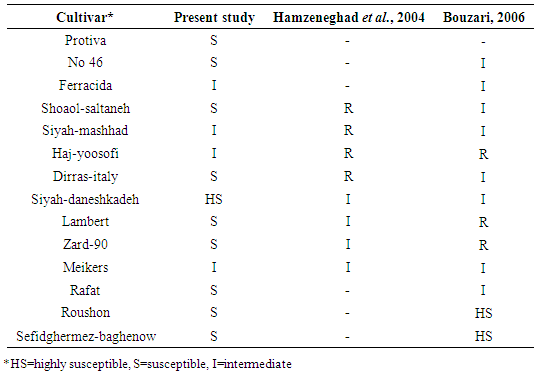
[7]  Buzari N, 2006. Evaluation of cherry cultivars for bacterial canker resistance. Proc 17th Plant Protection Congress of Iran, Karaj. 376 (in Persian).
[8]  Cameron, H.R. 1971. Effect of root or trunk stock on susceptibility of orchard trees to Pseudomonas syringae. Plant Disease Reporter. 55. 421-423.
[9]  Garrett, C.M.E. 1986. Influence of rootstock on the susceptibility of sweet cherry scions to bacterial canker, caused by Pseudomoinas syringae pv. morspronorum and syringae. Plant Pathology. 35. 114-119.
[10]  Crosse, J.E., Garrett, C.M.E .1966. Bacterial canker of stone-fruits. VII. Infection experiments with Pseudomonas morsprunorum and P. syringae. Annals of Applied Biology. 58. 31-41.
[11]  De Vries, D.P. 1965. Field resistance to bacterial canker in some cherry seedling populations. Euphytica. 14. 78-82.
[12]  Elahinia, S.A., Rahimian, H. 1992. Identification angent bactrerial canker os stoem fruit tree in summer area Mazandaran. Proc 11th Plant Protection Congress of Iran. 213 (in Persian).
[13]  Fischer, M. and Hohlfed, B. 1998. Resistance tests in sweet cherries. Acta Horticulturae 468: 87-94.
[14]  Fuchs, A. and De Vries, D.P. 1964. Optreden,bestrijding en voorkomen van bacteriekanker. Kersen, Meded Dir Tuinb. 27. 546-56.
[15]  Fuchs, A.1957. Bakteriekanker bij steenvruchten. 11. De identiteit van Pseudomonas morsprunorum Wormald en Pseudomonas syringae van Hall. Tijdschr PlZiekt 63. 45.
[16]  Geibel, M., Gross, D.C., Mo, Y.Y., Bonsal, R.F. and Geiger, H. 1994. Identification of flavonol glycosides from Prunus avium leaves which induce the production of syringomycin by Pseudomonas syringae pv. syringae. Acta Horticulturae. 381. 662-666.
[17]  Gerritsen, C.J. and Slits, J.A. 1959. Verslag over het kersenproefveld te Uden. Wageningen IVT, stencil, 11p.
[18]  Gilbert, V., Planchon, V., Legras, F, Maraite, H. and Bultreys, A. 2010. Pathogenicity and aggressiveness in populations of P. syringae from Belgian fruit orchards. European Journal of Plant Pathology. 126. 263-277.
[19]  Grubb, N.H. 1949. Cherrioes: Crosby lockwood $ Sons Ltd. London: 34-37.
[20]  Hamzeneghad, P., Rahimian, H. Ghasemi, A. and Mahmudpur, M. 2004. Investigation of commercial cherry cultivars to bacterial canker and evaluation of polyphenil oxidase and peroxidase as biochemical markers. Proc 16th Plant Protection Congress of Iran. 414 (in Persian).
[21]  Janse, J.D. 2006. Phytobacteriology, Principles and Practice. CABI Publishing, Wallinford, UK.
[22]  Kaluzna, M., Pulawska, J. and Sobiczewski, P. 2010. The use of PCR melting profile for typing of Pseudomonas syringae isolates from stone fruit trees. European Journal of Plant Pathology. 126. 437-443.
[23]  Karimi-Kurdistani, G. and Harighi, B. 2008. Phenotypic and molecular properties of Pseudomonas syringae pv. syringae the causal agent of bacterial canker of stone fruit trees in Kurdistan province. Plant Pathology. 90. 81-86.
[24]  Kennelly, M.M., Cazorla, M., de Vincente, A., Ramos, C. and Sundin, G.W. 2007. Pseudomonas syringae diseases of fruit trees, progress toward understanding and control. Plant Disease. 91. 4-17.
[25]  Le Lezec, M., Pauline, J.P. and Lecomte, P. 1987. Shoot and blossom susceptibility to fireblight of apple cultivars. Acta Horticulturae. 217. 311-316.
[26]  Lelliott, R.A. and Stead, D.E. 1087. Methods for the Diagnosis of Bacterila Disease of Plants. Blackwell Scientific Publications, Oxford, London, UK, pp 169-199.
[27]  Lyskanowska, K. and Rejman, A. 1978. Bacterial canker of sweet cherry in Poland. III. Degree of susceptibility of wild cherry seedlings. Plant Disease Reports. 62. 500-503.
[28]  Matthews, P. 1959. Bacterial canker of cherries. Ann Rep John Innes Hortic Institution. 50. 14.
[29]  Matthews, P. 1979. Progress in breeding cherries for resistance to bacterial canker. Proc Eucarpia Fruit Section Symposium on Tree Fruit Breeding, Angers, pp. 157-174.
[30]  Mo, Y.Y., Geibel, M.. Bonsal, R.F. and Gross, D.C. 1995. Analysis of sweet cherry leaves for plant signal molecules that activate the syrB gene required for synthesis of the phytotoxin syringomycin, by Pseudomonas syringae pv. syringae. Plant Physiology. 107. 603-612.
[31]  Muranty, H., Schermann, N., Santi, F. and Dufour, J. 1998: Genetic parameters estimated from a wild cherry diallel: consequences for breeding. Silvae Genetics. 47. 249-257.
[32]  Nicoll, F.J. 1993. Genetic improvement of cherry for farm woodlands. Quaternar Journal of Forestry. 8. 187-194.
[33]  Renick, L.J., Cogal, A.G. and Sundin, G.W. 2008. Phenotypic and genetic analysis of epiphytic Pseudomonas syringae populations from sweet cherry in Michigan. Plant Disease. 92. 372-378.
[34]  Roche, M.M. 2001. Development of an in vitro and modification of an in vivo bioassay to screen cherry genotypes for response to inoculation with Pseudomonas syringae pv. syringae. MS thesis, Oregon State University, Corvallis.
[35]  Roche, M. and Azarenko, A.N. 2005. An in vitro bioassay to evaluate sweet cherry response to inoculation with Pseudomonas syringae pv. syringae. Acta Horticulturae 667: 503-508.
[36]  Santi, F., Russell, K., Me´nard, M. and Dufour, J. 2004. Screening wild cherry (Prunus avium) for resistance to bacterial canker by laboratory and field tests. Forest Pathology. 34. 349-362.
[37]  Schaad, N.W., Jones, J.B. and Chun, W. 2001. Laboratory Guide for Identification of Plant Pathogenic Bacteria. APS Press, St. Paul, Minnesota, USA.
[38]  Scortichini, M., Biocca, M. and Rossi, M.P. 1995. Pseudomonas syringae pv. morsprunorum on wild cherry for timber production: outbreak and field susceptibility. European Journal of Plant Pathology. 25. 343-350.
[39]  Shams-Bakhsh, M. and Rahimian, H. 1989. Characterization of stone fruits bacterial canker in Mazandaran. Proc 9th Plant Protection Congress of Iran. 13 (in Persian).
[40]  Spotts, R.A., Facteau, T.J., Cervantes, L.A. and Chestnut, N.E. 1990. Incidence and control of cytospora canker and bacterial canker in young sweet cherry orchard in Oregaon. Plant Disease. 74. 577-580.
[41]  Spotts, R.A., Wallis, K.M., Serdani, M. and Azarenko, A.N. 2010. Bacterial canker of sweet cherry in Oregon- Infection of horticultural and natural wounds and resistance of cultivar and rootstock combination. Plant Disease. 94. 345-350.
[42]  Thornton, G. and Nugent, J. 2002. Bacterial canker suppression. www.maes.msu.edu/nwmi hort.
[43]  Vicente, J.G., Alves, J.P., Russel, K. and Roberts, S.J. 2004. Identification and discrimination of P syringae from wild cherry in englammd. European Journal of Plant Pathology. 110. 337-351.
[44]  Vicente, J.G. and Roberts, S.J. 2007. Discrimination pf Pseudomonas isolates from sweet and wild cherry using Rep-PCR. European Journal of Plant Pathology. 117. 383-392.
[45]  Webster, A.D. 1980. Dwarfing rootstocks for plums role in cherry canker. Acta Horticulturae. 114. 201-207.
[46]  Wilson, E. 1933. Bacterial canker of stone-fruit trees in California. Hilgardia. 8. 83.
[47]  Wilson, E. 1953. Bacterial canker of stone fruits. The Yearbook of Agriculture and Plant Diseases. USDA. 722-729.
[48]  Young, J.M. 1991. Pathogenicity and identification of the lilac pathogen, Pseudomonas syringae pv. syringae van Hall 1902. Annlals of Applied Biology. 118. 283-298.
[49]  Zamze, S. 1983. The structure of lipopolysaccharide from Pseudomonas syringae pv. morsprunorum and its role in canker decay. PhD Thesis, Council for and cherries. National Academic Awards, Thames Polytechnic.