-
Paper Information
- Paper Submission
-
Journal Information
- About This Journal
- Editorial Board
- Current Issue
- Archive
- Author Guidelines
- Contact Us
Journal of Health Science
p-ISSN: 2166-5966 e-ISSN: 2166-5990
2018; 8(1): 1-3
doi:10.5923/j.health.20180801.01

The Verrucous Butterfly Tattoo
 Abstract
 Reference
 Full-Text PDF
 Full-text HTML
Asma G. Sadeq1, Ghadah M. Al-Rubaya1, Farah Alzahrani2, Iqbal A. Bukhari3
1College of Medicine, Imam Abdulrahman Bin Faisal University (IAU), Dammam, Kingdom of Saudi Arabia
2College of Medicine, Arabian Gulf University (AGU), Bahrain
3Dermatology Department, College of Medicine, Imam Abdulrahman Bin Faisal University (IAU) and King Fahd Hospital of the University, Dammam, Kingdom of Saudi Arabia
Correspondence to: Iqbal A. Bukhari, Dermatology Department, College of Medicine, Imam Abdulrahman Bin Faisal University (IAU) and King Fahd Hospital of the University, Dammam, Kingdom of Saudi Arabia.
| Email: |  |
Copyright © 2018 Scientific & Academic Publishing. All Rights Reserved.
This work is licensed under the Creative Commons Attribution International License (CC BY).
http://creativecommons.org/licenses/by/4.0/

Tattoo is a popular cosmetic decoration worldwide. A number of cutaneous and systemic side effects related to tattoo procedure have been reported in the medical literature. Here we report a case of a 48-year-old Arabic female presented with hyperpigmented verrucous plaque of 20 mm in diameter size on the anterolateral aspect of the lower third of the right leg following cosmetic tattoo procedure. It was treated with four sessions of cryotherapy two week apart with complete resolution.
Keywords: Tattoo, Verrucae, Viral, Infection, Wart
Cite this paper: Asma G. Sadeq, Ghadah M. Al-Rubaya, Farah Alzahrani, Iqbal A. Bukhari, The Verrucous Butterfly Tattoo, Journal of Health Science, Vol. 8 No. 1, 2018, pp. 1-3. doi: 10.5923/j.health.20180801.01.
1. Introduction
- Tattoos are becoming popular in the world among teenagers and young adults as a cosmetic and decorative body art. Recently, there has been increasing number of reported cases with tattoo related cutaneous side effects including infections such as bacteria, viral and fungal, allergic reactions and cutaneous malignant tumors due to the absence of clear legislation for the safety of tattoos. It is important to increase awareness in the young generation regarding risks of tattooing and tattoo artists should be educated about the importance of using proper aseptic tattoo procedures. [1] Besides, public health organizations should have a good control of the licensed tattoo parlors in the country.
2. The Case
- A 48-year-old Arabic female presented to the dermatology clinic of King Fahd Hospital of the University with dry hyperkeratotic skin lesion on the lower third of the right leg. Six years back the patient had a blue red color decorated tattoo on that area done at a professional tattoo parlor. In the following month, she developed numerous slightly itchy, small, skin-coloured, dry papules in the area of the tattoo. That time she was seen by a dermatologist who gave her topical steroid cream and reassured her. However, the lesion continued to grow and expand in size with rough surface. There was no other area affected with similar lesion. The patient is a known case of discoid lupus erythematousus affecting her chin and left ear lobe for the past 6 years, which resolved with treatment. On examination there was hyperpigmented large verrucous plaque of 20 mm in diameter size following the tattoo pattern on the anterolateral aspect of the lower third of the right leg (figure1a). There was no other area that had similar lesion in the skin. Blood investigations and chest radiograph were normal ruling out any systemic illnesses specifically sarcoidosis. Skin biopsy was planned but due to the location of the lesion the patient preferred not to take biopsy. So, we diagnosed the patient clinically as a case of verrucae vulgaris complicating tattoo procedure. We treated the lesion with four sessions of cryotherapy two week apart with complete resolution and postinflammatory depigmentation (figure 1b).
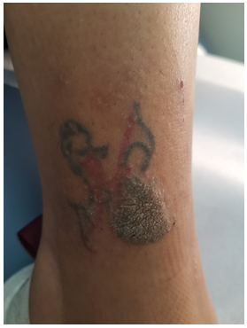 | Figure 1a. Large verrucous plaque of 20 mm in diameter following the tattoo pattern on the anterolateral aspect of the lower third of the right leg |
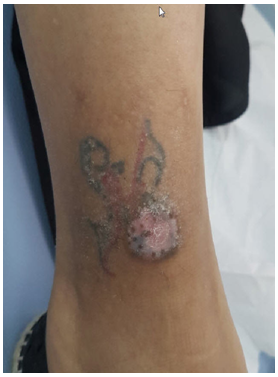 | Figure 1b. Complete resolution of the condition with residual postinflammatory depigmentation |
3. Discussion
- Tattoo is an ancient practice and has been performed for therapeutic, cultural identification, cosmetic, and punishment purposes. In Saudi Arabia the concept of decorative permanent tattooing is not encouraged with certain artwork. It is usually done by unlicensed amateur tattoo artists or outside the country due to the absence of official licensed tattoo parlors. However, only non-permanent henna tattoo is widely used. Modern tattoo uses an electric pen which punctures the skin with needles at a rate of 50-3,000 rpm, depositing ink into the dermis. The tattoo inks are suspensions of metal salts and organic pigments in water, alcohol, or glycerin. Licensed tattoo artists in licensed tattoo parlors using proper aseptic technique should perform the procedure. However, the rate of complications from tattooing has been estimated as high as 2%, [2] which include: inflammatory, infectious, and neoplastic complications.Inflammatory tattoo reactions:Inflammatory tattoo reactions begin anywhere from days to decades after tattooing with any tattoo ink including: metal salts, red mercury or cinnabar–containing salts, nickel, as well as chromium. [3] The reaction is usually a delayed type of hypersensitivity. Introduction into the skin or exposure to ultraviolet (UV) light could alter the chemical component of tattoo inks, making them more allergenic due to photodecomposition. [4] The role of patch testing in tattoo reactions was explored in one study and found a generally low incidence of positive reactions even when a sample of the same ink presumed to cause the reaction was applied, suggesting that haptenization plays a role in tattoo reactions. [5] Granulomatous tattoo reactions are among the most common inflammatory tattoo reactions and can be foreign body-like or sarcoidal or granuloma annulare- or necrobiosis lipoidica-like reaction. [6] All patients presenting with suspected tattoo reactions, regardless of the duration of the tattoo, should reasonably undergo questioning about eye or pulmonary symptoms, complete skin examination looking for other skin lesions, and skin biopsy to confirm the diagnosis. In addition to reactions to permanent tattoos, reactions to temporary tattoos can occur, such as allergic contact dermatitis to henna tattoo which is often mixed with paraphenylenediamine (PPD), a well-known contact allergen.Infectious tattoo reactions:Majority of infections occurring within tattoo include bacterial (e.g., acute pyogenic, mycobacterial), viral (e.g., HPV) and mycotic (e.g., dermatophytosis) infections. [7-23] A possible causality is contamination during tattooing via the instruments, the colors used, or even the tattooist handling hygiene. [13] Specifically HPV infection could be explained by the possible induction of replication through mechanical impairment of the skin barrier since cutaneous HPV are excessively activated in people suffering from psoriasis. [23-25] Besides, the hair follicle is regarded as the natural reservoir for the beta HPVs and it may be released from the hair follicle by the act of tattooing. [27-29] Neoplasms occurring within tattoo:Tattoo pigments have been reported to contain aromatic amines, phthalates, polycyclic aromatic hydrocarbons, and even highly bioavailable nanoparticles. [30] It is not yet known whether tattoo inks act as a primary carcinogen or as a co-carcinogen together with sunlight or trauma. A review of the literature revealed that a few basal cell carcinomas developed in tattoos located on sun-protected areas, which suggests that other factors such as trauma are associated with the occurrence of basal cell carcinoma. Other reported neoplasms include: pseudocarcinomatous hyperplasia, keratoacanthoma and squamous cell carcinoma. Treatment of tattoo reactions:The treatment of tattoo reactions should be individualized to the patient and type of tattoo reaction. For bacterial infections antibiotics are used, for fungal antifungals are used and for viral infections cryotherapy, curettage, podophyllotoxin or topical imiquimod. Most inflammatory reactions, including lichenoid and granulomatous reactions, superpotent topical or intralesional steroids are reasonable first-line therapies. Surgical excision may be needed for recalcitrant or recurrent reactions. [31] Our patient was treated with cryotherapy sessions every two weeks for 8 weeks which completely resolved with postinflammatory hypopigmentation.
4. Conclusions
- We are documenting the 28th case of verruca vulgaris complicating decorative tattoo in the medical literature which was successfully treated with cryotherapy. Although tattoos obtained at a professional tattoo parlor should be generally safe, unanticipated complications can arise, even decades after tattooing. In our country, the complications could be more due to absence of official licensed tattoo parlors. Dermatologists should also educate patients to only obtain tattoos in licensed tattoo parlors by licensed tattoo artists to minimize their risk of complications.