-
Paper Information
- Previous Paper
- Paper Submission
-
Journal Information
- About This Journal
- Editorial Board
- Current Issue
- Archive
- Author Guidelines
- Contact Us
Journal of Health Science
p-ISSN: 2166-5966 e-ISSN: 2166-5990
2016; 6(6): 90-91
doi:10.5923/j.health.20160606.02

Giant Cutaneous Horn that Surprisingly Fell Down without Intervention
 Abstract
 Reference
 Full-Text PDF
 Full-text HTML
Sahar Alsharif1, Mazin Aljabri2, Khalid Al hawsawi2, Ahmad Kurdi3
1PGY at the Dermatology Residency Program, Western region, Saudi Arabia
2Dermatology Department, King Abdul Aziz Hospital, Makkah, Saudi Arabia
3Dermatology Department, Ministry of Health, Madinah, Saudi Arabia
Correspondence to: Sahar Alsharif, PGY at the Dermatology Residency Program, Western region, Saudi Arabia.
| Email: |  |
Copyright © 2016 Scientific & Academic Publishing. All Rights Reserved.
This work is licensed under the Creative Commons Attribution International License (CC BY).
http://creativecommons.org/licenses/by/4.0/

Cutaneous horn is a conical projection of cornified epithelial cells. Rarely exceeding 1 to 2 cm in length. It usually appears the sun-exposed areas. It is more common in white races. Here we present a case of right foot giant cutaneous horn that fell down spontaneously.
Keywords: Cutaneous horn, Giant, Foot, Fell down
Cite this paper: Sahar Alsharif, Mazin Aljabri, Khalid Al hawsawi, Ahmad Kurdi, Giant Cutaneous Horn that Surprisingly Fell Down without Intervention, Journal of Health Science, Vol. 6 No. 6, 2016, pp. 90-91. doi: 10.5923/j.health.20160606.02.
1. Introduction
- A cutaneous horn is a conical projection of hyperkeratotic epidermis. These lesions are uncommon and may arise from different epidermal lesions, which may be benign or malignant in nature. They are usually found on sun-exposed areas and they tend to have a conical shape.It is most common in Caucasian patients and relatively less known in other races. This racial tendency can be endorsed to the relative protection of pigmented skin to ultraviolet sun rays [1]. It has been reported to affect more men than women, and occurs in the seventh decade of life. The lesions have been reported to be benign in 60% of the cases.Rarely has cutaneous horn been described to occur in the lower extremities [2]. We report a patient with a cutaneous horn on an unusual site, the dorsal aspect of his foot [3].
2. Case Report
- An 81- year- old man presented to our dermatology clinic with bilateral lower limbs pitting edema and asymptomatic horn like projection over dorsum of his right foot for a long time. The lesion had increased in size over the last three months. The patient was unable to determine when the lesion started to grow and denied any trauma to the area. There was no history of similar lesion elsewhere on the body. Systemic review was unremarkable. On examination: (Figure 1 A)Over the dorsal aspect of the right foot, there is a solitary firm, painless, irregular horn like projection measuring 5 cm at the base and 8 cm in length. Surrounded by thick dry skin and edema. There is also onychogryphosis of the big toe of the right foot. There was no regional lymphadenopathy. No similar cutaneous horn elsewhere in the body.All laboratory parameters were within the normal range. We diagnosed patient as a case of cutaneous horn and we did skin biopsy from the base of the lesion and the histopathological results (Figure 2 A & B) showed hyperkeratosis with minimal superficial perivascular inflammation, there was no evidence of malignancy.Next follow up was within one month and the cutaneous horn has been falling off spontaneously without history of trauma or any intervention. (Figure 1 B)We did second biopsy from the center of the base of the previous horn and the histopathological studies (Figure 3 A & B) showed atypical keratinocytes with dyskeratotic cells and horn cyst.We followed up the patient and we refer him to vascular surgery to role out lower limbs venous insufficiency which may be the cause of the edema.
 | Figure 1. (A) Cutaneous horn at presentation (B) After the cutaneous horn fell down |
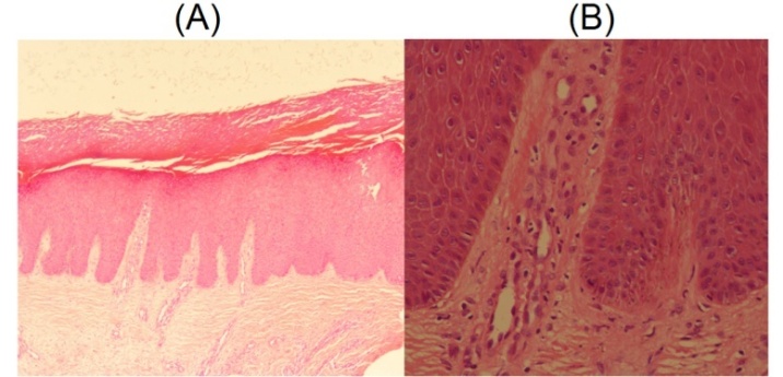 | Figure 2. (A) (H&E ×4) Epidermal acanthosis with hyperkeratosis (B) (H&E ×20) Perivascular lymphocytic infiltration |
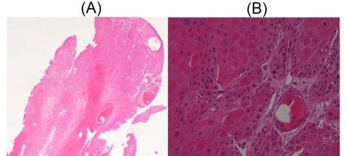 | Figure 3. (A) (H&E ×4) Showing hyperparakeratosis (B) (H&E ×20) Atypical keratinocytes, dyskeratotic cells, and horn cyst |
3. Discussion
- Cutaneous horn is a conical projection of cornified epithelial cells. Rarely exceeding 1 to 2 cm in length [4]. They are thought to result from underlying benign, premalignant or malignant causes. It usually appears in the sun-exposed parts of the body like face and upper extremities. Less common to be arise over feet. As mentioned in Olugbenga study, cutaneous horn is more common in white races, but reportedly rare in black descent [1]. He attributed that to the pigmented skin that has relative protection from ultraviolet sun rays. Although that, we report a case of giant cutaneous horn that arise over the dorsal surface of foot of patient with a black skin. In reviewing the literature, we found a similar case of cutaneous horn that arise over the dorsal surface of foot of patient with a black skin but it was smaller in size [3]. However, it appears to be no previous mention in the literature of a giant cutaneous horn that fell down without any intervention. This may be the first such a case report.
4. Conclusions
- The possibility of malignancies in any cutaneous horn should be kept in mind and skin biopsy from the base of the horn is mandatory even if the cutaneous horn fall down spontaneously.