Food and Public Health

p-ISSN: 2162-9412    e-ISSN: 2162-8440

2014;  4(4): 204-208

doi:10.5923/j.fph.20140404.05

Toxicity Test of Alginate from Sargassum and Padina on the Liver of Mice

Wahyu Mushollaeni, Nonok Supartini, Endang Rusdiana

Agroindustrial Technology, Tribhuwana Tunggadewi University, Malang, East Java, 65144, Indonesia

Correspondence to: Wahyu Mushollaeni, Agroindustrial Technology, Tribhuwana Tunggadewi University, Malang, East Java, 65144, Indonesia.

Email:

Copyright © 2014 Scientific & Academic Publishing. All Rights Reserved.

Abstract

This research aimed to study the toxicity of alginate Sargassum and Padina with animal (mice) testing. This research is an experimental approach to post-test only control group design. Wistar rats used as an object of alginate toxicity testing. The mice were grouped into 5, i.e. control group (no alginate diet), 2 groups treated by Sargassum 0,75% and 1%, 2 groups treated by Padina 0,75% and 1%, respectively. Based on the research, it was concluded that alginate from Sargassum and Padina not give toxic effects on Wistar mice and no significant effect on liver cell damage.

Keywords: Sargassum, Padina, Alginate, Mice, Toxicity

Cite this paper: Wahyu Mushollaeni, Nonok Supartini, Endang Rusdiana, Toxicity Test of Alginate from Sargassum and Padina on the Liver of Mice, Food and Public Health, Vol. 4 No. 4, 2014, pp. 204-208. doi: 10.5923/j.fph.20140404.05.

1. Introduction

Alginate has an important role in the food industry. The use of alginate as a food additive in the food industry with regard to the nature of the rough, such as a thickener [16] [18] [26], so that the product is more stable [8] [10] [19], stabilize mixtures, dispersions and emulsions with regard to its nature as a gelling and viscosity increase [24].
Although alginate has been known and used in various applications, especially in food purposes, but there is no toxicity studies that examine the effects of toxicity associated with the possible presence of harmful chemical residues present in the extract alginate applied to food products. Toxicity and health effects of using test animals that Wistar rats or mice (Rattus norvegicus). The toxicity of a substance can be determined by using the test animals are male or female white rat strain Whistar [2][3][30]. The liver is an organ that did the metabolism and detoxification of toxins from the entry of foreign material into the body [12].
Dietary fiber particularly one which is soluble in water, is known to play a role in lowering plasma cholesterol levels [5]. Alginate has a high potential in lowering blood cholesterol by inhibiting cholesterol absorption in the intestine [39]. The effect of sodium alginate, performed with mice test [2][3]. An important part of the subchronic toxicity testing is liver and kidney histopathology [40].
The liver is an important organ that detoxifies many substances functioning digestive tract digestive results [37]. The main function of the liver is to metabolize and detoxify toxins [12]. Clinical features and diagnosis of drug-induced hepatotoxicity according to the Common Toxicity Criteria, covering grades 0-4 from increased alkaline phosphatase, increased bilirubin, increased GGT, hepatomegaly, hypoalbuminemia, clinical signs of liver dysfunction, decreased portal venous flow or retrograde, increased SGOT, and increased SGPT. One test that is often performed to determine the liver function is testing serum transaminase SGPT and SGOT are. Both serum is a sensitive indicator of damage to the liver cells. The damage to the liver cells can be characterized by the levels of enzymes SGOT (Serum Glutamic Oxaloasetate transaminase) and SGPT (Serum Glutamic Piruvate transaminase) were increased. SGOT-SGPT are two transaminase enzymes produced by the liver cells [14]. An increase in The enzyme aspartate aminotransferase (AST) and alanine aminotransferase (ALT) indicate liver cell damage compared with other liver enzymes, because this enzyme increases both first and increases dramatically when compared with other enzymes in the event of damage to the liver cells [7]. This research aimed to study the toxicity of alginate Sargassum and Padina with animal (mice) testing. Toxicity testing on food safety aspects of alginates in this study, using male mice strains Wistar.

2. Materials and Methods

2.1. Materials

The raw materials used are 2 types of alginat i.e. Sargassum and Padina, derived from the rocky coast of South Mountain area of Yogyakarta. The chemicals used include distilled water, CaCl2 1% and 74%, HCl 1, 3, 5 and 35%, 0.5 and 90% KOH, Na2CO3 2.25; 10 and 95%, NaOCl 10 and 12%, IPA 95%, HNO3.

2.2. Population and Sample

This study is divided into three stages, i.e: Extraction of alginates from brown seaweed Sargassum and Padina, Treatment period to wistar alloxan diabetic mice for 38 days, and Termination stage, blood sampling and blood test for SGPT and SGOT levels at the end of treatment.
The population were mice (wistar specification). Samples were obtained in consecutive random sampling, with (a) the inclusion criteria of male Wistar mice, aged 3 months, weight 140-155 grams, healthy conditions (active and not disabled), and (b) exclusion criteria that mice experiencing pain, mice decreased weight (less than 140 grams), the mice died within the study period.
Alginate to be tested, applied in the form of mackerel fish balls, which will be administered orally every day for 90 days. The concentration of alginate in mackerel fish ball is 0.75% (1) and 1% (2) for each of the 2 types of alginate. Control treatment (K) treated by mackerel fish balls (B) but without alginates.

2.3. Data Analysis

Data analysis performed using specific program analysis for windows 16:00. Hypothesis testing using parametric One Way test (p <0.05).

2.4. Methods

Alginate is applied as stabilizer in the form of a mice feeding mackerel fish balls. Parameter analysis in this study include SGPT and SGOT blood levels and body weight. Fish balls containing alginate administered orally every day for 90 days. The concentration of alginate in the mackerel fish ball is 0.75% (1) and 1% (2) for each of the two types of the alginate Sargassum (S) and Padina (P). Control treatment (K) is the mackerel fish balls without alginates. In this study using 25 mice and divided into 5 mice in each treatment.

3. Results and Discussions

3.1. Alginate Diet Effect on Weight

Based on the results of the study with 25 mice, both alginate diet of brown seaweed Sargassum and Padina maintenance for 90 days, showed no toxic effect on the physiological and biochemical conditions. Before treatment, there was no significant difference in body weight of rats due to a homogeneous sample. Overall both control mice and mice that were given feed containing alginate, are in good condition and there were no deaths. The behaviour of all the mice in the control and four treatment looks healthy, active, and there are no signs of poisoning. All mice that fed a diet containing alginate of both these types, showed no effect on their body weight. This is in accordance with another research that if the repeated administration of a particular substance with the highest or the maximum dose and showed no signs of toxic or abnormality, it can be said that the substance is safe for long term use [6]. There is no weight loss at all Wistar mice were given feed containing alginate. But it has increased body weight, with a mean increase of 49-60 grams (Figure 1).
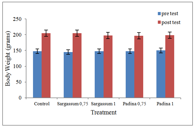
Figure 1. Effect of treatment by alginate from Sargassum and Padina on body weight of wistar mice at the pre and post test
Weight gain of mice treated by alginate from Sargassum, providing greater value than diet by alginate from Padina. This suggests that although the two of them are not toxic on body weight of mice, but the type of alginat e from Sargassum showed increased body weight better and more possible not toxic. Based on previous research, a diet using alginate from Sargassum and Padina that contain antioxidants, have an impact on reducing free radicals and may help protect cells from free radical damage caused by exposure [46]. A diet product that serves as a catcher hydroxyl radicals, can help protect cells from free radical damage caused by exposure [22] [27] [28] [29] [36]. In addition, there is research that includes bayleaf extract flavonoid which have glycosidic sugar groups are also reactive to capture hydroxyl radicals [4] [25]. Alginate constituent group is a group consisting of sugar guluronic acid and mannuronic acid, which allows this sugar moiety is able to capture the hydroxyl radical. Therefore, with low effects of free radicals that can damage cells and the immune system, the body remains in a stable condition and body weight of mice was not affected.
Weight gain of Wistar mice also indicate the absorption of nutrients from food substances that better nutritional intake is converted into a more maximum weight. Feed a diet containing alginate has been made possible also metabolized by the body and mice with either suspected because there was no damage to the liver that serves as a detoxifying organ of the influence of foreign substances that may be toxic to the body. If the feed of rats was not changed to the maximum weight, the suspected food is not metabolized completely and suspected liver damage that affects the body weight of rats [47]. The liver is an organ that did the metabolism and detoxification of toxins from the entry of foreign material into the body [12]. Changes in body weight that is not normal in mice, may show signs of clinical symptoms that are not good [48]. Increase or decrease in body weight Wistar mice, also caused by the absorption rate and amount of feed nutrients from the feed. The amount of feed consumed by the test mice, implies a growth rate of body weight. The number of calories from carbohydrate ration given has been replaced by a number of seaweed, which affects the absorption of nutrients [17]. Seaweed is an important component of high fibre foods [23]. The existence of these fibre, resulting nutrients are released slowly into the small intestine or reduce the absorption rate of glucose, so that the blood glucose levels will rise slowly [11] [13] [21] [31] [33] [41] [44] [45]. The presence of the polymer contained in dietary fibre, can affect physiological functions [9].

3.2. Alginate Diet Effects on Levels of ALT and AST

Tests on blood and serum biochemical testing of liver cell damage due to the possibility of a toxic effect, carried out tests on the levels of SGPT and SGOT enzymes, which can be seen in Figure 2.
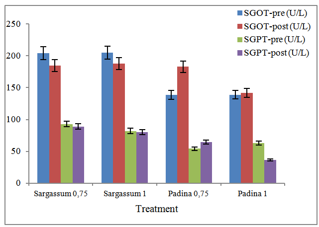
Figure 2. Levels of SGOT and SGPT of wistar mice
The results showed that the levels of SGPT and SGOT was highest on Sargassum alginate’s diet. Dietary dose of 0.75% alginate Sargassum provide the highest levels of alanine aminotransferase 89 U / L, followed by dietary Sargassum alginate 1% that is 80 U / L. While the diet of the type Padina alginate, have ALT levels lower end compared to alginate from Sargassum. The ALT levels at a dose of 0.75% was 64.4 U / L and 1% was 36.4 U / L. However, high levels of alanine aminotransferase end of this Sargassum alginate diet, is lower than the initial ALT levels before treatment. While the diet of the type Padina alginate at a dose of 0.75%, giving a final ALT levels (64.4 U / L) were higher than the levels at the beginning of treatment (54.2 U / L). This suggests that the tendency of alginate type Padina consumption of a dose equal to or above 0.75%, has the potential to result in changes in the cells of the liver so that it can raise the levels of ALT. While the use of alginate from Sargassum diets are safe to eat until the dose limit of 1% for not showed signs of liver cell damage as indicated by the absence of an increase in the activity of the enzyme alanine aminotransferase. The enzyme ALT is an indicator of liver cell damage. Impaired hepatic cells and cell function, may result in elevated levels of ALT and AST enzymes. In the first month there is a relationship between cell damage in the form of hydropic degeneration of the liver, and fat necrosis with elevated levels of the enzyme [35]. This final SGPT levels remained in the normal range for the levels of alanine aminotransferase control mice used in this study was 71-91 U / L.
At the end of treatment with dietary Sargassum alginate, the mean levels of SGOT end has a lower value than the levels at the start of treatment, as well as having lower levels of SGOT levels than the control group of mice. Control mice AST was 205 U / L. While the test mice were given diets Padina alginate types, have the final AST levels greater than the levels at the beginning of treatment, but the levels of SGOT end is still lower than the levels of SGOT control. Based on this, it has been demonstrated that the use of dietary alginate with Padina types can provide potential toxic to the liver because it has been able to increase the levels of SGOT enzyme doses ranging from 0.75% to 1%. Therefore, the toxic potential has been demonstrated by the use of alginate diet Padina types with concentrations equal to or higher than the 0.75%.
The immune system of mice are not the same between mice within and among groups, resulting in the final state becomes non-uniform treatment [20] [32]. It also affects the levels of SGPT and SGOT of test mice, demonstrated by both levels of this enzyme at the end of treatment. High levels of SGPT and SGOT at the end of the dietary treatment of type Padina alginate showed a toxic effect of the use of alginate starting dose of 0.75%. The liver is the organ metabolic, secretory and immunological who did all the metabolism of foreign substances, and drug substance. The presence of liver damage can be detected with the two kinds of aminotransferase enzymes namely aspartate aminotransferase (AST) also called AST and alanine aminotransferase (ALT), also called SGPT. If there is damage to the liver, transaminase enzymes released into the blood from the cytosol and organelles such as mitochondria subsel, lysosomes, and the nucleus [1][34][49]. Measurement of the concentration of enzyme in the blood with AST and ALT tests, can provide important information about the level of liver dysfunction. Transaminase activity in the liver can be detected even in very small quantities. Liver cells are the major networks are the main objectives of the effect of free radicals because the liver is the site of the metabolic process xenobiotic compounds. The damage to the liver cells, resulting in the release of enzymes found in the liver as blood serum GPT and GPT activity will be found high in the blood [9].
Diet alginate from Sargassum, resulted in lower SGPT and SGOT levels than the control. This is possible due to the antioxidants contained by alginate. Using seaweed diet that contain antioxidants, have an impact on reducing the formation of free radicals and may help protect cells from damage caused by free radical exposure [22] [27-29] [36]. The body's defense system in the form of antioxidant enzymes control system regulates the formation of free radical reactions necessary and neutralize excess free radicals that are formed as a result of food intake which can damage tissues in the body [42]. The condition of the formation of free radicals, increased levels of hydrogen peroxide and superoxide. Using seaweed diet that contain antioxidants, have an impact on the reduction of free radical formation due to the effects of free radicals can be reduced with a diet product that serves as a hydroxyl radical catcher [46]. The brown algae contain a chemical compound chlorophyll-a and-c, β-carotene, and fukosantin violasantin, pirenoid and filakoid, laminarin, cellulose and algin. Carotene pigments are known to have antioxidant properties that can act as antioxidants and free radical scavengers in the body's metabolic processes results [9].

4. Conclusions

Based on the results of a study of the levels of SGPT and SGOT of Wistar rats, it was concluded that alginate from Sargassum at a dose of 0.75% and 1% did not give a real effect on the occurrence of liver cell damage, with evidence of ALT and AST levels at the end of treatment is lower than the control. Diet alginate Padina types have a tendency to result in damage to the liver cells with doses ranging from 0.75%.

ACNOKLEDGEMENTS

The authors thank to Directorate of Research and Community Service, Kopertis 7, East Java Republic of Indonesia 2014.

References

[1]  A. L. Lehninger, Basics Bichemistry, Jakarta, 2008.
[2]  A. Mirshafiey A, S. Cuzzocrea, B. Rehm, E. Mazzon, F. Saadat and M. Sotoude, Sodium Alginate as A Novel Therapeutic Option in Experimental Colitis. Scand J Immunol 61:435–441, 2005.
[3]  A. Razavi, A. Khodadadi, M. B. Eslami, S. Eshraghi and A. Mirshafiey, Therapeutic Effect of Sodium Alginate in Experimental Chronic Ulcerative Colitis. Iran J Allergy Asthma Immunol 7: 13 – 18R. E. Sorace, V. S. Reinhardt, and S. A. Vaughn, “High-speed digital-to-RF converter,” U.S. Patent 5 668 842, Sept. 16, 1997.
[4]  B. A. Nugroho and E. Puwaningsih, Pengaruh Diet Ekstrak Rumput Laut (Eucheuma sp.) terhadap Kadar Glukosa Darah Tikus Putih (Rattus norvegicus) Hiperglikemik, Media Medika Indonesia, 39(3): 60-154, 2004.
[5]  B. O. Schneeman and J. Tietyen, Dietary fiber. In: M. E. Shils, J. A. Olson, M. Shike (ed). Modern Nutrition in Health and Disease. Philadelphia, 1994.
[6]  C. D. Lasanti, Sub Chronic Toxicity Study Of Ethanolic Extract Of Binahong [Anredera Cordifolia (Ten.) V. Steenis] Leaves In Wistar Rats [online]. Available http://digilib.pharmacy.itb.ac.id/gdl.php?mod=browse&op=read&id=jbptitbsf-gdl-citradewis-206, 2014.
[7]  D. F. Calbreath, Clinical Chemistry. WB. Saunder Company. USA, 1992.
[8]  D. Gomez-Diaz and J. M. Navaza, Rheology of Food Stabilizers Blends. J Food Eng 64: 143 – 149, 2004.
[9]  D. M. W. Anderson, W. G. Brydon, M. A. Eastwood and D. M. Sedgwick, Dietary Effects of Sodium Alginate in Humans. Food Addit Contam 8: 237 – 248, 1991.
[10]  D. Paraskevopoulou, D. Boskou, and A. Paraskevopoulou, Oxidative Stability of Olive Oil–Lemon Juice Salad Dressings Stabilized with Polysaccharides. Food Chem 101: 1197–1204, 2006.
[11]  Dietary Fiber Association. Carbohydrates and dietary fiber [Internet]. [cited 2009 February 5]. Available from: http://www.naturopathicvermont.com/CarbohydratesandDietary_Fiber.Pdf
[12]  F. C. Lu, Basic Toxicology. UI-Press, Jakarta, 2006.
[13]  G. Wilson, Fiber dynamics part 1 [Internet]. [cited 2009 February 5]. Available from: http://www.abcbodybuilding. com/fiberdynamics1.pdf
[14]  Gmikro, SGOT-SGPT. [serial on line]. [16 February 2007], 2006
[15]  H. Anderson, Effects Of Cabohydrates On The Excretion Of Bile Acids, Cholesterol, And Fat From The Small Bowel. Am J Clin Nutr 59 (suppl): 785, 1994.
[16]  H. S. Gujral, P. Sharma, N. Singh and D. S. Sogi, Effect of Hydrocolloids on the Rheology of Tamarind Sauce. J Food Sci Technol 38: 314 – 318, 2001.
[17]  Hardoko, The Consumption Effect of Gel and Solution Types of Eucheuma cottonii Seaweeds on Hypercholesterolemic of Blood Wistar Rat. J. Teknol dan Industri Pangan. Vol XIX (2): 97-104, 2008.
[18]  I. A. Brownlee , A. Allen, J. P. Pearson, P. W. Dettmar, M. E. Havler, M. R. Atherton and E. Onsoyen, Alginate as a source of dietary fiber. Crit Rev Food Sci Nutr 45: 497–510, 2005.
[19]  I. M. P. L. V. Ferreira, K. Jorge, L. C. Nogueira, F. Silva and L. C. Trugo, Effects of the Combination of Hydrophobic Polypeptides, Iso-Alpha Acids, and Malto-Oligosaccharides on Beer Foam Stability. J Agric Food Chem 53: 4976– 4981, 2005.
[20]  I. N. Suarsana, B. P. Priosoeryanto, M. Bintang and T. Wresdiyati, Profile of blood glucose and ultrastucture of beta cells pancreatic is let in alloxan compound induced rats. JITV 15(2): 118-123, 2010.
[21]  J. Gray, Dietary Fibre Definition, Analysis, Physiology And Health. In: International Life Sciences Institute (ILSI) Europe. 2006 [cited 2009 February 5], Available http://europe.ilsi.org/NR/rdonlyres/0603B318-9329-4F54-9029-F7D27A EF3272 /0/DietaryFibre.pdf
[22]  J. P. Crandall, Diabetus mellitus (DM). August. [online], Available http://www.merck./mmpe/sec12/ch158/ ch158b. html, 2007.
[23]  K. A. Kang, H. D Bu, D. S Park, G. M Go, Y. Jee, T. Shin and J. W. Hyun, 2005, Antioxidant Activity of Ethanol Extract of Callophyllis japonica. Phytother Res 19:506-510, 2005.
[24]  K. Toft, H. Grasdalen and O. Smidsrod, Synergistic Gelation of Alginates and Pectins, ACS Symp Ser 310:117-132, 1986.
[25]  L. M. Dorfman and G. E. Adams, National Standard Reference Data System, NBS, vol 4, pp. 1-59, 1973.
[26]  Mancini F, L. Montanari, D. Peressini, P. Fantozzi, Influence of Alginate Concentration and Molecular Weight on Functional Properties of Mayonnaise. LWT Food Sci Technol 35: 517 – 525, 2002.
[27]  P. Suptijah, Seaweed: Prospects and Challenges, [cited 2013 August 06]. Available from: http://tumoutou.net/ 702_04212/pipih_suptijah.htm, 2009.
[28]  R. Dianitami, The effect of Eucheuma sp. Seaweed on Platelet Count of Wistar Rats injected by Alloxant, [cited 2013 August 05]. Available from: http://eprints.undip.ac.id/ 7761/1/ratna_dia nitami.pdf, 2009.
[29]  R. Gitawati, Free Radicals - The nature and role of the Potential Damage / Death Cells, Cermin Dunia Kedokteran, 102 : 33, 1995.
[30]  R. Hendriani, E. Yulinah and J. I. Sigit, Subchronic Toxicity Test Combination Ethanol Extract Noni Fruit (Morinda citrifolia Linn.) And Elephant Ginger rhizome (Zingiber officinale Rosc.) In Wistar Rats. [online]. Available http://bahan-alam.fa.itb.ac.id, 2006.
[31]  R. Mason, Natural diabetes cure, curing blood sugar disorders without drugs [Internet].[cited 2009 January 23]. Available from: http://www.youngagain.org/books/diabetes.pdf.
[32]  R. S. Shila, The effect of Eucheuma sp. Seaweed on Platelet Count of Wistar Rats injected by Alloxant, [online], [cited 2013 August 05]. Available from: http://eprints.undip. ac.id/7764/, 2009.
[33]  S. Dalimartha, Traditional herb for Treatment of Diabetes Mellitus, Penebar Swadaya, Jakarta, pp. 28, 2002.
[34]  S. Mustofa, Effect of Extract on Liver Function Tempe and Rat Liver Cell Damage Induced White Paracetamol, [online], Available http://juke.kedokteran.unila.ac. id/index.php/juke/, 2013.
[35]  S. Sawitri, The Correlation between histopatologic pattern, Value of some Liver Enzymes and Value of liver bilirubin of Wistar mouse which has been given Ganoderma lucidum Extract, [online] Available http://eprints.undip.ac.id /20361/1/Sawitri.pdf, 2006.
[36]  S. Schroder, W. Palinski, G. W. Schmid-Schonbein, Activated Monocytes and Granulocytes, Capillary Nonperfusion, and Neovascularization in Diabetic Retinopathy. American Journal of Pathology, 1991 July; 139(1): 81–100, 1991.
[37]  Section of Pharmachologist, Pharmacology and Therapy, Medical Faculty of Indonesian University, Jakarta, 1999.
[38]  T. A. A. Wibowo, The Diet Effect of Eucheuma sp. on Monocyte Count in a Wistar Rat Injected by Alloxan, [online], Available http://eprints.undip.ac.id/7768/1/Tjues AryoAgung Wibowo.pdf, 2009.
[39]  T. Suzuki, K. Nakai, Y. Yoshie, T. Shirai, T. Hirano, Effect of sodium alginates rich in guluronic and mannuronic acids on cholesterol levels and digestive organs of high-cholesterol -fed rats. Nippon Suisan Gakkaishi 59:545- 551FLEXChip Signal Processor (MC68175/D), Motorola, 1996.
[40]  T. Szkudelski, The Mechanism of Aloksan and Streptozotocin Action in Β Cells of the Rat Pancreas. J Physiol. Res. 50:536-546 “PDCA12-70 data sheet,” Opto Speed SA, Mezzovico, Switzerland, 2001.
[41]  T. Wikanta, 2000, Alginate from brown algae extract and Utilization in Industry and Health, Faculty of Pharmacy, Universitas 17 Agustus 1945. Jakarta, 4 Nopember 2000. 16 pp.
[42]  T. Wikanta, A. Riyadi and L. Rahayu, Effect of Sodium Alginate Feeding on The Reduction of Blood Total Cholesterol Level and Body Weight of Rats. J. Octopus. Networking on Marine Fisheries and Environment. Jakarta. 4(1):1-13, 2000.
[43]  T. Wikanta, R. Damayanti and L. Rahayu, Effect of κ-carrageenan and ί-carrageenan feeding on the reduction of hyperglicemic rat blood glucose level, Jurnal Pascapanen dan Bioteknologi Kelautan dan Perikanan. 3(2): 131-138, 2008.
[44]  T. Wresdiyati, A. B. Hartanta and M. Astawan, Seaweed Eucheuma Cottonii Increased The Level Of Superoxide Dismutase [Sod] In Kidney Of Hypercholesterolemic Rats. Jurnal Veteriner. 12(2): 126-135, 2011.
[45]  Tensiska, Dietary Fiber, [cited 2013 August 06]. Available from: http://pustaka.unpad.ac.id/wp-content /uploads/2009/ 05/serat_makanan_1.pdf, 2008.
[46]  W. Mushollaeni and E. Rusdiana, Test of Hypoglikemic Effect of Brown Seaweeds Extract (Padina and Sargassum binderi) on Mice Induced by Alloxan, J. Agric. Food. Tech., 3(7)5-11, 2013.
[47]  W. Oliviany W, Effect of Dietary Seaweed Eucheuma sp throuhg the number of lymphocytes with alloxan diabetic Wistar rats, [online], [cited 2013 August 05]. Available from: http://eprints.undip.ac.id/7770/, 2009.
[48]  Y. Kuroiwa, A. Nishikawa, T. Imazawa, Y. Kitamura, K. Kanki, Y. Ishii, T. Umemura and M. Hirose, A subchronic toxicity study of dunaliella carotene in F344 rats. J Food and Chemical Technology 44:138–145, 2006.
[49]  Y. Sulistyowati, The effect of lycopene supplementa tion on antioxidant status (Vitamin C, Vitamin E and Gluthathione Peroxidase) in Hypercholesterolemic Rats (Rattus norvegicus strain Sprague Dawley), [online], [cited 2013 August 06]. Available from: http://eprints.undip.ac.id/17528/1/YENY_SULISTYOWATI.pdf, 2006.