Food and Public Health

p-ISSN: 2162-9412    e-ISSN: 2162-8440

2013;  3(1): 37-51

doi:10.5923/j.fph.20130301.05

Bioactivities of Brazilian Fruits and the Antioxidant Potential of Tropical Biomes

Veridiana Vera de Rosso

Department of Bioscience, Federal University of São Paulo - UNIFESP, Silva Jardim Street, 136, Santos, São Paulo State, ZIP Code: 11015-020, Brazil

Correspondence to: Veridiana Vera de Rosso , Department of Bioscience, Federal University of São Paulo - UNIFESP, Silva Jardim Street, 136, Santos, São Paulo State, ZIP Code: 11015-020, Brazil.

Email:

Copyright © 2012 Scientific & Academic Publishing. All Rights Reserved.

Abstract

The increased consumption of fruits rich in antioxidants has been associated with reduced risk of several chronic diseases caused by oxidative stress. The biological properties of these fruits have been largely attributed to their high levels of various phenolic compounds, carotenoids and ascorbic acid. Brazil is a country with appropriate climatic conditions for growing a large number of underexploited native and exotic fruit species. This fruits have great potential to the agricultural industry and can offer a future source of income for local peoples. The study of their bioactivities has been very promising and became advisable to incorporate some of these fruits in the diet of the Brazilian.

Keywords: Antioxidant Activity, Bioactivities, Brazilian Fruits

Cite this paper: Veridiana Vera de Rosso , Bioactivities of Brazilian Fruits and the Antioxidant Potential of Tropical Biomes, Food and Public Health, Vol. 3 No. 1, 2013, pp. 37-51. doi: 10.5923/j.fph.20130301.05.

1. Introduction

Epidemiological studies have shown that dietary patterns are significantly associated with the development or prevention of chronic degenerative diseases such as cancer, diabetes, coronary heart disease and Alzheimer's[1-3]. Under normal conditions, human metabolism maintains a balance between oxidants and antioxidants, an important process in maintaining appropriate physiological conditions[1],[4]. An overproduction of reactive oxygen species (ROS) and reactive nitrogen species (RNS) can cause an imbalance, leading to oxidative and nitrosative stress, which are important factors in the development of chronic and degenerative diseases[5-8].
There is growing interest in fruit consumption primarily due to their nutritional value and medicinal properties. Fruits contain bioactive compounds such as phenolic compounds, anthocyanins, carotenoids, and ascorbic acid, among others. These compounds act by combating reactive oxygen and nitrogen species, stimulating the immune system, regulating the expression of genes involved in cell proliferation or apoptosis and modulating hormone metabolism[4-8]. They also have antibacterial and antiviral action[3],[9]. In the human body, these actions reduce the incidence of cancer, inflammation, cataracts, macular degeneration and cardiovascular disease[3],[10-14].
Brazilian native fruits or those adapted to the tropical climate are surprisingly rich in bioactive compounds and have significant antioxidant activity[15-20]. Additionally, they are distributed across the six terrestrial biomes of Brazil. Although the majority of studies to date have focused on the fruits of the Amazon Forest, Atlantic Forest and Cerrado biomes.
The Amazon is the largest reserve of biodiversity in the world and is also the largest Brazilian biome, occupying almost half of Brazil (49.3%). It is dominated by a warm, humid climate, with an average temperature of 25 °C and torrential rains that are well distributed throughout the year. The Amazon biome is characterised by the Amazon River basin, which drains 20% of the world's fresh water, and the characteristic vegetation is tall trees. It is estimated that this biome harbours more than half of all species living in Brazil[21]. Because of these features, the Amazon is home to numerous fruit species that are consumed by the local population and distributed through the local economy at trade fairs. However, most of these fruit species are unknown to the wider Brazilian population.
The biome known as Cerrado (a type of savannah) spans approximately 2 million km2 across central Brazil, representing approximately 23% of the land area of the country. In terms of area, it is only smaller than the Amazon rainforest, which covers approximately 3.5 million km2. Its rich flora has only just begun to be studied, and to date includes approximately 1000 species of trees, 3000 species of herbs and shrubs and approximately 500 vine species[22 -24]. Over the past 30 years, agricultural mechanisation and the ease of cleaning and fertilising the land has contributed to accelerated devastation of native Cerrado vegetation, and it is estimated that approximately 40% of the biome has already been deforested[24]. The consumption of fruit produced from the Cerrado is quite limited in national terms. Only the fruits of a palm known as “pequi” are widely consumed in other Brazilian regions, namely, the midwest and parts of the north-eastern and south-eastern regions.
The Atlantic Forest biome is a complex environment that includes mountain ranges, valleys, plateaus and plains along the Brazilian continental Atlantic margin and continues from the Southern Plateau (“Planalto Meridional”) to the State of Rio Grande do Sul, covering 1.1 million km2 of Brazilian territory. Its main vegetation type is dense rain forest, usually composed of tall trees. Some fruits from the Atlantic Forest are well known, such as the “jaboticaba” (Myrciaria cauliflora) and “pitanga” (Eugenia uniflora). However, numerous fruit species are poorly known, but have begun to be marketed in the form of frozen pulp, such as “uvaia” (Eugenia pyriformis), “grumixama” (Eugenia brasiliensis) and “jussara” (Euterpe edulis).

2. Reactive Oxygen and Nitrogen Species

In the last decade, much research has been conducted to elucidate the mechanism of formation and action of ROS and RNS in organisms. ROS and RNS are products of normal cellular metabolism, and their presence and concentration is a biological paradox[5, 25]. These radicals serve to prevent diseases, helping the immune system mediate cellular signalling and cellular regulation and acting in apoptosis. However, they can also cause significant damage to cell macromolecules and thus play an important role in carcinogenesis and the development of cardiovascular disease[26-28].
The overproduction of reactive species is controlled by an efficient antioxidant system that can control and restore the pro- and anti-oxidant balance. However, when this system is unbalanced with a predominance of oxidants, oxidative and/or nitrosative stress occurs. In biological systems, the cell membrane is one of the main sites of action of reactive species, along with the membranes of intracellular organelles such as the mitochondria, endoplasmic reticulum and the nucleus[6],[7],[26]. Table 1 shows the main reactive species of oxygen and nitrogen, their characteristics and origin.
With regard to the potential reactivity of ROS and RNS in the biological environment, the hydroxyl radical (HO•) is the most reactive and, in theory, can oxidise any organic molecule[26]. Thus, it is likely that the radical will react very close to the site where it was generated[26],[29]. Typically, reactions with guanine and thymine occur by the addition of HO• to the aromatic rings, which induce breaks in the DNA chain[26]. HO• is primarily responsible for the initiation of lipid oxidation due to its ability to remove an allylic hydrogen from the double bond of unsaturated lipids[8]. Hydrogen peroxide is more stable than the hydroxyl radical and can permeate membranes, allowing reactions to occur with biological targets in compartments distant from the site of formation. In the presence of transition metals, HO• is generated via the Fenton reaction (reaction 1).
(1)

3. Antioxidant Compounds Present in Fruits

Exposure to reactive oxygen and nitrogen species from different sources has led organisms to develop a series of defence mechanisms, one of which uses antioxidants[27, 29]. To neutralise the attack of ROS and RNS, cells have biological enzymatic antioxidants that convert ROS and RNS into less reactive species. For example, the superoxide anion radical is converted into molecular oxygen and hydrogen peroxide by superoxide dismutase, and H2O2 is converted into water and molecular oxygen by catalase. There is also a non-enzymatic antioxidant system comprised of compounds synthesised by the human body such as bilirubin, ceruloplasmin, melatonin and others ingested through the diet or through supplementation, such as ascorbic acid, α-tocopherol, phenolic compounds and carotenoids, which deactivate reactive species[26],[30].
Antioxidants can act in different ways to protect biomolecules, such as the removal of ROS and RNS by enzymatic action, reducing the formation of reactive species, physically suppressing ROS and deactivation of ROS and RNS by so-called “agents of sacrifice”, which are molecules that are preferentially oxidised, preserving biomolecules of great biological importance[7],[26].Compounds such as carotenoids and phenolic compounds serve this function and are degraded when they deactivate some ROS and RNS[31].The physical deactivation mechanism of singlet oxygen by the action of carotenoids is an exception to this rule, as it involves only the transfer of energy and thus does not alter the structure of the carotenoid[32],[33].

3.1. Carotenoids

Carotenoids are a group of pigments that are widely distributed in nature, occur naturally in large quantities and are known for their structural diversity and the various biological functions that they serve[34].
In general, the basic structure of carotenoids is one tetraterpene with 40 carbon atoms, consisting of eight isoprenoid units, which are connected such that the molecule is linear with inverted symmetry in the centre[35]. This chain has a series of conjugated double bonds that generate a system of π electrons that move about the whole polyene chain via resonance, providing these compounds with high chemical reactivity and absorption in the visible region[35]. Due to this absorption of light, the colouration of carotenoids ranges from yellow to red.
The high chemical reactivity provided by the system of conjugated double bonds makes these compounds able to deactivate various reactive species and therefore confers antioxidant properties both in biological systems and in food.
Table 1. Characteristics of the main reactive species of oxygen and nitrogen
     
The following three mechanisms have been suggested for the deactivation of radicals such as ROO• and HO• by carotenoids: 1) electron transfer; 2) allylic hydrogen abstraction; and 3) the addition of the radical to the conjugated double bonds. Which mechanism occurs depends on the characteristics of the reaction system, the solvent polarity and the carotenoid structure[36],[37].

3.2. Phenolic Compounds

Phenolic compounds are secondary metabolites synthesised by plants during normal development and in response to stress, such as that caused by infections, wounds and UV radiation. These compounds are present in all plants and constitute a diverse photochemical group[38],[39].
Plants contain a variety of phenolic compounds, including the following: simple phenols (resorcinol), phenolic acids (p-hydroxybenzoic acid), hydroxycinnamic acids (caffeic acid), coumarins, flavonoids (quercetin), stilbenes (resveratrol), condensed tannins (procyanidin), lignans (matairesinol) and lignins. The major phenolic compounds in the human diet are phenolic acids, tannins and flavonoids, which occur in food in amounts of approximately 1-3 mg/kg. The daily intake of total flavonoids in American adults was estimated to be 189 mg/day[40] and in Brazilians the daily intake varies between 60 and 106 mg, with oranges as the main source[41].
Flavonoids are the largest group of phenolic compounds in plants[42],[43]. These compounds are characterised by having 15 carbon atoms in their centre, which is composed of two aromatic rings joined by a chain of three carbon atoms that may or not form a third ring (C6-C3-C6). The major subclasses of flavonoids may be distinguished based on the oxidation of the central pyran ring and by the position of the B ring in flavonols, flavones, flavanol or flavan-3-ol, flavanones, isoflavones and anthocyanins[44],[45].
Individual compounds within each subclass can be differentiated by the number and position of the hydroxyl and methoxyl groups. Flavonoids occur as aglycones or are linked to sugar molecules which are known as the glycosylated form, often occurring as O-glucoside and more rarely as C-glucoside[38, 39],[44, 45].
Phenolic compounds are known to be potent deactivators of reactive species. The following two main mechanisms for this action have been observed: 1) the transfer of a hydrogen atom; and 2) the transfer of one electron. With the reduction in levels of ROS and RNS, phenolic compounds can act by modulating signal pathways that depend on the redox potential of the cells, thus modulating gene expression[46, 47].

3.3. Vitamin C

In biological systems (pH 7.4), 99.9% of Vitamin C is in the form of ascorbate, which is the form that acts as an antioxidant by donating an H• to one radical[8]. Ascorbate is able to disable ROS and RNS in the aqueous biological environment, resulting in the formation of the semidehydroascorbic radical anion or the minimally reactive ascorbyl. It can act directly on cell membranes, preventing lipid oxidation, or indirectly by regenerating vitamin E, which acts as an antioxidant on the lipophilic surface of the membrane[8],[48].

3.4. Vitamin E

Vitamin E is the common name of two different families of lipophilic compounds, tocopherols and tocotrienols. α-tocopherol is the most potent compound and is generally the predominant form found in foods. Structurally, tocopherols and tocotrienols differ only in their side chains and are subdivided into the α, β, γ, and δ forms, depending on the number and position of the methyl groups on the chromanol ring[6],[49]. Tocopherol is a liposoluble antioxidant that acts by blocking the propagation step of lipid peroxidation of unsaturated fatty acids in membranes and lipoproteins[8]. It intercepts the peroxyl radical (RO2•), resulting in the formation of the tocopheryl radical, which can be regenerated to tocopherol by ascorbate or glutathione[8],[50].

4. Methods Used to Determine Fruit Antioxidant Activity

A wide variety of chemical methods are used to determine the antioxidant activity of fruits, such as the oxygen radical absorbance capacity (ORAC)[51],[52]; inhibition of lipoprotein oxidation induced by cupric ion (CUPRAC - cupric ion reducing antioxidant capacity)[53-55]; the total capacity of oxyradical deactivation (TOSC - total oxidant scavenging capacity)[56]; the reduction of the ferric ion (FRAP - ferric reducing ability of plasma)[52],[57],[58]; ABTS/TEAC (Trolox equivalent antioxidant capacity)[52],[58],[59 ]; and TRAP (total radical trapping antioxidant parameter)[58]. The methods used to evaluate the antioxidant activity are classified according to the mechanism of deactivation of reactive species, which is either based on electron transfer (ET) and hydrogen atom transfer (HAT). In some cases these two mechanisms cannot be differentiated[60].

4.1. Methods Based on Hydrogen Atom Transfer (HAT)

The HAT methods measure the ability of a compound or antioxidant extract to disable free radicals (usually peroxyl radicals) by donating a hydrogen atom, as shown in the reaction 2.
(2)
The aryloxy radical (ArO•) is formed from the reaction of a phenolic antioxidant and peroxyl radical and is stabilised by resonance. The effectiveness of a phenolic antioxidant comes from reacting with free radicals faster than biomolecules, thus protecting them from oxidation or nitration[60].
ORAC (which measures the absorption capacity of the hydrogen radical) is one of the principal methods employing this mechanism. It is based on the use of β-phycoerythrin as an indicator of oxidative damage caused by free radicals from 2,2-azobis-amidinopropane dihydrochloride (ABAP), a peroxyl radical generator, and Trolox as an antioxidant reference substance[61].
The addition of an extract from tissue, serum or food samples leads to the inhibition of oxidative processes (delayed fluorescence decay over time), measured in terms of μmol of Trolox equivalent (TE)/litre or gram of tissue. Adaptations of the method have been developed that employ fluorescein instead of β-phycoerythrin[62],[63]. Other methods such as FRAP (which measures oxygen consumption), PCL (a photochemiluminescence assay measuring the chemical luminescence of the luminol radical) and the β-carotene/linoleic system (measures the discoloration of β-carotene) are also based on the deactivation of reactive species by the HAT mechanism.

4.2. Methods Based on Electron Transfer (ET)

Among the methods that employ the ET mechanism, ABTS (2,29-azinobis-(3-ethylbenzothiazoline-6-sulfonic acid) and DPPH(di(phenyl)-(2,4,6-trinitrophenyl)iminoazanium) are the most widely used. The ABTS method, first suggested by Miller et al.[64] is based on metmyoglobin acting as a peroxidase in the presence of hydrogen peroxide and forming the ferryl myoglobin radical, which oxidises ABTS, forming ABTS•+. However, Rice-Evans et al.[65] reported another way to generate the ABTS•+ radical by chemical reduction using manganese dioxide. Re et al.[59] proposed a modification of this method where the radical is directly generated by potassium persulfate (reaction 3). Although the main advantage of this method is its simplicity, as it can be performed in any laboratory, the results express the ability of the extract or antioxidant substance to react with the ABTS•+ radical and not the ability to inhibit an oxidative process[66]. This is also a limitation of the DPPH method.
(3)
(4)
The use of the DPPH method was initially reported by Brand-Williams et al.[67] and involves the loss of purple colour (λ=515 nm) by deactivation of the DPPH radical by the action of antioxidant compounds or extracts according to reaction 5.
(5)
The FRAP method measures the ability of an antioxidant compound or extract to reduce the Fe (III)TPTZ complex to the ferrous form, which shows intense blue staining[68]. Therefore, the increase in absorbance measured at λ=593 nm is due to the reduction of the ferric form to the ferrous form of the complex employed in the reaction system at a low pH (reaction 6).
(6)
The CUPRAC method was developed from the principle of reducing the cupric ion and measures the formation of an orange-yellow complex (λ=450 nm) by reduction of the chromogenic compound bis(neocuproine)copper (II) chelate to bis(neocuproine)copper (I) chelate as in reaction 7[54],[55].
2nCu(Nc)22+ + Ar(OH)n → 2n Cu(Nc)2+ + Ar(=O)n + 2nH+
The efficiency of the reduction of this complex by phenolic compounds depends on the number and position of the hydroxyl groups and the degree of conjugation of the molecule.

5. Bioactivity of Fruits in Brazilian Biomes

5.1. Amazon Forest

Recently, several studies have been conducted to assess the presence of and quantify the bioactive compounds in fruits native to the Amazon rainforest. The antioxidant potential of these fruits and the health benefits that their consumption can bring were also evaluated.
One of the most studied fruits that has reached the status of “super fruit” is “açaí” (Euterpe oleracea), which, due to its sweet taste and energetic properties, has gained worldwide recognition in recent years. The major bioactive components of acai are polyphenols, specifically anthocyanins[15],[69],[70], with the total content ranging from 252.9 to 303.7 mg C3G.100g-1[69],[71]. The total phenolic and carotenoid content is shown in Table 2.
Table 2. Bioactive compounds from acai (Euterpe oleracea)
     
Using the ORAC method, Kang et al.[16] evaluated the antioxidant activity of acai against several species of reactive oxygen and nitrogen species. The results showed that acai extract had excellent activity against the hydroxyl radical (1357.3 μmol TE.g-1, DW) and the superoxide anion radical (169.0 3 μmol TE.g-1, DW), which are two extremely reactive radicals. It also showed activity against the following radicals: peroxyl (1014 μmol TE.g-1, DW), peroxynitrite (37.2 μmol TE.g-1, DW) and singlet oxygen (71.6 μmol TE.g-1, DW), totalling 2649.1 μmol TE.g-1. The antioxidant activity measured in this study was much higher than that reported in previous studies for other dark-coloured berries.
Other authors have also evaluated the antioxidant capacity of acai. Gordon et al.[72] submitted an extract of acai to a system employing the stable radical ABTS•+, and the TEAC values obtained were 17.0 and 2.78 μmol TE.100g-1 DM for unripe and ripe acai, respectively. The antioxidant activity of the same extracts was also measured against peroxyl and peroxynitrite radicals, employing the TOSC method. The values obtained for the peroxyl radical were 12.1 mg DM.100 mL-1 for unripe acai and 24.0 for ripe acai, while for the radical peroxynitrite the values obtained were 46.4 and 87.2 mg DM.100 mL-1 for unripe and ripe acai, respectively.
The use of cellular models to evaluate the antioxidant activity of fruit extracts is becoming more common and may give more reliable results than chemical models. Kang et al.[16] evaluated the inhibition of oxidative stress by CAP-e assays (cell-based antioxidant protection in erythrocyte), in which an aqueous extract containing the antioxidant is introduced into living cells to quantify the protection it offers against the oxidative damage caused by peroxyl radicals. The IC50 of acai extract was quantified as 0.167 g.L-1.
Studies using animal models have also demonstrated the positive effect of diets supplemented with acai pulp on oxidative stress biomarkers. Oliveira de Souza et al.[73] found that a hypercholesterolemic diet supplemented with 2% (dry matter wt/wt) of acai for 6 weeks led to a decrease in total cholesterol (33%) and non-HDL cholesterol (34%) in Fischer rats when compared to animals fed only the hypercholesterolemic diet. Acai supplementation also led to a significant reduction in the activity of superoxide dismutase (30%) and increased activity of PON-arylesterase (39%), indicating that there may be a decrease in oxidative stress caused by the hypercholesterolemic diet. In addition to these results, a decrease in food intake was also observed in the group that consumed a normal diet supplemented with acai, suggesting that polyphenols may modulate appetite.
A reduction in oxidative stress was also observed in a study by Ribeiro et al.[71], who investigated the genotoxic and antigenotoxic effects of acute and subacute treatments with acai pulp in Swiss albino rats. It was found that acai protected the DNA of liver and kidney cells from damage induced by the drug doxorubicin, with harm reduction values reaching 98.1% in the kidneys and 92.7% in the livers of rats that underwent the subacute treatment for 14 days, with acai administered at 16.67 g.kg-1 bw.
There are few studies evaluating the effect of consuming acai in humans. One was performed by Jensen et al.[74] and evaluated the antioxidant activity and lipid peroxidation levels in 12 volunteers in a randomised, placebo-controlled study. Blood samples were taken before and after (1 and 2 hours) ingestion of 120 ml of a juice whose main component was acai (Mona Vie). The levels of the serum antioxidant and lipid peroxidation assays were analysed using CAP-e and TBARS (thiobarbituric acid reactive substances). The results showed that the levels of antioxidants in the serum increased significantly in 11 of the 12 volunteers at 2 hours after ingestion of Mona Vie. A significant decrease in lipid peroxidation was also observed 2 hours after administration of acai.
Another fruit native to the Amazon region that has been studied due to its elevated quantity of phytochemicals, notable for their actions related to promoting health, is the “camu-camu” (Myrciaria dubia). The bioactive composition of this fruit is shown in Table 3.
Table 3. Bioactive compounds from camu-camu (Myrciaria dubia)
     
The camu-camu contains a large variety of bioactive compounds, including ascorbic acid, at varying levels depending on the maturation stage. According to Chirinos et al.[77] the ascorbic acid contents in ripe camu-camu was found 2,010 ± 65 mg.100 g-1 FM, while levels in unripe fruits reached 2,280 ± 65 mg.100 g-1 FM. Other authors also reported similar values for the ascorbic acid content in the camu-camu, including 2,061 mg.100 g-1 FM at the ripe stage and 1,910 mg.100 g-1 FM at the unripe stage[78]. The ascorbic acid content of camu-camu was found to be 1.5 and 11 times that of acerola and cashew, two other Brazilian fruits, respectively[19].
Regarding the presence of natural pigments, it has been found that anthocyanins and carotenoids are present mostly in the fruit exocarp[75],[76]. The content of anthocyanins in the fresh peel of camu-camu ranged from 30 to 54 mg.100 g-1 in fruit collected from Iguape and Mirandópolis, respectively[75]. Two studies have reported total carotenoid levels in camu-camu, and the fresh peel content quantified by Zanatta and Mercadante[76] ranged from 354.8 to 1,095 μg.100 g-1 in fruit also collected from Iguape and Mirandópolis. Rufino et al.[19] found similar carotenoid levels (400 μg.100 g-1 FM).
Together with ascorbic acid, phenolic compounds make up the highest concentrations of bioactive compounds in camu-camu. In a recent study, Chirinos et al.[77] reported that the stage of ripening influenced the total levels of phenolic compounds. Unripe fruits showed values of 1,120 mg GAE.100g-1 FM, while the partially ripe and fully ripe fruits showed levels of 1,420 and 1,320 mg GAE.100g-1 FM, respectively. Similar content was reported by Rufino et al.[19] for camu-camu in natura (1,176 ± 14.8 mg GAE.100g-1 FM), and dry pulp levels have been quantified at 11,615 ± 384 mg GAE.100 g-1 by Rufino et al.[19] and 10,100 ± 25 mg GAE.100 g-1 by Reynertson et al.[79].
The major phenolic compounds identified in camu-camu include the following: ellagic acid (45 mg.100 g-1 DM), quercetin (24 mg.100 g-1 DM), quercitrin (6 mg.100 g-1 DM) and rutin (13 mg.100 g-1 DM)[79]. Kaempferol (0.4 to 2.5 mg.100 g-1 DM) and quercetin (42 mg.100 g-1 DM) have also been quantified in camu-camu fruit[80].
Different methods have been used to establish the antioxidant activity of camu-camu, including bleaching of the ABTS•+ radical, DPPH, ORAC and FRAP. Rufino et al.[19] found that camu-camu antioxidant activity measured by the DPPH method was IC50= 42.6 g DM.g-1, suggesting a positive association between the antioxidant activity and the ascorbic acid content. Chirinos et al.[77] also using the DPPH method, demonstrated that ascorbic acid is responsible for 70% of the antioxidant capacity of camu-camu (153 – 167 μmol TE.g-1 FM). Another determination of the IC50 for camu-camu by the DPPH method gave 57.2 ± 5.61 μg.mL-1[79]. Genovese et al.[18] showed that the antioxidant activity of camu-camu pulp was the highest of the 5 fruits and 7 pulps studied. Those authors also found that the antioxidant capacity was 10 times higher when compared with pulps from other exotic fruits such as “cagaita” (Stenocalyx dysentericus), which had the second highest antioxidant activity.
De Souza Schmidt Gonçalves et al.[80] found that the antioxidant capacity of camu-camu measured by ORAC and DPPH was the highest among 16 fruits analysed, and a positive correlation between the total polyphenol content and antioxidant capacity was also observed for ORAC (r = 0.795; p <0.001) and DPPH (r = 0.989; p <0.001). The high correlation between the Folin-Ciocalteau and DPPH methods was attributed to the similarity of the mechanisms of action of the two methods, which are based on electron transfer. The ORAC method is based on the transfer of a hydrogen atom from the antioxidant to the peroxyl radical formed by the thermal decomposition of AAPH (2,2'-azobis(2-amidinopropane) dihydrochloride).
Using the TOSC method to establish the antioxidant capacity of camu-camu juice, Rodrigues et al.[81] found that its action against peroxyl and peroxynitrite radicals was significantly higher than that of apple, acai, blueberry and orange juice. This study also noted the significant contribution of ascorbic acid to the antioxidant activity of camu-camu juice.
In addition to the potent chemical antioxidant activity attributed to the high levels of phenolic compounds and ascorbic acid, camu-camu showed a significant antigenotoxic effect on the blood cells of mice exposed to subacute, acute and chronic treatments with camu-camu juice at concentrations of 25, 50 and 100%[82]. The results of the comet assay (ex vivo analysis using H2O2) showed a significant decline in the damage index for all groups receiving acute treatment with camu-camu juice by gavage, regardless of the concentration of the juice. When subacute treatment was used, there was a significant decrease in the damage index in the group treated with 50% juice. In the case of chronic treatment, the damage index was reduced only in the groups treated with 100% juice. These results were attributed to the phytochemical compounds of the juice, which could have acted through two distinct mechanisms depending on the treatment the animals underwent. In the case of acute treatment, the antigenotoxic effect was attributed to the deactivation of free radicals by bioactive compounds. In the longer subacute and chronic treatments, it is possible that the antigenotoxic effect was due to the induction of antioxidant enzymes triggered by the presence of the anthocyanin flavonoids present in the juice[82].
Inoue et al.[83] reported that camu-camu juice (70 mL containing 1,050 mg of vitamin C) showed a significant effect in reducing markers of oxidative stress and anti-inflammatory activity in humans. Urine (8-hydroxy-deoxyguanosine) and 10 proinflammatory cytokines present in the blood, including interleukin 6 and 8, were analysed in 20 smokers who consumed the juice for 7 days. In this study, it was also shown that the control group (who received a 1,050 mg vitamin C supplement) did not exhibit the same effects as those individuals supplemented with the juice. Thus, the observed effect was attributed to other antioxidant substances present in the juice.
Due to the promising results obtained in studies of the bioactive compounds from acai and camu-camu, other fruits from the Amazon biome are being studied, including the “pequiá” (Caryocar villosum). Recent studies have investigated the bioactive composition of pequiá[84], its ability to deactivate reactive oxygen and nitrogen species[20] and its antigenotoxic effect in multiple mouse organs[85].
The major phenolic compounds identified and quantified by HPLC-PDA-MS/MS in pequiá were gallic acid (182.4 μg.g-1 FM), ellagic acid (104 μg.g-1 FM) and ellagic acid-rhamnose (107 μg.g-1 FM)[84]. As for carotenoid levels, Chisté & Mercadante[84] reported that the major components were all-trans-antheraxanthin (3.4 μg.g-1 FM), all-trans-zeaxanthin (2.9 μg.g-1 FM) and lutein-like (2.8 μg.g-1 FM). Using the ORAC method, Chisté & Mercadante[84] showed that the antioxidant activity of pequiá pulp (3.74±1.09 mmol TE.100 g-1) is greater than the average value (2.7 mmol TE.100 g-1) reported for both 41 fruits consumed in the United States[86] and two Amazonian fruits[87], B. crassifolia (2.65 mm TE.100 g-1) and I. edulis (2.34 mmol TE.100 g-1). However, these values were much lower than the activity of freeze-dried acai pulp (Euterpe oleracea Mart) (99.7 mmol TE.100 g-1)[88].
An evaluation of the potential of pequiá to deactivate reactive oxygen and nitrogen species, Chisté et al.[20] demonstrated that aqueous (aq.) and aqueous/alcoholic extracts (aq/alc.) contained a high content of phenolic compounds, totalling 5,163 and 1,745 μg.g-1 of the extract, respectively. In turn, the antioxidant activity of these extracts against superoxide radicals (IC50=15±5 μg.mL-1 aq.; 37 ±8 μg.mL-1 aq/alc.), hydrogen peroxide (IC50=19±4 μg.mL-1 aq.; 23±2 μg.mL-1 aq/alc.), hypochlorous acid (IC50=6.3±2.3 μg.mL-1 aq.; 3.6±1.0 μg.mL-1 aq/alc.), singlet oxygen (IC50=156±11 μg.mL-1 aq.; 74±8 μg.mL-1 aq/alc.), nitric oxide (IC50=4.8±0.6 μg.mL-1 aq.; 2.8±0.8 μg.mL-1 aq/alc.) and peroxynitrite (IC50=17±0.8 μg.mL-1 aq.; 4.8±8 μg.mL-1 aq/alc.) was positively correlated with the content of phenolic compounds. Additionally, the extracts (ethanol, ethanol/ethyl acetate and ethyl acetate) that primarily contained carotenoids had less or no antioxidant activity when compared to other aqueous and alcoholic extracts[20].
The ingestion of pequiá pulp also resulted in a significant reduction in DNA damage induced by doxorubicin in the liver, kidney, heart, and bone marrow cells of rats[85]. Furthermore, it was observed that small pulp doses of 75 mg.kg-1 bw administered by gavage for 14 days caused an reduction in DNA damage of 81.1% in liver cells, 56.5% in kidney cells and 42.8% in heart cells. Those authors also reported that higher doses of pequiá pulp (150 and 300 mg.kg-1 bw) resulted in a smaller reduction in DNA damage, showing an inverse dose-response relationship[85].

4.2. Cerrado

Bioactive compounds from the Cerrado may be the least explored of the Brazilian biomes, especially when compared with Amazonian fruits. However, some studies have evaluated the bioactive compounds and antioxidant activity of fruits such as “marolo”, “jenipapo”, “murici”, soursop, sweet passion fruit[89], araticum/marolo[90],[91], “lobeira”, “cagaita”, “banha de galinha”[91] and pequi (Caryocar brasiliense)[91],[92]. Some results of these studies are shown in Table 4.
Among the fruits studied by De Souza et al.[89], the pulp of araticum or marolo showed the greatest antioxidant potential (131.58 μmol TE.g-1), which was attributed to the high content of polyphenols (739.37 mg GAE.100 g-1) and ascorbic acid (59.05 mg.100 g-1) present in this fruit. Araticum is a member of the Anonnaceas family, which contains a variety of exotic fruit-producing species and whose fruits have a rustic appearance and the characteristic shape of the conde fruit (Annona squamosa). This family also contains the soursop (Annona muricata), which had an antioxidant activity 3.5 lower than araticum. These two fruits are eaten by the local population in natura, but can also be used to prepare juices, ice creams and jams.
Among the fruit studied by Roesler et al.[91], the best antioxidant activity results as measured by the DPPH method were found for the peel of pequi (IC50 of 9.44 μg.mL-1 for the ethanol extract and 17.98 μg.mL-1 for the aqueous extract). However, when comparing the edible fractions of the fruit (pulp, peel+pulp, pulp+seed), it was found higher antioxidant activity in araticum (IC50 of 148.82 μg.mL-1 for the ethanol extract) and lobeira (IC50 of 162.97 μg.mL-1 for the ethanol extract). This study also reported a significant correlation between IC50 values and the total phenolic compound content in fruits, with an r = 0.9966[91].
The pequi is the Cerrado fruit that is most consumed by the local population. This fruit is an important part of local dishes such as chicken with pequi, and it is also eaten with rice, beans and “farinha” (yucca flour). The characteristic colour of dishes cooked with this fruit comes from the high concentration of carotenoids (7.25 mg.100 g-1)[93] mostly violaxanthin, lutein and zeaxanthin, plus smaller amounts of neoxanthin, β-cryptoxanthin and β-carotene[94]. Roesler et al.[92] reported that ethanol extracts of the pequi peel (IC50 0.78 μg.mL-1) inhibited lipid peroxidation of rat liver microsomes at concentrations similar to those reported using the same TBARS method for highly reactive polyphenols such as gallic acid (IC50 1.01 μg.mL-1) and quercetin (IC50 1.18 μg.mL-1). The powerful antioxidant action of the pequi peel was attributed to the polyphenols gallic acid, quinic acid, quercetin and quercetin 3-arabinoside, which were identified in the alcoholic extract of the fruit by ESI-MS fingerprinting[92]. Aqueous and organic extracts obtained from pequi pulp demonstrated a protective effect against oxidative damage to DNA caused by two antineoplastic drugs, indicating the ability to inhibit chemical mutagenesis in vivo[95]. However, this same study found that the aqueous extract of pequi led to greater lipid peroxidation in rats of both sexes in vivo, as measured by the TBARS method, while the organic extract caused an increase in lipid peroxidation only in male rats.
A paradoxical effect was observed by Aguilar et al.[96] in rats treated for 6 weeks with a hypercholesterolemic diet supplemented with 7% soybean oil in the control group and 7% pequi oil in the treated group. The group treated with pequi showed a greater atherogenic lipid profile and more advanced atherosclerotic lesions in the aortic root than the control group. However, the pequi group also had less advanced lesions in the aorta and showed less lipid peroxidation in the liver. These results suggest that a diet rich in pequi oil slows atherogeneses in the early stages, possibly due to the antioxidant activity of pequi oil, which is extremely rich in carotenoids. However, the increased serum cholesterol induces a prominent migration of LDL through the inside of the arteries, increasing the advance of atherosclerotic plaque[96].

5.3. Atlantic Forest

Jaboticaba (Myrciaria cauliflora) belongs to the Myrtaceae family, is native to Brazil and is distributed throughout the Atlantic Forest biome. Recent studies have demonstrated the presence of bioactive compounds with strong antioxidant activity. Abe et al.[97] reported that jaboticaba had the highest levels of total ellagic acid (3.11±0.19 g.kg-1 FW) when compared with the 34 other fruits analysed. Variation in the levels of total and free ellagic acid was also evaluated in the pulp, peel and seeds of jaboticaba in relation to the ripening stage. It was observed that in the unripe stage, the levels of total ellagic acid were higher in all fruit parts. In ripe fruits, the seeds showed the highest level (40.18±0.60 g.kg-1 FW) followed by the peel (22.5±1.3 g.kg-1 FW). The evaluation of the antioxidant activity of the 10 fruits richest in ellagic acid against the DPPH radical demonstrated that jaboticaba exhibits excellent activity (62±6 mmol TE.kg-1 FW), second to only camu-camu (141±6 mmol TE.kg-1 FW), which had a high concentration of ascorbic acid[97].
Table 4. Bioactive compounds and antioxidant activity from Brazilian Cerrado fruits
     
Table 5. Bioactive compounds and antioxidant activity from Atlantic Rainforest fruits
     
In another study, Wu et al.[98] determined the composition of phenolics in fresh fruit extracts and the juice of jaboticaba using HPLC-PDA-HR-ESI-TOF-MS, successfully identifying twenty-two compounds. In the extract, the phenolics identified were mainly cyanidin 3-glucoside (29.8±1.73 mg.10 g-1 FW), quercetin (11.57±0.66 mg.10 g-1 FW), delphinidin 3-glucoside (7.36±0.64 mg.10 g-1 FW) and gallic acid (5.07±0.48 mg.10 g-1 FW). The majors constituents found in the juice were casuarictin (7.08±0.30 mg.10 g-1 FW) and casuarinin (2.49±0.09 mg.10 g-1 FW), both of which are derived from gallic acid and were reported in jaboticaba for the first time[98]. The same authors also found that processed products such as juice and jam lacked the anthocyanins cyanidin 3-glucoside and delphinidin 3-glucoside, which are major compounds of the fresh jaboticaba fruit. Another interesting point was the detection of citric acid and tributyl citrate only in processed products. The authors also compared the antioxidant activity of fresh fruit and processed jaboticaba products against the ABTS and DPPH radicals. The results showed that the extracts from fresh fruits were more active against the DPPH radical (IC50= 0.282±0.009 mg.mL-1 in fruit extract, 0.453±0.00 mg.mL-1 in juice, 0.618±0.023 mg.mL-1 in jam). The results obtained by the ABTS method showed higher antioxidant activity in the juice than in fresh fruit extract and jam. The difference between the results obtained by each method was attributed to overlapping bands of DPPH and anthocyanins in the UV-Vis spectra[98].
The extraction of bioactive compounds from the peel of jaboticaba was studied by Santos et al.[99] who evaluated levels of total phenolic compounds, monomeric anthocyanins and antioxidant activity of extracts obtained using ultrasound, orbital shaking, combining ultrasound and orbital shaking, soxhlet with ethanol and soxhlet with acidified ethanol (pH 3.0). The highest level of total phenolic compounds was obtained by the soxhlet with acidified ethanol method (35.85 mg GAE.g-1), while the highest content of monomeric anthocyanins was obtained by the orbital shaking method (6.18 mg C3G.g-1). When the antioxidant activity of the extracts was compared, all types of extract showed less activity than the reference substances used (butylated hydroxytoluene (BHT) and quercetin). As expected, the extract with the highest concentration of phenolic compounds showed the highest antioxidant activity, measured by the method of β-carotene/linoleic acid discoloration. However, the per cent inhibition of oxidation by the extracts was significantly different, and thus the authors indicated that the establishment of the composition of the extracts was fundamental for the choice of the extraction method to be used[99].
The “pitanga” (Eugenia uniflora) is a tree native to the Atlantic Forest of southern and south-eastern Brazil, where the fruit is eaten in natura and is also consumed in the form of juices, jams and ice creams. The study by Celli et al.[100] found that the levels of phenolic compounds and antioxidant activity against DPPH decreased as the cherry ripened in both the red and purple varieties; however, even in the intermediate stages of ripening these values were much lower than in the unripe stage. When the two varieties were compared (Table 5), the purple variety had a higher antioxidant activity than the red; however, this difference was not significant.
Another study compared the levels of bioactive compounds and antioxidant activity of three varieties of pitanga grown in the State of Rio Grande do Sul[101]. It was found that the antioxidant activity against the DPPH radical was similar among the three varieties (37 mmol TE.100 g-1 in the purple variety and 41 mmol TE.100 g-1 in the red and orange varieties). However, against the FRAP radical, the purple variety (8.2 mmol TE.100 g-1) had twice the antioxidant activity of the orange (4.4 mmol TE.100 g-1) and red (4.2 mmol TE.100 g-1) varieties. The difference between the results was attributed to the higher concentration of anthocyanins in the purple variety (136±6 mg.100 g-1) than in the red (69±3 mg.100 g-1) and orange (25±1 mg.100 g-1) varieties. The antioxidant activity (determined by DPPH and FRAP) was positively correlated with the levels of phenolic compounds, but not with the levels of carotenoids, and lycopene was the major carotenoid identified in the red (166±7 μg.g-1) and orange (151±30 μg.g-1) varieties[101].
The Surinam cherry has huge potential as a source of carotenoids through the use of supercritical extraction. Furthermore, the use of different processing conditions (temperature, pressure and using ethanol as cosolvent) enabled the selective extraction of the following major carotenoids: all-trans-rubixanthin, all-trans-lycopene and all-trans-β-cryptoxanthin[102]. At 40°C and 300 bar, rubixanthin was mostly extracted, and at 60°C and 250 bar lycopene was the carotenoid extracted at the highest concentration. However, the study did not evaluate the antioxidant potential of the extracts.
Among the fruits native to the Atlantic Forest, jussara has begun to gain prominence. The fruit has a round shape and dark purple colour due to the presence of anthocyanins. It has nutritional and organoleptic properties similar to those of acai and can be consumed the same way. Some studies have evaluated its bioactive composition. De Brito et al. 103] reported the HPLC-MS identification and quantification of six anthocyanins (cyanidin 3-sambubioside, cyanidin 3-glucoside, cyanidin 3-rutinoside, perlagonidin 3-glucoside, perlagonidin 3-rutinoside and cyanidin 3-rahmnoside) in jussara pulp from the State of São Paulo. Da Silva Campelo Borges et al.[104] tentatively identified and quantified four phenolic acids (ferulic, gallic, protocatechuic and p-coumaric) and three flavonoids (catechin, epicatechin and quercetin) in fruit pulp from the state of Santa Catarina. Rufino et al.[19] reported the quantification of ascorbic acid (186 ± 43.3 mg.100 g-1), total anthocyanins (192 ± 43.2 mg.100 g-1), yellow flavonoids (375 ± 87.6 mg.100 g-1), total carotenoids (1.9 ± 0.5 mg.100 g-1) and chlorophyll (21.5 ± 4.1 mg.100 g-1) in jussara pulp from the Sate of São Paulo. In addition to establishing the composition of some bioactive compounds, Rufino et al.[19] and Da Silva Campelo Borges et al.[104] also evaluated the antioxidant activity of juçara against the ABTS and DPPH radicals (Table 5).
The bioactive levels and antioxidant activity of other fruits such as “uvaia”[105], “guabiroba”[105], and jambolão[106] have also been analysed. The results are shown in Table 05.

6. Conclusions

The results of studies investigating Brazilian tropical fruits demonstrate the incredible potential of the biodiversity in the different biomes in terms of the variety and quantity of bioactive compounds. It is also worth noting that the methods employed to measure the antioxidant activity of these compounds and extracts from fruits are still very limited when trying to establish relationships with their activity in vivo. Despite the extensive use of chemical methods, there are still several questions regarding their results, as most of the studies used radicals that are not found in biological systems, did not reproduce the physiological conditions of the cells and did not consider the bioavailability and metabolism of the bioactive compounds.
Biological systems are much more complex than the chemical mixtures applied in the chemical methods employed. In addition, antioxidant compounds can act through diverse mechanisms. Animal models and human studies are the best methods to determine the actual antioxidant activity of bioactive compounds in the body, but such studies are expensive and time-consuming. Therefore, more effective methods, such as Cellular Antioxidant Activity, are being developed to determine the antioxidant potential of foods and their ingredients. Furthermore, the great chemical diversity of antioxidant compounds complicates the separation and evaluation of individual antioxidant activity. Thus, most methods measure the total antioxidant capacity of the fruit using extracts prepared with different aqueous or organic solvents that contain antioxidant species.
It is therefore necessary to use methods that measure the antioxidant activity in cell systems and in vivo to establish the effect of diet (i.e., the matrix of food items) on the bioavailability of these compounds. When combined, the results from these two areas may facilitate the recommendation of fruit types and quantities in the Brazilian diet, taking into account the benefits that each variety can offer.

ACKNOWLEDGEMENTS

The author acknowledges financial support from FAPESP (process 2009/53884-8).

References

[1]  Norman J. Temple, “Antioxidant and Disease: More Questions Than Answers”, Elsevier Ltd, Nutrition Research, vol. 30, no. 3, pp. 449-459, 2000.
[2]  Walter C. Willett, “Balancing Life-Style and Genomics Research for Disease Prevention”, American Association for the Advancement of Science, Science, vol. 296, no. 5568, pp. 695-698, 2002.
[3]  Agnieszka Szajdek, E. J. Borowska, “Bioactive Compounds and Health-Promoting Properties of Berry Fruits: a Review”, Springer, Plant Foods for Human Nutrition, vol. 63, no.4, pp. 147-156, 2008.
[4]  Lilian U. Thompson, “Antioxidant and Hormone-Mediated Health Benefits of Whole Grains”, Taylor & Francis, Critical Reviews in Food Science and Nutrition, vol. 34, no. 5-6, pp. 473-497, 1994.
[5]  Harold E. Seifried, Darrell E. Anderson, Evan I. Fischer, John A. Milner, “A Review of the Interaction among Dietary Antioxidants and Reactive Oxygen Species” Elsevier Ltd, Journal of Nutritional Biochemistry, vol. 18, no. 9, pp. 567-579, 2007.
[6]  Sandra Mary Lima Vasconcelos, Marília Oliveira Fonseca Goulart, José Benedito de França Moura, Vanusa Manfredini, Mara da Silveira Benfato, Lauro Tatsuo Kubota, “Reactive Oxygen and Nitrogen Species, Antioxidants and Markers of Oxidative Damage in Human Blood: Main Analytical Methods for Their Determination”, Sociedade Brasileira de Química, Química Nova, vol. 30, no. 5, pp.1323-1338, 2007.
[7]  Sônia Machado Rocha Ribeiro, José Humberto de Queiroz, Maria do Carmo Gouveia Pelúzo, Neuza Maria Brunoro Costa, Sérgio Luis Pinto da Matta, Maria Eliana Lopes Ribeiro de Queiroz, “The Formation and The Effects os the Reactive Oxygen Species in Biological Media”, Bioscience Journal, vol. 21, no. 3, pp. 133-149, 2005.
[8]  Eunok Choe, David B. Min, “Chemistry and Reactions of Reactive Oxygen Species in Foods”, Institute of Food Technologists, Journal of Food Science, vol. 70, no. 9, pp. R142-R159, 2005.
[9]  Rui Hai Liu, “Supplement Quick Fix Fails to Deliver”, Food Technology International, vol. 1, no. 1, pp. 71-72, 2002.
[10]  Michelle L. Fraser, Andy H. Lee, Colin W. Binns, “Lycopene and Prostate Cancer: Emerging Evidence”, Expert Review Anticancer Therapy, vol. 5, no. 5, pp. 847-854, 2005.
[11]  Wilhelm Stahl, Helmut Sies, “Bioactivity and protective effects of natural carotenoids”, Elsevier Ltd, Biochimica et Biophysica Acta - Molecular Basis of Disease, vol. 1740, no. 2, pp. 101-107, 2005.
[12]  Norman I. Krinsky, John T. Landrum, Richard A. Bone, “Biologic Mechanisms of the Protective Role of Lutein and Zeaxanthin in the Eye”, Annual Reviews, Annual Review of Nutrition, vol. 23, pp. 171-201, 2003.
[13]  Hong Wang, Guohua Cao, Ronald L. Prior, “Total Antioxidant Capacity of Fruits”, American Chemical Society, Journal of Agricultural and Food Chemistry, vol. 44, no. 3, pp. 701-705, 1996.
[14]  Navindra P. Seeram, Muraleedharan Nair, “Inhibition of Lipid Peroxidation and Structure-Activity-Related Studies of the Dietary Constituents Anthocyanins, Anthocyanidins, and Catechins”, American Chemical Society, Journal of Agricultural and Food Chemistry, vol. 50, no. 19, pp. 5308-5312, 2002.
[15]  Michael Heinrich, Tasleem Dhanji, Ivan Casselman, “Açai (Euterpe oleracea Mart.) – A Phytochemical and Pharmacological Assessment of the Species Health Claims”, Elsevier Ltda, Phytochemistry Letters, vol. 4, no.1, pp. 10-21, 2011.
[16]  Jie Kang, Keshari M. Thakali, Chenghui Xie, Miwako Kondo, Yudong Tong, Boxin Ou, Gitte Jensen, Marjorie B. Medina, Alexander G. Schauss, Xianli Wu, “Bioactivities of Açaí (Euterpe precatoria Mart.) Fruit Pulp, Superior Antioxidant and Anti-Inflammatory Properties to Euterpe olerace Mart.”, Elsevier Ltd, Food Chemistry, vol. 133, no. 3, pp. 671-677, 2012.
[17]  Mst. Sorifa Akter, Sejong Oh, Jong-Bang Eun, Maruf Ahmed, “Nutritional Compositions and Health Promoting Phytochemicals of Camu-Camu (Myrciaria dubia) fruit: A Review”, Elsevier Ltd, Food Research International, vol. 44, no. 7, pp. 1728-1732, 2011.
[18]  Maria I. Genovese, Maria da Silva Pinto, Any Elisa de Souza Schmidt Gonçalvez, Franco Maria Lajolo, “ Bioactive Compounds and Antioxidant Capacity of Exotic Fruits and Commercial Frozen Pulps from Brazil”, Sage Publications, Food Science and Technology International, vol. 14, no. 3, pp.207-214, 2008.
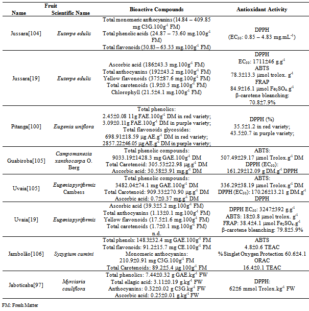
[19]  Maria do Socorro M. Rufino, Ricardo E. Alves, Edy S. de Brito, Jara Pérez-Jiménez, Fulgêncio Saura-Calixto, Jorge Mancini-Filho, “Bioactive Compounds and Antioxidant Capacities of 18 non-traditional tropical fruits from Brazil”, Elsevier Ltd, Food Chemistry, vol. 121, no. 4, pp. 996-1002, 2010.
[20]  Renan Campos Chisté, Marisa Freitas, Adriana Zerlotti Mercadante, Eduarda Fernandes, “The Potential of Extracts of Caryocar villosum Pulp to Scavenge Reactive Oxygen and Nitrogen Species”, Elsevier Ltd, Food Chemsitry, vol. 135, no. 3, pp. 1740-1749, 2012.
[21]  Online Available: http://www.biomasdobrasil.com/
[22]  Roberta Cunha de Mendonça, Jeanine Maria Felfini, Bruno Machado Teles Walter, Manoel Claudio da Silva, Alba Valéria Rezende, Tarciso S. Filgueiras, Paulo Ernane Nogueira, “Flora Vascular do Bioma Cerrado” in S. M. Sano, S. P. Almeida (Eds.), Cerrado, Ambiente e Flora, Embrapa, Brazil, pp. 289-556, 1998.
[23]  Carolyn Proença, Rafael S. Oliveira, Ana Palmira Silva, “Flores e Frutas do Cerrado”, Editora Rede Sementes do Cerrado, 2ª ed, Brazil, 2006.
[24]  J. A. Ratter, J. F. Ribeiro, S. Bridgewater, “The Brazilian Cerrado Vegetation and Threats to its Biodiversity”, Oxford Journals, Annals of Botany, vol. 80, no. 3, pp. 223-230, 1997.
[25]  A. K. Glyan’ko, G. G. Vasil’eva, “Reactive Oxygen and Nitrogen Species in Legume-Rhizobial Symbiosis: A review”, Springer, Applied Biochemistry and Microbiology, vol. 46, no. 1, pp. 15-22, 2010.
[26]  Barry Halliwell, John M. C. Gutteridge, “Free Radicals in Biology and Medicine”, OUP Oxford, 4a ed., United States, pp. 280-525, 2007.
[27]  Marian Valko, Dieter Leibfritz, Jan Moncol, Mark T. D. Cronin, Milan Mazur, Joshua Telser, “Free Radicals and Antioxidants in Normal Physiological Functions and Human Disease”, Elsevier Ltd, International Journal of Biochemistry e Cell Biology, vol. 39, no. 1, pp. 44-84.
[28]  Christine C. Winterbourn, “Reconciling the Chemsitry and Biology of Reactive Oxygen Species”, Nature Publishing Group, Nature Chemical Biology, vol. 4, no 5, pp. 278-286, 2008.
[29]  Marian Valko, C. J. Rhodes, Jan Moncol, M. Izakovic, Milan Mazur, “Free Radicals, Metals and Antioxidants in Oxidative Stress-Induced Cancer”, Elsevier Ltd, Chemico_Biological Interactions, vol. 160, no. 1, pp. 1-40, 2006.
[30]  Dejian Huang, Boxin Ou, Ronald L. Prior, “The Chemistry Behind Antioxidant Capacity Assays”, American Chemical Society, Journal of Agricultural and Food Chemistry, vol. 53, no. 6, pp. 1841-1856.
[31]  Marjolaine Roche, Claire Dufour, Nathalie Mora, Olivier Dangles, “Antioxidant Activity of Olive Phenols: Mechanistic Investigation and Characterization of Oxidation Products by Mass Spectroscopy”, The Royal Society of Chemistry, Organic & Biomolecular Chemistry, vol. 03, pp. 423-430, 2005.
[32]  Paolo Di Mascio, Stephan Kaiser, Helmut Sies, “Lycopene as the Most Efficient Biological Carotenoid Singlet Oxygen Quencher”, Elsevier Ltd, Archives of Biochemistry and Biophysics, vol. 274, no. 2, pp. 532-538, 1989.
[33]  Mariana A. Montenegro, Alessandro de Oliveira Rios, Adriana Zerlotti Mercadante, Mônica A. Nazareno, Claudio Dario Borsarelli, “Model Studies on the Photosensitized Isomerization of Bixin” Americal Chemical Society, Journal of Agricultural and Food Chemistry, vol. 52, no. 2, pp.367-373, 2004.
[34]  Semih Otles, Ozlen Çagindi, “Carotenoids as Natural Colorants” in Carmen Socaciu (Ed.), Food Colorants: Chemical and Functional Properties, CRC Press, United States, pp. 51-70, 2007
[35]  George Britton, Synn Liaaen-Jensen, Hanspeter Pfander, Carotenoids Handbook. Birkhäuser, Switzerland, 2004.
[36]  Ali El-Agamey, Gordon M. Lowe, David J. McGarvey, Alan Mortensen, Denise M. Phillip, T. George Truscott, Andrew J. Young, “Carotenoid Radical Chemistry and Antioxidant/Pro-oxidant Properties, Elsevier Ltd, Archives of Biochemistry and Biophysics, vol. 430, no. 1, pp. 37-48, 2004.
[37]  Jian-Jhih Guo, Ching-Han Hu, “Mechanism of Chain Termination in Lipid Peroxidation by Carotenes: a Theoretical Study”, American Chemical Society, The Journal of Physical Chemistry B, vol. 114, no. 50, pp. 16948-16958, 2010.
[38]  Wilfred Vermerris, Ralph Nicholson, Phenolic Compound Biochemistry, Springer Science + Business Media BV, United States, 2008.
[39]  Marian Naczk, Fereidoon Shahidi, “Extraction and Analysis of Phenolic in Food”, Elsevier Ltd, Journal of Chromatography A, vol. 1054, no. 1-2, pp. 95-111, 2004.
[40]  Ock Kyoung Chun, Sang Jin Chung,Won O. Song, “Estimated Dietary Flavonoid Intake and Major Food Sources of U.S. Adults”, American Society for Nutrition, The Journal of Nutrition, vol. 137, no. 5, pp. 1244-1252, 2007.
[41]  Paola R. Arabbi, Maria Inês Genovese, Franco Maria Lajolo, “Flavonoids in Vegetable Foods Commonly Consumed in Brazil and Estimated Ingestion by the Brasilian Population”, American Chemical Society, Journal of Agricultural and Food Chemistry, vol. 52, no. 5, pp. 1124-1131, 2004.
[42]  Obdúlio Benavente-García, Julían Castillo, Francisco R. Marin, Ana Ortuno, José A. Del Rio, “Uses and Properties of Citrus Flavonoids”, American Chemical Society, Journal of Agricultural and Food Chemistry, vol. 45, no. 12, pp. 4505-4515, 1997.
[43]  Øyvind M. Andersen, Kenneth R. Markham, “Flavonoids: Chemistry, Biochemistry and Applications”, CRC Press, United States, 2005.
[44]  Alan Crozier, Indu B. Jaganath, Michael N. Clifford, “Dietary Phenolics: Chemistry, Bioavailability and Effects on Health”, Royal Society of Chemistry, Natural Products Report, vol. 26, no. 8, pp. 1001-1043, 2009
[45]  P. C. H. Hollman, M. B. Katan, “Dietary flavonoids: intake, health effects and bioavailability”, Elsevier, Food and Chemical Toxicology, vol. 37, no. 9-10, pp. 937-942, 1999.
[46]  Fujiki Hirota, Kazue Imai, Kei Nakachi, Masahita Shimizu, Hisataka Moriwaki, Massami Suganuma, “Challenging the Effectiveness of Green Tea in Primary and Tertiary Cancer Prevention”, Springer, Journal of Cancer Research Clinical Oncology, vol. 138, no. 8, pp. 1259-1270, 2012.
[47]  Jurgen F. Leikert, Thomas R. Räthel, Paulus Wohlfart, Véronique Cheynier, Angélika M. Vollmar, Verena M. Dirsch, “Red Wine Polyphenols Enhance Endothelial Nitric Oxide Synthase Expression and Subsequent Nitric Oxide Release From Endothelial Cells, American Heart Association, Circulation, vol. 106, no. 13, pp. 1614-1617, 2012.
[48]  Volker Spitzer, Florian Schweigert, “Vitamin Basic, The Facts about Vitamins in Nutrition”, 3a Ed, DSM Nutritional Products Ltd, Germany, 2007.
[49]  Andre Theriault, Jun-Tzu Chao, Qi Wang, Adbul Gapor, Khosrow Adeli, “Tocotrienol: a Review of its Therapeutic Potential”, Elsevier, Clinical Biochemistry, vol. 32, no. 5, pp. 309-319, 1999.
[50]  Francesca Mangialasche, Weili Xu, Miia Kivipelto, Emanuela Costanzi, Sara Ercolani, Martina Pigliautile, Roberta Cecchetti, Mauro Baglioni, Andrew Simmons, Hilkka Soininen, Magda Tsolaki, Iwona Kloszewska, Bruno Vellas, Simon Lovestone, Patrizia Mecocci, “Tocopherols and Tocotrienols Plasma Levels are Associated with Cognitive Impairment”, Elsevier, Neurobiology of Aging, vol. 33, no. 10, pp. 2282-2290, 2012.
[51]  Hong Wang, Guohua Cao, Ronald L. Prior, “Oxygen Radical Absorbing Capacity of Anthocyanins”, American Chemical Society, Journal of Agricultural and Food Chemistry, vol. 45, no. 2, pp. 304-309, 1997.
[52]  Anna R. Proteggente, Ananth Sekher Pannala, George Paganga, Leo van Buren, Eveline Wagner, Sheila Wiseman, Frans van de Put, Clive Dacombe, Catherine A. Rice-Evans, “The Antioxidant Activity of Regularly Consumed Fruit and Vegetable Reflects Their Phenolic and Vitamin C Composition”, Free Radical Research, vol. 36, no. 2, pp. 217-233, 2002.
[53]  Joe A. Vinson, Xuehui Su, Ligia Zubik, Pratima Bose, “Phenol Antioxidant Quantity and Quality in Foods: Fruits”, American Chemical Society, Journal of Agricultural and Food Chemistry, vol. 49, no. 11, pp. 5315-5321, 2001.
[54]  Reşat Apak , Kubilay Güçlü , Mustafa Özyürek , Saliha Esin Karademir, “Novel Total Antioxidant Capacity Index for Dietary Polyphenols, Vitamins C and E, using Their Cupric Ion Reducing Capability in the Presence of Neocuproine: CUPRAC Method” American Chemical Society, Journal of Agricultural and Food Chemistry, vol. 52, no. 26, pp. 7970-7981, 2004.
[55]  Reşat Apak , Kubilay Güçlü , Mustafa Özyürek , Saliha Esin Karademir, Mehmet Altun, “Total Antioxidant Capacity Assay of Human Serum Using Copper(II)-neocuproine as Chromogenic Oxidant: The CUPRAC Method”, Free Radical Research, vol. 39, no. 9, pp. 949-961, 2005.
[56]  Jie Sun, Yi-Fang Chu, Xianzhong Wu, Rui Hai Liu, “Antioxidant and Antiproliferative Activities of Common Fruits”, American Chemical Society, Journal of Agricultural and Food Chemistry, vol. 50, no. 25, pp. 7449-7954, 2002.
[57]  Bente L. Halvorsen, Kari Holte, Mari C. Myhrstad, Ingrid Barikmo, Erlend Hvattum, Siv Fagertun Remberg, Anne-Brit Wold, Karin Haffner, Halvard Baugerod, Lene Frost Andersen, Ø Moskaug, David Jacobs, Rune Blomhoff, “A Systematic Screening of Total Antioxidants in Dietary in Plants”, American Society of Nutrition, The Journal of Nutrition, vol. 132, no. 3, pp.461-471, 2002.
[58]  Nicoletta Pellegrini, Mauro Serafini, Barbara Colombi, Daniele Del Rio, Sara Salvatore, Marta Bianchi, Furio Brighenti, “Total Antioxidant Capacity of Plant Foods, Beverages and Oils Consumed in Italy Assessed by Three Different in Vitro Assays”, American Society of Nutrition, The Journal of Nutrition, vol. 133, no. 9, pp. 2812-281, 2003.
[59]  Roberta Re, Nicoletta Pellegrini, Anna Proteggente, Ananth Pannala, Min Yang, Catharine Rice-Evans, “Antioxidant Activity Applying an Improved ABTS Radical Cation Decolorization Assay”, Elsevier Ltd, Free Radical Biology and Medicine, vol. 26, no. 9, pp. 1231-1237, 1999.
[60]  Reşat Apak , Esma Tütem, Mustafa Özyürek, Kubilay Güçlü , “Antioxidant Activity/Capacity Assay Methods Applied to Fruit and Cereals” in Fruit and Cereal Bioactives – Sources, Chemistry, and Applications, CRC Press, United States, 2011.
[61]  Guohua Cao, C. P. Verdon, H. A. Wu, Hong Wang, Ronald L. Prior, “Automated Assay of Oxygen Radical Absorbance Capacity with the COBAS FARA II ”, American Association for Clinical Chemistry, Clinical Chemistry, vol. 41, no. 12, pp. 1738-1744, 2005.
[62]  Boxin Ou, Maureen Hampsch-Woodil, Ronald L. Prior, “Development and Validation of an Improved Oxygen Radical Absorbance Capacity Assay Using Fluorescein as the Fluorescent Probe”, American Chemical Society, Journal of Agricultural and Food Chemistry, vol. 49, no. 10, pp. 4619-4626, 2001.
[63]  Ronald L. Prior, Ha Hoang, Liwei Gu, Xianli Wu, Mara Bacchiocca, Luke Howard, Maureen Hampsch-Woodill, Dejian Huang, Boxin Ou, Robert Jacob, “Assays for Hydrophilic Antioxidant Capacity (Oxygen Radical Absorbance Capacity (ORAC(FL))) of Plasma and other Biological and Food Samples”, American Chemical Society, Journal of Agricultural and Food Chemistry, vol. 51, no. 11, pp. 3273-3279, 2003.
[64]  Nicholas J. Miller, Catherine A. Rice-Evans, M. J. Davies, V. Gopinathan, A. Wilner, “A novel method for measuring antioxidant capacity and its application to monitoring the antioxidant status in premature neonates”, Clinical Science, vol. 84, no. 4, pp. 407-412, 1993.
[65]  Catherine A. Rice-Evans, Nicholas J. Miller, George Paganga, “Structure Antioxidant Activity Relationships of Flavonoids and Phenolic Acids”, Elsevier, Free Radical Biology and Medicine, vol. 20, no. 7, pp.933-956, 1996.
[66]  Vitaly Roginski, Eduardo A. Lissi, “Review of Methods for Determine Chain-Breaking Antioxidant Activity in Food”, Elsevier, Food Chemistry, vol. 92, no. 2, pp. 235-254, 2005.
[67]  W. Brand-Willians, M. E. Cuvelier, C. Berset, “Use of a Free Radical Method to Evaluate Antioxidant Activity”, Elsevier, LWT-Food Science and Technology, vol. 28, no. 1, pp. 25-30, 1995.
[68]  Iris F. F. Benzie, J. J. Strain, “The Ferric Reducing Ability of Plasma (FRAP) as a Measure of ‘‘Antioxidant Power’’: The FRAP Assay”, Academic Press Inc, Analytical Biochemistry, vol. 239, no. 1, pp. 70-76, 1996.
[69]  Lisbeth A. Pacheco-Palencia, Christopher E. Duncan and Stephen T. Talcott, “Phytochemical composition and thermal stability of two commercial açai species, Euterpe oleracea and Euterpe precatoria”, Elsevier, Food Chemistry, vol. 115, no. 4, pp. 1199–1205, 2009.
[70]  Veridiana Vera De Rosso, Silke Hillebrand, Elyana Cuevas Montilla, Florianda O. Bobbio, Peter Winterhalter, Adriana Zerlotti Mercadante, “Determination of Anthocyanins from Acerola (Malpighia emarginata DC.) and Açai (Euterpe oleracea Mart.) by HPLC-PDA and HPLC-MS, Elsevier, Journal of Food Composition and Analysis, vol. 21, no. 4, pp. 291-299, 2008.
[71]  Juliana Carvalho Ribeiro, Lusânia Maria Greggi Antunes, Alexandre Ferro Aissa, Joana D’arc Castania Darin, Veridiana Vera De Rosso, Adriana Zerlotti Mercadante, Maria de Lourdes Pires Bianchi, “Evaluation of the genotoxic and antigenotoxic effects after acute and subacute treatments with açai pulp (Euterpe oleracea Mart.) on mice using the erythrocytes micronucleus test and the comet assay”, Elsevier Ltd, Mutation Research. Genetic Toxicology and Environmental Mutagenesis, vol. 695, no. 1-2, p. 22-28, 2010.
[72]  André Gordon, Ana Paula Gil Cruz, Lourdes Maria Corrêa Cabral, Sidinéa Cordeiro de Freitas, Cristina Maria Araujo Dib Taxi, Carmen Marino Donangelo, Rafaella de Andrade Mattietto, Mirko Friedrich, Virgínia Martins da Matta, Friedhelm Marx, “Chemical Characterization and Evaluation of Antioxidant Properties of Açaí fruits (Euterpe oleraceae Mart.) During Ripening”, Elsevier, Food Chemistry, vol. 133, no. 2, pp. 256-263, 2012.
[73]  Melina Oliveira de Souza, Maísa Silva, Marcelo Eustáquio Silva, Riva de Paula Oliveira, Maria Lucia Pedrosa, “Diet Supplementation with Acai (Euterpe oleracea Mart.) Pulp Improves Biomarkers of Oxidative Stress and the Serum Lipid Profile in Rats”, Elsevier, Nutrition, vol. 26, no. 7-8, pp. 804-810, 2010.
[74]  Gitte S. Jensen, Xianli Wu, Kelly M. Patterson, Janelle Barnes, Steve G. Carter, Larry Scherwitz, Robert Beamam, John R. Endres, Alexandre Schauss, “In Vitro and in Vivo Antioxidant and Anti-Inflammatory Capacities of an Antioxidant-Rich Fruit and Berry Juice Blend. Results of a Pilot and Randomized, Double-Blinded, Placebo-Controlled, Crossover Study”, American Chemical Society, Journal of Agricultural and Food Chemistry, vol. 56, no. 18, pp. 8326-8333, 2008.
[75]  Cinthia Fernanda Zanatta, Elyana Cuevas, Florinda O. Bobbio, Peter Winterhalter, Adriana Zerlotti Mercadante, “Determination of Anthocyanins from Camu-Camu (Myrciaria dubia) by HPLC-PDA, HPLC-MS, and NMR”, American Chemical Society, Journal of Agricultural and Food Chemistry, vol. 53, no. 24, pp. 9531-9535, 2005.
[76]  Cinthia Fernanda Zanatta, Adriana Zerlotti Mercadante, “Carotenoid Composition from the Brazilian Tropical Fruit Camu-Camu (Myrciaria dubia)”, Elsevier, Food Chemistry, vol. 101, no. 4, pp. 1526-1532, 2007.
[77]  Rosana Chirinos, Jorge Galarza, Indira Betalleluz-Pallardel, Romina Pedreschi, David Campos, “Antioxidant Compounds and antioxidant Capacity of Peruvian Camu Camu (Myrciaria dubia (H.B.K.) McVaugh) Fruit at Different Maturity Stages”, Elsevier, Food Chemistry, vol. 120, no. 4, pp. 1019-1024, 2010.
[78]  Ricardo Elesbão Alves, Heloisa Almeida Cunha Filgueiras, Carlos Farley Hebster Moura, Nágela Cristina Costa Araújo, Adriano Silva Almeida, “Camu-camu (Myrciaria dubia McVaugh): A Rich Natural Source of Vitamin C”, in Proceedings of the InterAmericam Society for Tropical Horticulture”, vol. 46, pp. 11-13, 2002.
[79]  Kurt A. Reynertson, Hui Yang, Bei Jiang, Margaret J. Basile, Edward J. Kennelly, “Quantitative Analysis of Antiradical Phenolic Constituents from Fourteen Edible Myrtaceae Fruits”, Elsevier, Food Chemistry, vol. 109, no. 4, pp. 883-890, 2008.
[80]  Any Elisa De Souza Schmidt Gonçalves, Franco Maria Lajolo, Maria Inês Genovese, “Chemical Composition and Antioxidant/Antidiabetic Potential of Brazilian Native Fruits and Commercial Pulps”, American Chemical Society, Journal of Agricultural and Food Chemistry, vol. 58, no. 8, pp. 4666-4674, 2010.
[81]  Roberta Belandrino Rodrigues, Menelaos Papagiannopoulos, José Guilherme Soares Maia, Kaoru Yuyama, Friedhelm Marx, “Antioxidant Capacity of Camu Camu (Myrciaria dubia (H.B.K.) McVaugh) Pulp”, Ernährung/Nutrition, vol. 30, no. 9, pp. 357-362, 2006.
[82]  Francisco Carlos da Silva, Andrelisse Arruda, Alexandre Ledel, Cíntia Dauth, Nathalia Faria Romão, Rafaele Nazário Viana, Alexandre de Barros Ferraz, Jaqueline Nascimento Picada, Patrícia Pereira, “Antigenotoxic Effect of Acute and Chronic Treatments with Amazonian Camu-Camu (Myrciaria dubia) Juice on Mice Blood Cells”, Elsevier, Food and Chemical Toxicology, vol. 50, no. 7, pp. 2275-2281, 2012.
[83]  Teruo Inoue, Hiroshi Komoda, Toshihiko Uchida, Koichi Node, “Tropical Fruit Camu-Camu (Myrciaria dubia) has Anti-Oxidative and Anti-Inflammatory Properties”, Elsevier, Journal of Cardiology, vol. 52, no. 2, pp. 127-132, 2008.
[84]  Renan Campos Chisté, Adriana Zerlotti Mercadante, “Identification and Quantification, by HPLC-DAD-MS/MS, of Carotenoids and Phenolic Compounds from the Amazonian Fruit Caryocar villosum”, American Chemical Society, Journal of Agricultural and Food Chemistry, vol. 60, no. 23, pp. 5884-5892, 2012.
[85]  Mara Ribeiro Almeida, Joana D’Arc Castania Darin, Lívia Cristina Hernandes, Alexandre Ferro Aissa, Renan Campos Chisté, Adriana Zerlotti Mercadante, Lusânia Maria Greggi Antunes, Maria Lourdes Pires Bianchi, “Antigenotoxic Effects of Pequiá (Caryocar villosum) in Multiple Rat Organs”, Springer, Plant Foods for Human Nutrition, vol. 67, no. 2, pp. 171-177, 2012.
[86]  Xianli Wu, Gary R. Beecher, Joanne M. Holden, David B. Haytowitz, Susan E. Gebhardt, Ronald L. Prior, “Lipophilic and Hydrophilic Antioxidant Capacities of Common Foods in the United States”, American Chemical Society, Journal of Agricultural and Food Chemistry, vol. 52, no. 12, pp. 4026-4037, 2004.
[87]  Jesus N. S. Souza, Evaldo M. Silva, Adeline Loir, Jean-François Rees, Hervé Hogez, Yvan Larondelle, “Antioxidant Capacity of Four Polyphenol-Rich Amazonian Plant Extracts: A Correlation Study using Chemical and Biological in Vitro Assays”, Elsevier, Food Chemistry, vol. 106, no. 1, pp. 331-339, 2008.
[88]  Alexandre G. Schauss, Xianli Wu, Ronald L. Prior, Boxin Ou, Dejian Huang, Jonh Owens, Amit Agarwal, Gitte S. Jensen, Aaron N. Hart, Edward Shanbrom, “Antioxidant Capacity and Other Bioactivities of the Freeze-Dried Amazonian Palm Berry, Euterpe oleracea Mart. (Acai)”, American Chemical Society, Journal of Agricultural and Food Chemistry, vol. 54, no. 22, pp. 8604-8610, 2006.
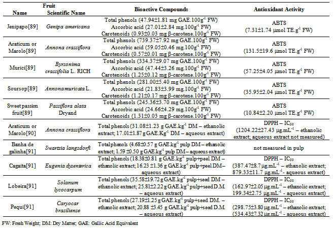
[89]  Vanessa Rios de Souza, Patrícia Aparecida Pimenta Pereira, Fabiana Queiroz, Soraia Vilela Borges, João de Deus Souza Carneiro, “Determination of Bioactive Compounds, Antioxidant Activity and Chemical Composition of Cerrado Brazilian Fruits”, Elsevier Ltd, Food Chemistry, vol. 134, no. 1, pp. 381-386, 2012.
[90]  Roberta Roesler, Luciana G. Malta, Luciana C. Carrasco, Gláucia Pastore, “Evaluation of the Antioxidant Properties of the Brazilian Cerrado Fruit Annona crassiflora (Araticum)”, Institute of Food Technologists, Journal of Food Science, vol. 71, no. 2, pp. C102-C107, 2006.
[91]  Roberta Roesler, Luciana Gomes Malta, Luciana Cristina Carrasco, Roseane Barata Holanda, Clélia Alves Socorro Sousa, Glaucia Maria Pastore, “Atividade Antioxidante de Frutas do Cerrado” Sociedade Brasileira de Ciência e Tecnologia de Alimentos, Ciência e Tecnologia de Alimentos, vol. 27, no. 1, pp. 53-60, 2007.
[92]  Roberta Roesler, Rodrigo R. Catharino, Luciana G. Malta, Marcos N. Eberlin, Glaucia Maria Pastore, “Antioxidant Activity of Caryocar brasiliense (pequi) and Characterization of Components by Electrospray Ionization Mass Spectrometry”, Elsevier, Food Chemistry, vol. 110, no. 3, pp. 711-717, 2008.
[93]  Alessandro de Lima, Ana Mara de Oliveira e Silva, Reginaldo Almeida Trindade, Rosângela Pavan Torres, Jorge Macini-Filho, “Chemical Composition and Bioactive Compounds in the Pulp and Almond of Pequi Fruit (Caryocar brasiliense, Camb.)”, Revista Brasileira de Fruticultura, vol. 29, no. 3, pp. 695-698, 2007.
[94]  Cristiane H. Azevedo-Meleiro, Delia B. Rodriguez-Amaya, “Confirmation of the Identity of the Carotenoids of Tropical Fruits by HPLC-DAD and HPLC-MS”, Elsevier, Journal of Food Composition and Analysis, vol. 17, no. 3-4, pp. 385-396, 2004.
[95]  Ana L. Miranda-Vilela, Inês S. Resck, Cesar K. Grisolia, “Antigenotoxic Activity and Antioxidant Properties of Organic and Aqueous Extracts of Pequi Fruit (Caryocar brasiliense Camb.) Pulp”, Sociedade Brasileira de Genética, Genetics and Molecular Biology, vol. 31, no. 4, pp. 956-963, 2008.
[96]  E.C. Aguilar, T.L. Jascolka, L.G. Teixeira, P.C. Lages, A.C.C. Ribeiro, E.L.M. Vieira, M.C.G. Peluzio, J.I. Alvarez-Leite, “Paradoxical Effect of a Pequi Oil-Rich Diet on the Development of Atherosclerosis: balance Between Antioxidant and Hyperlipidemic Properties”, Brazilian Journal of Medical and Biological Research, vol. 45, pp. 601-609, 2012.
[97]  Lucile Abe, Franco M. Lajolo, Maria Inês Genovese, “Potential Dietary Sources of Ellagic Acid and other Antioxidants Among Fruits Consumed in Brazil: Jabuticaba (Myrciaria jaboticaba (Vell.) Berg)”, Society of Chemical Industry, Journal of Science Food and Agriculture, vol. 92, no. 8, pp.1679-1687, 2012.
[98]  Shi-Biao Wu, Keyvan Dastmalchi, Chunlin Long, Edward J. Kennelly, “Metabolite Profiling of Jaboticaba (Myrciaria cauliflora) and other Dark-Colored Fruit Juices”, American Chemical Society Publications, Journal of Agricultural and Food Chemistry, vol. 60, no. 30, pp. 7513-7525, 2012.
[99]  Diego T. Santos, Priscilla C. Veggi, M. Angela A. Meireles, “Extraction of Antioxidant Compounds from Jabuticaba (Myrciaria cauliflora) skins: Yield, Composition and Economical Evaluation”, Elsevier Ltd, Journal of Food Engineering, vol. 101, no. 1, pp. 23-31, 2010.
[100]  Giovana Bonat Celli, Adaucto Bellarmino Pereira-Neto, Trust Beta, “Comparative Analysis of Total Phenolic Content, Antioxidant Activity, and Flavonoids Profile of Fruits from Two Varieties of Brazilian Cherry (Eugenia uniflora L.) Throughout the Fruit Development Stages”, Elsevier, Food Research International, vol. 44, no. 8, pp. 2442-2451, 2011.
[101]  Milena Bagetti, Elizete Maria Pesamosca Facco, Jaqueline Piccolo, Gabriela Elisa Hirsch, Delia Rodriguez-Amaya, Cintia Nanci Kobori, Márcia Vizzotto, Tatiana Emanuelli, “Physicochemical Characterization and Antioxidant Capacity of Pitanga Fruits (Eugenia uniflora L.)”, Sociedade Brasileira de Ciência e Tecnologia de Alimentos, Ciência e Tecnologia de Alimentos, vol. 31, no. 1, pp.147-154, 2011.
[102]  Genival Lopes Filho, Veridiana Vera De Rosso, Maria Angela A. Meireles, Paulo de Tarso da Rosa, Alexandra L. Oliveira, Adriana Z. Mercadante, Fernando A Cabral, “Supercritical CO2 Extraction of Carotenoids from Pitanga Fruits (Eugenia uniflora L.)”, Elsevier, The Journal of Supercritical Fluids, vol. 46, no. 1, pp. 33-39, 2008.
[103]  Edy Sousa De Brito, Manuela Cristina Pessanha de Araújo, Ricardo Elesbão Alves, Colleen Carkeet, Beverly A. Clevidence, Janet A. Novotny, “Anthocyanins Present in Selected Tropical Fruits: Acerola, Jambolão, Jussara, and Guajiru”, American Chemical Society Publications, Journal of Agricultural and Food Chemistry, vol. 55, no. 23, pp. 9389-9394, 2007.
[104]  Graciele da Silva Campelo Borges, Francilene Gracieli Kunradi Vieira, Cristiane Copetti, Luciano Valdemiro Gonzaga, Rui Carlos Zambiazi, Jorge Macini Filho, Roseane Fett, “Chemical Characterization, Bioactive Compounds, and Antioxidant Capacity of Jussara (Euterpe edulis) Fruit from the Atlantic Forest in Southern Brazil”, Elsevier, Food Research International, vol. 44, no. 7, pp. 2128-2133, 2011.
[105]  Marina C. Pereira, Rosana S. Steffens, André Jablonski, Plinho F. Hertz, Alessandro de O. Rios, Márcia Vizzoto, Simone H. Flores, “Characterization and Antioxidant Potential of Brazilian Fruits from the Myrtaceae Family”, American Chemical Society Publications, Journal of Agricultural and Food Chemistry, vol. 60, no. 12, pp. 3061-3067, 2012.
[106]  Adelia F. Faria, Marcella C. Marques, Adriana Z. Mercadante, “Identification of Bioactive Compounds from Jambolão (Syzygium cumini) and Antioxidant Capacity Evaluation in Different pH Conditions”, Elsevier, Food Chemistry, vol. 126, no. 4, pp. 1571-1578, 2011.