Food and Public Health

p-ISSN: 2162-9412    e-ISSN: 2162-8440

2013;  3(1): 12-20

doi:10.5923/j.fph.20130301.02

Extraction of Polyphenols and Anthocyanins from the Jambul (Syzygium cumini) Fruit Peels

Diego T. Santos 1, Rodrigo N. Cavalcanti 1, Maurício A. Rostagno 1, Carmen L. Queiroga 2, Marcos N. Eberlin 3, M. Angela A. Meireles 1

1LASEFI/DEA/FEA (School of Food Engineering)/UNICAMP, University of Campinas, 13083-862, Campinas, São Paulo, Brazil

2CPQBA/UNICAMP, Universityof Campinas, 13083-970, Campinas, São Paulo, Brazil

3Thomson Mass Spectrometry Laboratory, University of Campinas, 13083-970, Campinas, São Paulo, Brazil

Correspondence to: M. Angela A. Meireles , LASEFI/DEA/FEA (School of Food Engineering)/UNICAMP, University of Campinas, 13083-862, Campinas, São Paulo, Brazil.

Email:

Copyright © 2012 Scientific & Academic Publishing. All Rights Reserved.

Abstract

In this work, the feasibility of using ultrasound alone and in combination with other techniques for the extraction of polyphenols and anthocyanins from jambul (Syzygiumcumini) peels was evaluated. The results were compared with conventional techniques (agitated bed and Soxhlet extraction techniques) based on total extract yields, total phenolic and monomeric anthocyanin concentrations in the extracts and the relative yields of these compounds. Ultrasound-assisted extraction was more efficient and selective for the extraction of anthocyanins than both conventional methods. When a combination of ultrasound and agitated bed extraction was used, a significant increase in total yields, and both polyphenol and anthocyanin concentrations was achieved. Electrospray Ionization Tandem Mass Spectrometry analysis indicated that diglycosideanthocyanins (cyanidin-3,5-diglycoside, peonidin-3,5-diglycoside, delphinidin-3,5-diglycoside, petunidin-3,5-diglycoside and malvidin-3,5-diglycoside) were the main polyphenols present in the samples.

Keywords: Polyphenols, Anthocyanins, Jambul, SyzygiumCumini, Ultrasound Assisted Extraction, Combined Extraction Techniques

Cite this paper: Diego T. Santos , Rodrigo N. Cavalcanti , Maurício A. Rostagno , Carmen L. Queiroga , Marcos N. Eberlin , M. Angela A. Meireles , Extraction of Polyphenols and Anthocyanins from the Jambul (Syzygium cumini) Fruit Peels, Food and Public Health, Vol. 3 No. 1, 2013, pp. 12-20. doi: 10.5923/j.fph.20130301.02.

1. Introduction

In the last years, studies have increasingly associated the intake of fruits and vegetables with reduced risk for the development of chronic diseases such as cancer, diabetes type II, neurodegenerative disorders and cardiovascular diseases[1]. This phenomenon is likely influenced by biologically active molecules that are naturally present in those foods[2-4]. The interest in polyphenols is mainly due to their antioxidant properties; however, their mechanisms of action are known to be much more complex and to depend on several factors[5],[6]. Phenolic compounds are widely distributed in plants, although they are found in relatively high amounts in some plants, seeds and fruits, such as tea, yerba mate, cocoa, coffee, and grapes[7-10]. Many studies have been conducted in order to evaluate the biological activities of extracts of several usual and unusual sources of polyphenols. The major studies have investigatedantioxidant activity from extracts of tea[11],[12], curry[13] and fenugreek leaves[14], citrus peels[15] valerian (Valerianaofficinalis) root, lavender (Lavandulaofficinalis) and lemon balm (Melissa officinalis) leaves, (Alceakurdica) and (Stachyslavandulifolium) flowers[16], Aeglemarmelos leaves[17], wheat bran[18], among others. However, additional biological functions attributed to plant extracts have been analyzed such as antibacterial and antifungal[19], and anti-tumor[20],[21] activities. Polyphenols may also be present in high amounts in other less studied samples, such as other types of fruits available in certain countries. Among these fruits, jambul may represent a rich source of these compounds[22-25].
Jambul (Syzygiumcumini L.), also known as jambu, jamblon, jambula, jamboola, Java plum, jamun,jaam/kalojaam, jamblang, jambolan, blackplum, Damsonplum, Duhat plum, Jambolan plum, or Portuguese plum, belongs to the Myrtaceae family and has a small, egg-shaped purple fruit when ripe, with the pulp surrounding a single large seed. Several different phenolic classes have been reported in the fruits in high amounts[22-25]. Among these compounds, anthocyanins are one of the main polyphenols present, especially in the peels of the fruits. Anthocyanins are the largest and most important group of water-soluble and vacuolar pigments in nature. They are glycosylated polyhydroxy and polymethoxy derivatives of 2-phenylbenzopyrylium cation (Figure 1), i.e., the flavyliumcation (anthocyanidin group)[26]. Anthocyanins are of special interest because their antioxidant activity and their potential use in the food industry as natural colorants and preserving agents[27],[28].
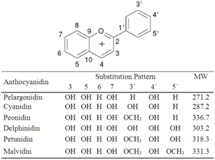
Figure 1. Chemical structure of the main anthocyanidins found in nature
Extraction of polyphenols from the fruit of the jambul is usually conducted using methods that were adapted from different sample matrices. However, the sample matrix can play a significant role in extraction kinetics and influence extraction efficiency because interactions between polyphenols and proteins are known to exist[29],[30]. Therefore, it is necessary to evaluate the extraction of polyphenols for each sample type and, if possible, using different techniques and methods. In general, conventional extraction techniques such as stirring/shaking or Soxhlet extraction have long extraction time and use high amount of solvent[22-25]. The long extraction times are partially due to the use of non-specific methods using extraction techniques that result in low extraction efficiencies and can lead to the degradation of some polyphenols[28-31].
In this context, ultrasound is increasingly being used as an alternative to conventional extraction methods. Cavitation has been suggested to be one of the main mechanisms by which ultrasound can improve extraction efficiency. Briefly, cavitation produced by the passage of ultrasonic waves through the bulk media exerts mechanical effects in the sample, improving mass transfer between the sample and solvent[30],[32],[33]. In addition to its higher efficiency, ultrasound can be easily combined with other techniques that can significantly improve extraction yields. In fact, one of the latest trends in sample preparation is the combination and/or hyphenation of extraction techniques to develop faster and more efficient methods[34]. There are several applications of this technique and its combination with other techniques for the extraction of phenolics from other samples[34-39]. Therefore, the objective of this work was to evaluate the feasibility of enhancing the extraction of polyphenols and anthocyanins from the peels of jambul by the use of high-frequency ultrasound alone or in combination with other techniques and to compare the efficiencies with conventional methods.

2. Materials and Methods

2.1. Raw Material and Sample Preparation

Jambul (Syzygiumcumini) fruits were obtained from the Institute of Biology at the University of Campinas (UNICAMP), Campinas, Brazil.The separation of peels, pulps and seeds was performed by crushing the whole fruit while avoiding damage to the seeds. Peels were washed with water to eliminate residual pulp. Samples were stored at -18℃ until submitted to the extraction procedures.

2.2. Chemical Reagents

Ethanol (99.5%), methanol (99.5%), glacial acetic acid (99.7%) and sodium carbonate (99.5%) were obtained from Ecibra (Santo Amaro, Brazil). Ethyl acetate (99.5%) was purchased from Merck (Darmstadt, Germany). Hydrochloric acid (36.5-38%), sodium acetate (99%) and potassium chloride (99%) were obtained from Synth (Diadema, Brazil). Formic acid (98%) was purchased from Vetec (Rio de Janeiro, Brazil). Folin-Ciocalteu reagent was obtained from Dinâmica (Diadema, Brazil). Anthocyanin reference standards (cyanidin-3-glycoside chloride, delphinidin-3- glycoside chloride, peonidin-3-glycoside chloride, malvidin-3-glycoside chloride, pelargonidin-3- glycoside chloride and petunidin-3-glycoside chloride) and gallic acid were acquired from Extrasynthese (Genay, France). Reference standards of all analyzed compounds were HPLC grade with purities greater than 95%.

2.3. Extraction Procedures

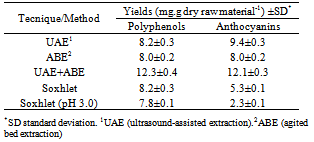
The samples were weighed and extracted using one of the different methods (Soxhlet, ultrasound, agitated bed and a combination of ultrasound and agitated bed). Soxhlet extractions were performed using 5 g of sample that was extracted with 120 mL of ethanol or acidified ethanol (acidified to pH 3 with HCl) (sample:solvent ratio of 1:25) for 8 hours at the boiling point of the solvent. The ultrasound-assisted extractions (UAE) were performed in an ultrasonic bath (40 kHz/81 W) (Thornton, T 1440, São Paulo, Brazil) using 2.5 g of the sample extracted with 25 mL of ethanol (sample:solvent ratio of 1:10) for 2 hours at room temperature. In a similar way, the agitated bed extractions (ABE) were performed in a rotary shaker (Marconi, MA 420, Piracicaba, Brazil) using 2.5 g of sample and 25 mL of ethanol (sample:solvent ratio of 1:10). Extractions were performed with agitation (150 rpm) for 2 hours at 30℃. Finally, a combination of ultrasound and agitated bed extraction (UAE + ABE) was used to improve the yields. Initially, 2.5 g of sample was extracted with 25 mL of ethanol (sample:solvent ratio of 1:10) during 10 minutes by UAE and submitted afterward to the same conditions of the ABE protocol. Independently of the method, the extracts were filtered, the solvent evaporated under vacuum (Laborota 4001, Heidolph Instruments GmbH, Viertrieb, Germany) at 40℃ and stored protected from light at -18℃ until its use in the chemical analysis.

2.4. Chemical Analyses

2.4.1. Total Phenolic Content(TPC)
The content of total phenolic content(TPC) was determined using the Folin-Ciocalteau method, based on the colorimetric oxidation/reduction reaction of phenols[40]. Briefly, 1 mL of extract was mixed with 1 mL of Folin-Ciocalteu reagent. After 3 minutes, 1 mL of a saturated solution of sodium carbonate (50 %, w/w) was added to the mixture and the volume was adjusted to 10 mL with distilled water. The reaction mixture was kept protected from light for 90 minutes at room temperature, and afterwards, the absorbance was measured at 725 nm using a UV-Vis spectrophotometer (Hitachi, model U-3010, Tokyo, Japan). A blank was prepared using 1 mL of distilled water instead of the extract. The results were calculated based on the calibration curve of gallic acid (GA) (10.9-86.8 mg. L-1) and expressed as milligrams of Gallic Acid Equivalents (GAE) per gram of extract in dry base (d.b.) (mg GAE. g-1, d.b.). All determinations were made in triplicate.
2.4.2. Total Monomeric Anthocyanins (TMA)
The total monomeric anthocyanin (TMA) content of the samples was determined using the pH differential method[41], which relies on the structural transformation of the anthocyanin chromophore as a function of pH. A UV-Vis spectrophotometer (Hitachi, model U-3010, Tokyo, Japan) and glass cuvettes of 1 cm in length were used to measure the maximum spectral absorption wavelength (512 nm and 700 nm) using distilled water as a blank. For this purpose, the sample was prepared with 20 mg of extract dissolved in 10 mL of distilled water. Two dilutions of this solution were prepared: one with hydrochloric acid/potassium chloride buffer (pH = 1.0) and the other with sodium acetate/acetic acid buffer (pH = 4.5). The pH values of the buffer solutions were measured using a pH meter (Digimed, model DM-22, Sao Paulo, Brazil) calibrated with buffers at pH 4.01 and 6.86, which were adjusted with HCl. Aliquots of the extract were measured and adjusted to pH 1.0 and 4.5 with the addition of buffer solutions. After 15 min, the absorbance of each equilibrated solution was measured at the maximum absorption wavelength and 700 nm for haze corrections using a 1 cm path length glass cell (l). The dilution factor (DF) was determined (the final volume divided by the original sample volume). The difference in absorbance values at pH 1.0 and 4.5 is directly proportional to the anthocyanin content. The total monomeric anthocyanin content (TMA) was calculated using cyanidin-3-glycoside as a reference (MW = 449.2 g mol-1 and ε = 26,900 L. mol-1.cm-1), and the results were expressed as mg cyanidin-3-glycoside equivalents/g of extract in dry base (d.b.) (mg cyn-3-gly equivalents g-1, d.b.). Relative standard deviations (RSDs) were lower than 8% for all samples. The absorbance of the diluted sample (A) and TMA were calculated with equations (1) and (2):
(1)
(2)
2.4.3. Thin-layer Chromatography (TLC)
For the determination of the qualitative chemical profile of the sample and its main polyphenols, the extract was fractionated using silica plates (20 × 20 cm, 1 mm height, Merck, Darmstadt, Germany). To identify the anthocyanins present in each extract, the mobile phase used was composed of ethyl acetate, glacial acetic acid, formic acid and distilled water (100:11:11:26), and no spray reagent was used[42]. Samples consisted of 10 mg of each extract dissolved in 1 mL of methanol. All anthocyanin standards were diluted in methanol to an approximate concentration of 5 mg. mL-1 and applied on the plates simultaneously with the sample.
2.4.4. Electrospray Ionization Tandem Mass Spectrometry (ESI-MS)
A Q-TOF system (Micromass, Manchester, United Kingdom) was used for Electrospray Ionization Tandem Mass Spectrometry (ESI-MS/MS) analysis. The conditions used were a source temperature of 100℃, capillary voltage of 2.1 kV and cone voltage of 40 V. All data obtained from ESI in the positive ion mode were treated using Mass Lynx software (v.3.5, Waters, Manchester, United Kingdom). The samples (1 mg) were dissolved in methanol:water (1:1) with 0.7% formic acid; 1 µL of each sample was injected.

3. Results and Discussion

3.1. Polyphenols Extraction

Extraction of the sample was performed using different techniques, namely ultrasound-assisted extraction (UAE), agitated bed extraction (ABE), a combination of UAE and ABE (UAE+ABE) and Soxhlet extraction (using two different solvents). Extraction yields (%, d.b.) and polyphenol concentrations (mg of GAE/ g of extract, d.b.) are graphically illustrated in Figure 2.
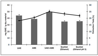
Figure 2. Total phenolic content and extraction yield from extracts of jambul peels (Syzygiumcumini) obtained through various methods of extraction
As can be seen in Figure 2, significant variations in the content of total phenolic compounds (TPC) and extraction yields were obtained with the use of different techniques for the extraction of polyphenols from the peels of jambul. The highest TPC of the sample (49.1 ± 0.3 mg GAE. g-1) was achieved using a combination of UAE and ABE, while the lowest was obtained using the traditional Soxhlet technique (35.2 ± 0.2 mg GAE. g-1), independent of the solvent used. The highest extraction yields were also collected from the UAE+ABE (25 ± 1%), while the lowest were retrieved using UAE (18.2 ± 0.8%). Although UAE produced the lowest yield, the samples derived from this technique had a higher TPC (44.1 ± 0.3 mg GAE. g-1) than conventional ABE and Soxhlet extractions.
In general, the utilization of ultrasound alone or in combination with other extraction techniques can improve the phenolic yields compared with conventional techniques. However, the extraction of phenolics from solid samples depends on several factors and interactions. For example, ultrasound frequency and intensity and sample characteristics can directly affect both extraction efficiency and yields (Santos et al., 2010). In this case, the low ultrasound intensity (81 W) might be the main factor responsible for the relatively low extraction yields obtained by UAE when compared with ABE. Furthermore, lower frequencies than we used (40 kHz) could be explored to enhance the effect of ultrasound in the sample matrix, facilitating the release of target compounds. On the other hand, the results also illustrate that UAE is more selective than ABE for polyphenols. Without a doubt, a cleaner sample may be useful in the identification of individual components using more sensitive detection techniques. However, we should also consider that the use of lower frequencies to improve the extraction of polyphenols, as previously suggested, might lead to losses in selectivity. The increase in structural damage of the sample matrix caused by lower frequency ultrasound may also increase the release and extraction rates of other sample components and therefore increase polyphenol concentrations in the samples[30],[32],[33].
It is also possible to observe that the effects of ultrasound are maximized when combined with another technique such as ABE. The combination of UAE and ABE resulted in higher TPCs and extraction yields than individual techniques. In fact, it achieved the highest phenolic yields (12.3 ± 0.4 mg GAE. g-1, d.b.) (Table 1) of all techniques tested. There are a few reports where submitting the sample to a short sonication treatment improved the yields of some phenolic compounds than using other techniques[34],[43], which supports our findings. The use of ultrasound can have several effects on the sample matrix. It may disrupt sample structure, improving contact between the sample and the solvent and releasing extractable analytes. These effects can enhance the extraction process[32-34], and may be partially responsible for the highest efficiency of the ABE performed after the UAE compared with techniques used alone. Thus, one of the main advantages of the use of ultrasound is that it may provide higher selectivity, even if combined with other techniques. Furthermore, it does not significantly reduce sample throughput time because the UAE time is relatively short (10 min) compared with the 2 hours of the ABE part of the method. It is also noteworthy that higher ultrasound intensities may drastically increase the effects on sample matrix structure and extraction kinetics and thus increase analytes and extract yields using this method either alone or in combination with other techniques.
In contrast, the Soxhlet extractions produced low yields and samples with low TPC, independently of the use of an acidified solvent. Even though higher temperatures, sample-to-solvent ratio (1:25) and longer extraction times were used in the Soxhlet technique, it was not possible to achieve higher extraction yields or TPC than the combination of UAE and ABE. Although Soxhlet extractions resulted in similar yields compared to UAE and ABE alone, it is less selective for polyphenols than these two techniques individually or combined. On the other hand, the lower TPC observed in the samples obtained by Soxhlet extraction may be partially caused by thermal degradation due to the high temperatures and long extraction times used[39],[44-47]. Thus, the longer extraction times and the temperature of this technique possibly increased yields, but thermal degradation reduced the concentration of these compounds in the final sample. Degradation of polyphenols can also explain the lower yields observed with the acidified solvent when compared to non-acidified solvent in the Soxhlet extractions because there is evidence indicating that some polyphenols may be sensitive to hydrolysis in acidic mediums[44-47]. A general overview of the results indicates that independent of the mechanism responsible for the relatively low yields with the Soxhlet extraction, the other techniques tested can be faster, more efficient and more selective for polyphenols than this technique, especially using a combinatorial approach.

3.2. Anthocyanins Extraction

Because anthocyanins have been reported to be the main polyphenols present in the peels of jambul[22],[24],[25], the influence of the extraction technique on the concentration of this compound class was also evaluated. Data for the concentrations of total monomeric anthocyanins (TMA) (mg of cyn-3-gly/ g of extract, d.b.) and the extraction yields (%, d.b.) obtained by different techniques are presented in Figure 3.
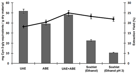
Figure 3. Total monomeric anthocyanin content and extraction yield from extracts of jambul peels (Syzygiumcumini) obtained through various methods of extraction
It can be observed in Figure 3 that the highest anthocyanin concentration of the sample was achieved for both treatments with ultrasound. These results suggest that the use of UAE alone or combined is not only more selective for polyphenols but is also more specific for anthocyanins present in the jambul peels. Furthermore, the anthocyanin concentration of the samples obtained with ultrasound (UAE and UAE+ABE) is similar to the TPC, and therefore, only relatively small amounts of other phenolic compounds (when compared to anthocyanins) can be expected to be present in the samples. This aspect is of critical importance when considering the separation and identification of the sample components because contaminants can overlap with anthocyanin peaks and interfere with their determination by more sensitive instrumental analytical techniques.
The slightly higher value of TMA compared to TPC observed for UAE can be explained by the influence of the reference substance used (gallic acid and cyanidin-3-glycoside equivalents) for the calibration curves of both methods (TMA and TPC). Because the extracts obtained are complex matrices, other anthocyanins that are present (see section 3.5) may have different calibration curve slopes and thus different concentration/response ratios. This is also the case for TPC, where values may be underestimated. Because anthocyanins are the main polyphenols present, using gallic acid as a reference may be causing a difference between the actual concentration and the estimated concentration. Nevertheless, a relative comparison of the effectiveness and selectiveness of different techniques tested can be made and would provide valuable information to be used by other researchers working with this type of sample.
In this context, the combination of UAE and ABE resulted in the highest anthocyanin yields. Illustratively, approximately 5-fold lower concentrations were found for Soxhlet extractions using acidified solvent than for UAE+ABE. As mentioned previously for the extraction yield of phenolic compounds, these results are due to the fact that ultrasonic and agitation can cause damage to the sample matrix structure, promoting the release of intracellular contents and facilitating the extraction procedure.
When comparing the results of UAE and UAE+ABE, the latter is capable of higher yields that compensate for the lower TMA concentration of the samples obtained with this technique, resulting in higher relative yields. However, the lower TMA concentration also implies that using UAE+ABE selectivity for anthocyanin can be reduced when compared to UAE alone. The lower selectivity can be attributed to the use of ABE because it produces samples with lower TMA concentrations than UAE and UAE+ABE.
Table 1. Total polyphenols and anthocyanin yields using different extraction techniques
     
In contrast, much lower TMA concentrations and yields were found in the samples obtained with either Soxhlet extraction method, indicating that this technique is not the most suitable for the extraction of these compounds. Furthermore, the use of an acidified solvent reduced the TMA concentration by approximately 2-fold, possibly due to an increased degradation rate compared to the use of non-acidified solvents. This is an intriguing observation because anthocyanins have been reported to be more stable at lower pHs. Most studies indicate that the anthocyanin flavilium ring is more stable at low pH levels because it is present in its ionized form[44-47]. However, the combination of high temperatures, long extraction times and low pHs may promote the hydrolysis of the glycosidic moiety. Therefore, based on the results presented, HCl acidified solvents should be avoided when using Soxhlet extractions because it may promote hydrolysis of the anthocyanins present in the jambulpeels.Table 1 shows the relative yields of polyphenols (mg of GAE. g of raw material-1, d.b.) and anthocyanins (mg of cyn-3-gly. g of raw material-1, d.b.) are shown in Table 1.

3.3. Anthocyanin Identification by Thin Layer Chromatography

In addition to the comparison of the extraction techniques, it is important to individually identify the main compounds present in the sample to understand the extraction process and possible degradation patterns. Therefore, a qualitative analysis of the anthocyanins present was conducted initially by Thin Layer Chromatography (TLC) and later by Mass Spectrometry (MS).
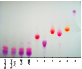
Figure 4. TLC plate for the extracts of the peels of jambul obtained by the different extraction techniques. Photograph was digitally enhanced to improve visualization by adjustment of color saturation and brightness/contrast as well as by the eliminate lens distortion. Anthocyanins: (1) cyaniding 3-glycoside; (2) delphinidin 3-glycoside; (3) peonidin3 - glycoside; (4) malvidin3-glycoside; (5) pelargonidin3-glycoside; (6) petunidin3-glycoside
Figure 4 shows the TLC plate for the extracts of the peels of jambul obtained by different extraction techniques. The only available standards were the mono-glycoside anthocyanins (cyanidin3-glycoside; delphinidin3-glycoside; peonidin3-glycoside; malvidin3-glycoside; pelargonidin3- glycoside; petunidin3-glycoside). As can be seen, only traces of some anthocyanins were found in the samples. However, the TLC results are consistent with the spectrophotometric analysis of the TMA concentration (Figure 3). In addition, it is feasible to assume that the major anthocyanins present may be di- or tri-glycosides due to the lower retention when compared to mono-glycoside anthocyanin standards.

3.4. AnthocyaninIdentification by Electrospray Ionization Tandem Mass Spectrometry (ESI-MS)

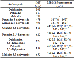
ESI-Q-TOF-MSwas used to support the evidence provided by the TLC analysis of the samples. The mass spectrum obtained with analysis of the Soxhlet extraction sample (with acidified solvent) is presented in Figure 5. Extracts obtained by acidified solvent were only evaluated to establish a trusted anthocyanin identification. It is well known that the hydrolysis of the glycosides to their respective aglycone forms occurs under acidic conditions. The composition of anthocyanins from jambulanalysed by ESI-Q-TOF-MS are presented in Table 2.The identified compounds present in the extract were mainly anthocyanin diglycosides (cyanidin3,5-diglycoside (MW=611), peonidin3,5-diglycoside (MW=625), delphinidin3 ,5- diglycoside (MW=627), petunidin3,5- diglycoside (MW=641), malvidin3,5- diglycoside (MW=655)).These results are consistent with those reported in previous studies with jambul fruits[22-25]. However, it was detected anthocyaninsmonoglycosides (petunidin 3-diglycoside (MW = 479) and malvidin 3-diglycoside (MW = 493)). Furthermore,anthocyanidins (anthocyanin aglycone form) were also detected in the extracts obtained by Soxhlet extraction with acidified solvent (peonidin (MW=301), delphinidin (MW=303), petunidin (MW=317), malvidin (MW=331)). This result suggests that hydrolysis of the diglycosides to their respective aglycone forms may be taking place under these drastic conditions (acid/heat/long incubation time). On the other hand, the presence of mainly diglycosides supports the difference between TPC and TMA concentration due to the use of cyaniding 3-glycoside as a reference standard. Neither cyaniding 3-glycoside (MW=449) nor delphinidin3-glycoside (MW=465) was detected in the sample.
Figure 5. Mass spectra data of sample from the Soxhlet with acidified solvent extractions
Table 2. Composition of anthocyanins from jambul obtained by ESI-Q-TOF-MS
     

4. Conclusions

The data presented in this manuscript suggest that ultrasound is an attractive alternative to conventional methods for the extraction of polyphenols and anthocyanins from the peels of jambul. Extraction using conventional methods usually results in lower yields of polyphenols and anthocyanins from the sample and may lead to the degradation of these compounds during the extraction process. The results also provide additional information about the improvement of polyphenol extraction processes with the use of ultrasound as a combinatorial technique. Results suggest that ultrasound used in combination with other techniques can improve the selectivity and efficiency of the extraction of polyphenols and anthocyanins. Such information can be useful in the overall effort to reduce the time required to extract these compounds from samples.Thus, these informations set jambul as a new alternative source of bioactive compounds, especially due to high polyphenols and anthocyanins content and rich-anthocyanin profile implicating in its potencial commercial aplication as food colorant or to production of nutraceuticals and pharmacological products with benefits to human health.

ACKNOWLEDGEMENTS

The authorsacknowledgethe financial supportfrom ConselhoNacional de DesenvolvimentoCientífico e Tecnológico (CNPq) and Coordenação de Aperfeiçoamento de Pessoal de Nível Superior (CAPES). Diego T. Santos would like to thank FAPESP (10/16485-5) for his postdoctoral fellowship.

References

[1]  S. C. S. R.Moura, P. E. R. Tavares, S. P. M.Germer, A. L. A. C. Nisida, A. B. Alves, A. S. Kanaan,“Degradation kinetics of anthocyanin of traditional and low-sugar blackberry jam”,Scientific & Academic Publishing, Food and Bioprocess Technology, vol. 5, pp. 2488-2496, 2012.
[2]  G. Trottier, P.J. Boström, N. Lawrentschuk, N.E. Fleshner, “Nutraceuticals and prostate cancer prevention: a current review”, Scientific & Academic Publishing, Nature Reviews Urology, vol. 7, pp. 21-30, 2010.
[3]  L.Dauchet, P.Amouyel, &, J. Dallongeville,“Fruits, vegetables and coronary heart disease”,Scientific & Academic Publishing, Nature Reviews Cardiology, vol. 6, pp. 599-608, 2009.
[4]  N.Okarter, R.H. Liu,“Health Benefits of Whole Grain Phytochemicals”, Scientific & Academic Publishing, Critical Reviews in Food Science and Nutrition, 50, 193-208, 2010.
[5]  A.García-Lafuente, E.Guillamón, A.Villares, M.A. Rostagno, J.A. Martínez,“Flavonoids as anti-inflammatory agents: implications in cancer and cardiovascular disease”,Scientific & Academic Publishing, Inflammation Research, vol. 58, pp. 537-552, 2009.
[6]  C.S. Yang, X. Wang, G. Lu, S.C. Picinich,“Cancer prevention by tea: animal studies, molecular mechanisms and human relevance”, Scientific & Academic Publishing, Nature Reviews Cancer, vol. 9, pp. 429-439, 2009.
[7]  C.Manach, A.Scalbert, C.Morand, C.Rémésy, L. Jiménez, “Polyphenols: food sources and bioavailability”, Scientific & Academic Publishing, American Journal of Clinical Nutrition, vol. 9, pp. 727-747, 2004.
[8]  M.G.Sajilata, P.R. Bajaj, R.S.Singhal, “Tea Polyphenols as Nutraceuticals”, Scientific & Academic Publishing, Comprehensive Reviews in Food Science and Food Safety, vol.7, pp.229-254, 2008.
[9]  J. Pérez-Jiménez, ,V.Neveu, , F.Vos, A. Scalbert,“Systematic Analysis of the Content of 502 Polyphenols in 452 Foods and Beverages: An Application of the Phenol-Explorer Database”, Scientific & Academic Publishing, Journal of Agricultural and Food Chemistry, vol.58, pp.4959-4969, 2010.
[10]  M.A.Rostagno, N.Manchón, M.D'Arrigo, E.Guillamón, A.Villares, A.García-Lafuente, A, Ramos, J.A. Martínez,“Fast and simultaneous determination of phenolic compounds and caffeine in teas, mate, instant coffee, soft drink and energetic drink by high-performance liquid chromatography using a fused-core column”. Scientific &Academic Publishing,AnalyticaChimicaActa, vol. 685, 204-211, 2011.
[11]  H. Chen, Y. Zhang, X. Lu, Z. Qu, “Comparative studies on the physicochemical and antioxidant properties of different tea extracts”, Scientific & Academic Publishing,Journal of Food Science and Technology, vol. 49, pp. 356-361, 2012.
[12]  A. Pȩkal, P. Dróżdż, M. Biesaga, K. Pyrzynska, “Screening of the antioxidant properties and polyphenol composition of aromatised green tea infusions”, Scientific & Academic Publishing, Journal of the Science of Food and Agriculture, vol. 92, pp. 2244–2249, 2012.
[13]  Ajay P. Singh, Ted Wilson, DevanandLuthria, Michelle R. Freeman, Rachel M. Scott, Dan Bilenker, Sneha Shah, Siva Somasundaram, NicholiVorsa, “LC-MS–MS Characterisation of Curry Leaf Flavonols and Antioxidant Activity”, Scientific & Academic Publishing, Food Chemistry, vol 127, pp. 80–85, 2011.
[14]  RamyaPremanath, J. Sudisha, N. Lakshmi Devi and S.M. Aradhya, “Antibacterial and Anti-oxidant Activities of Fenugreek (Trigonellafoenumgraecum L.) Leaves”, Scientific & Academic Publishing, Research Journal of Medicinal Plant, vol. 5, pp. 695-705, 2011.
[15]  M.B. Pashazanousi, M. Raeesi, S. Shirali, “Chemical composition of the essential oil, antibacterial and antioxidant activities, total phenolic and flavonoid evaluation of various extracts from leaves and fruit peels of Citrus limon” Scientific & Academic Publishing, Asian Journal of Chemistry, vol. 24, pp. 4331-4334, 2012.
[16]  J.Bouayed, K. Piri, H.Rammal, A.Dicko, F.Desor, C.Younos, R.Soulimani, “Comparative evaluation of the antioxidant potential of some Iranian medicinal plants”, Scientific & Academic Publishing, Food Chemistry, vol. 104, pp. 364–368, 2007.
[17]  S. Charoensiddhi, P. Anprung, “Bioactive compounds and volatile compounds of Thai bael fruit (Aeglemarmelos (L.) Correa) as a valuable source for functional food ingredients”, Scientific & Academic Publishing, International Food Research Journal, vol. 15, pp. 287-295, 2008.
[18]  Barinderjit Singh, Harish Kumar Sharma and Bhavesh Chandra Sarkar, “Optimization of extraction of antioxidants from wheat bran (Triticum spp.) using response surface methodology”, Scientific & Academic Publishing, Journal of Food Science and Technology, vol. 49, pp. 294-308, 2012.
[19]  Mohamed Trigui, Anis Ben Hsouna, Slim Tounsi, Samir Jaoua, “Chemical composition and evaluation of antioxidant and antimicrobial activities of Tunisian Thymelaeahirsuta with special reference to its mode of action”,Scientific& Academic Publishing, Industrial Crops and Products, vol. 41, pp. 150–157, 2013.
[20]  A. Fernandes, I. Fernandes, L. Cruz, N. Mateus, M. Cabral, V.DeFreitas, “Antioxidant and biological properties of bioactive phenolic compounds from QuercussuberL”, Scientific& Academic Publishing, Journal of Agricultural and Food Chemistry, vol. 57, pp. 11154-11160, 2009.
[21]  M.J. McCann, C.I.R. Gill, G. O’Brien, J.R. Rao, W.C. McRoberts, P. Hughes, R. McEntee, I.R. Rowland, “Anti-cancer properties of phenolics from apple waste on colon carcinogenesis in vitro”, Scientific & Academic Publishing, Food and Chemical Toxicology, vol. 45, pp. 1224–1230, 2007.
[22]  E.S. Brito, M.C.P.Araújo, R.E.Alves, C.Carkeet, B.A.Clevidence, J.A. Novotny,“Anthocyanins Present in Selected Tropical Fruits: Acerola, Jambolão, Jussara, and Guajiru”, Scientific & Academic Publishing, Journal of Agricultural and Food Chemistry, vol. 55, pp. 9389–9394, 2007.
[23]  J.M.Veigas, M.S. Narayan, P.M.Laxman, B. Neelwarne, “Chemical nature, stability and bioefficacies of anthocyanins from fruit peel of SyzygiumcuminiSkeels”, Scientific & Academic Publishing, Food Chemistry, vol. 105, pp. 619-627, 2007.
[24]  L. Li, Y.J. Zhang, N.P. Seeram,“Structure of anthocyanins from Eugenia jambolana fruit”, Scientific & Academic Publishing, Natural Product Communications, vol. 4, pp. 217-219, 2009.
[25]  A.F.Faria, M.C. Marques, A.Z. Mercadante,“Identification of bioactive compounds from jambolão (Syzygiumcumini) and antioxidant capacity evaluation in different pH conditions”, Scientific & Academic Publishing, Food Chemistry, vol. 126, pp. 1571-1578, 2011.
[26]  R. N.Cavalcanti, D. T. Santos, M. A. A. Meireles, “Non-thermal stabilization mechanisms of anthocyanins in model and food systems - an overview”, Scientific & Academic Publishing, Food Research International, vol. 44, pp. 499-509, 2011.
[27]  D.T. Santos, M.A.A. Meireles,“Jabuticaba as a source of functional pigments”, Scientific & Academic Publishing, Pharmacognosy Reviews, 3, 137-142, 2009.
[28]  A.Castañeda-Ovando, M.L. Pacheco-Hernández, M.E. Páez-Hernández, J.A. Rodríguez, , C.A. Galán-Vidal, “Chemical studies of anthocyanins: a review”, Scientific & Academic Publishing, Food Chemistry, vol. 113, pp. 859-871, 2009.
[29]  A.Papadopoulou, R.A. Frazier,“Characterization of protein-polyphenol interactions”, Scientific & Academic Publishing, Trends in Food Science and Technology, vol. 15, pp. 186-190, 2004.
[30]  M.A. Rostagno, , A. Villares, , E. Guillamón, , A. García-Lafuente,, J.A. Martínez,“Sample preparation for the analysis of isoflavones from soybeans and soy foods”, Scientific & Academic Publishing, Journal of Chromatography A, vol. 1216, pp. 2-29, 2009.
[31]  G. Woodward, P. Kroon, A. Cassidy, C. Kay,“Anthocyanin stability and recovery: implications for the analysis of clinical and experimental samples”, Scientific & Academic Publishing, Journal of Agricultural and Food Chemistry, vol. 75, pp. 5271-5278, 2009.
[32]  K.Vilkhu, R.Mawson, L. Simons, D. Bates,“Applications and opportunities for ultrasound assisted extraction in the food industry-A review”, Scientific & Academic Publishing, Innovative Food Science & Emerging Technologies, vol. 9, pp. 161-169, 2008.
[33]  A.C.Soria, M. Villamiel,“Effect of ultrasound on the technological properties and bioactivity of food: a review”, Scientific & Academic Publishing, Trends in Food Science and Technology, vol. 21, pp. 323-331, 2010.
[34]  M.A.Rostagno, , M.D`arrigo, &, J.A. Martínez,“Combinatory and hyphenated sample preparation for the determination of bioactive compounds in foods”, Scientific & Academic Publishing, Trends in Analytical Chemistry, vol. 29, pp. 553-561, 2010.
[35]  D.T. Santos, P.C.Veggi, M.A.A. Meireles,“Extraction of antioxidant compounds from Jabuticaba (Myrciariacauliflora) skins: Yield, composition and economical evaluation”, Scientific & Academic Publishing, Journal of Food Engineering, vol. 101, pp. 23-31, 2010.
[36]  F. Chen, Y. Sun, G. Zhao, X. Liao, X. Hu, J. Wu, Z. Wang,“Optimization of ultrasound-assisted extraction of anthocyanins in red raspberries and identification of anthocyanins in extract using high-performance liquid chromatography–mass spectrometry”, Scientific & Academic Publishing,Ultrasonicssonochemistry, vol. 14, pp. 767-778, 2007.
[37]  M.A.Rostagno, M. Palma, C.G. Barroso,“Ultrasound-assisted extraction of isoflavones from soy beverages blended with fruit juices isoflavones”, Scientific & Academic Publishing, AnalyticaChimicaActa, vol. 597, pp. 265-272, 2007.
[38]  K.Ghafoor, Y.H. Choi, J.Y.Jeon, I.H. Jo,“Optimization of ultrasound-assisted Extraction of phenolic compounds, antioxidants, and anthocyanins from grape (Vitisvinifera) seeds”, Scientific & Academic Publishing, Journal of Agricultural and Food Chemistry, vol. 57, pp. 4988-4994, 2009.
[39]  I.Ignat, I.Volf, V.I. Popa,“A critical review of methods for characterisation of polyphenolic compounds in fruits and vegetables”, Scientific & Academic Publishing, Food Chemistry, vol. 126, pp. 1821-1835, 2011.
[40]  V.L. Singleton, J.A.J. Rossi,”Colorimetry of total phenolics with phosphomolybdicphosphotungstic acid reagents”, Scientific & Academic Publishing, American Journal of Enology and Viticulture, vol. 16, pp. 144-157, 1965.
[41]  M.Giusti, R.E. Wrolstad, Current protocols in food analytical chemistry,John Wiley & Sons, New York, USA, 2001.
[42]  H. Wagner, S. Bladt, Plant Drug Analysis: A Thin Layer Chromatography Atlas, 2nd ed., Springer-Verlag Berlin Heidelbeg, New York, USA, 2001.
[43]  B.Klejdus, L.Lojková, M. Plaza, M.Šnóblová, D. Štěrbová,“Hyphenated technique for the extraction and determination of isoflavones in algae: Ultrasound-assisted supercritical fluid extraction followed by fast chromatography with tandem mass spectrometry”, Scientific & Academic Publishing, Journal of Chromatography A, vol. 1217, pp. 7956-7965, 2010.
[44]  H.M.Merken, G.R. Beecher,“Measurement of Food Flavonoids by High-Performance Liquid Chromatography: A Review”, Scientific & Academic Publishing, Journal of Agricultural and Food Chemistry, vol. 48, pp. 577-599, 2000.
[45]  M.Antolovich, P.Prenzler, K.Robards, D. Ryan,“Sample preparation in the determination of phenolic compounds in fruits”, Scientific & Academic Publishing, Analyst, vol. 125, pp. 989-1009, 2000.
[46]  M.Naczk, F. Shahidi, “Extraction and analysis of phenolics in food”, Scientific & Academic Publishing, Journal of Chromatography A, vol. 1054, pp. 95-111, 2004.
[47]  A.Kırca, M.Özkan, B. Cemeroğlu,“Effects of temperature, solid content and pH on the stability of black carrot anthocyanins”, Scientific & Academic Publishing, Food Chemistry, vol. 101, pp. 212-218, 2007.