International Journal of Food Science and Nutrition Engineering

p-ISSN: 2166-5168    e-ISSN: 2166-5192

2021;  11(2): 52-64

doi:10.5923/j.food.20211102.03

Received: Sep. 1, 2021; Accepted: Sep. 22, 2021; Published: Dec. 27, 2021


Improvement of Injera Shelf Life Through the Use of Silver (Ag) and Zinc Oxide (ZnO) Nano Particles

Oli Legassa1, Ashagrie Zewdu2, Eyobel Mulugeta3

1Food Science and Nutrition Directorate, Ethiopian Institute of Agricultural Research, Addis Ababa, Ethiopia

2School of Food Science and Nutrition, Addis Ababa University, Addis Ababa, Ethiopia

3Ministry of Innovation and Technology, Ethiopian Biotechnology Institute, Emerging Technology Center, Nanotechnology Directorate, Addis Ababa, Ethiopia

Correspondence to: Oli Legassa, Food Science and Nutrition Directorate, Ethiopian Institute of Agricultural Research, Addis Ababa, Ethiopia.

Email:

Copyright © 2021 The Author(s). Published by Scientific & Academic Publishing.

This work is licensed under the Creative Commons Attribution International License (CC BY).
http://creativecommons.org/licenses/by/4.0/

Abstract

About two-third of Ethiopian diet consists of Injera which is prepared from the flour of tef (Eragrostis tef (Zucc) Trotter), water and starter. Even though it is a nutritious food, the shelf life of Injera does not exceed 3 days. The aim of this study was to evaluate the effect of green synthesized silver (AgNPs) and zinc oxide (ZnONPs) nano particles in improving Injera shelf life. Ag and ZnONPS were synthesized from Eucalyptus globulus and Calpurnia aurea (Ait.) Benth extracts, respectively. Formation of nano particles were confirmed by characterization techniques (ultraviolet-visible spectroscopy, scanning electron microscopy and X-ray diffraction). Antifungal test was conducted using disc diffusion method. Both nano particles were coated on the interior and external surface of packaging materials by dip-coated method and Injera was packed in the plastic bags. The shelf life was determined as one day before mold growth appearance. Moisture content and pH was determined by oven and pH meter, respectively. Pour plate method was used for mold and yeast count. Finally, migration level was determined by microwave plasma - atomic emission spectroscopy. The results from UV-vis spectroscopy showed that characteristic peaks observed at 420 and 300 to 400 nm for silver and zinc oxide nano particles, respectively. Silver nano particles were mostly an irregular with few rod shaped whereas that of zinc oxide was mostly rod shaped. Both of them had significant antifungal effect. The shelf life of the stored Injera samples was increased significantly up to 24 days at higher concentrations. Both nano particles have no significant effect on moisture and pH of stored Injera. The colony forming unit per gram of molds and yeast decreased as the coating percentage increased. At 50 % concentration, the migration was 1.34 and 375 mg/Kg for silver and zinc oxide nano particles, respectively. Their migration was relatively high. Because, it needs further study to use them for Injera storage. Thus, it would be advisable to incorporate them as ingredients of plastics to decrease migration problem.

Keywords: Injera, Nano particles, Moulds and yeasts, Shelf life, Packaging, Storage

Cite this paper: Oli Legassa, Ashagrie Zewdu, Eyobel Mulugeta, Improvement of Injera Shelf Life Through the Use of Silver (Ag) and Zinc Oxide (ZnO) Nano Particles, International Journal of Food Science and Nutrition Engineering, Vol. 11 No. 2, 2021, pp. 52-64. doi: 10.5923/j.food.20211102.03.

1. Introduction

The shelf life of a food is described as the period for which it remains safe and suitable for consumption. The shelf life of baked foods is limited by different factors like water activity and microbiological spoilage [1]. Foods with pH value less than 4.5 are more susceptible to mold spoilage because of their tolerance to acid conditions than bacteria [2]. Injera is one of baked products with pH value less than this value. In addition to this, it has high moisture content that makes it more conducive for mold growth. As a result, shelf life of Injera does not usually exceed 3 days [3].
About two-third of Ethiopian diet [4] consists of Injera, a thin, fermented bread from flour of tef (Eragrostis tef (Zucc) Trotter). The three fungal species found to be responsible for Injera spoilage are Penicillium sp. and Rhizopus sp and Aspergillus niger [3]. Different preservation methods as reduction of available water and use of chemical preservatives are commonly used to elongate shelf life of bakery foods. Chemical preservatives (0.1% of sodium benzoates and benzoic acids) have prolonged the shelf life of Injera for up to 12 days [3]. However, they are applied as additives, which are not convenient for Injera preparation. Reduction of available water also does not work for Injera as its high moisture content is one of Injera quality.
Another recently growing food preservation method is the development of nano-enabled food packaging. Silver nano particles (AgNPs) and zinc oxide nano particles (ZnONPs) are metal’s nano particles used in packaging materials as antimicrobial agents [5]. They can be hosted in different polymers [6,7]. Such metal nano particles can be coated or directly incorporated in the synthesis processes of packaging materials [8].
Antimicrobial action may be obtained from the packaging by releasing the biocide directly into the food [9]. This can be exerted by both organic and inorganic materials [10]. The former ones are mostly organic acids and enzymes whereas the latter ones are nano particles of metals or metal oxides. The organic antimicrobial materials are less stable at high temperatures whereas metal and metal oxide nano particles withstand a harsher processing condition [11] which is additional advantage of using inorganic materials.
Migration of nano particles from packaging materials to food is reported in some researches. However, European Food Safety Authority and other national food safety authorities regulate the application of AgNPs in food packaging. It provided upper limits of Ag migration to less than 0.05 mg/kg in food [12]. It has also addressed that consumption of ZnONPs should not exceed 250 mg/kg, which is recommended daily intake (RDI) for Zn.
Different studies were conducted concerning the migration of silver nano particles from different types of nanocomposites (Low Density Poly Ethylene, 1,1 Diphenylethylene and polypropylene) into food simulants [13]. It was found that the Ag migration is well below the limits stated by the European Union legislation. For example, the research studied the effect of time and temperature on the migration of silver from polyethylene (PE) nanocomposites to boneless chicken breasts and found that migration of silver NPs was in a range from 0.003 to 0.005 mg/L [14]. Also, bread samples had recorded the lowest migration (<0.05 mg/L) level of AgNPs than other food samples [11]. Since Injera is somewhat similar products with bread, this type of antimicrobial packaging can also works for it. Therefore, the aim of this study was to evaluate the effects of green synthesized Ag and ZnONPs in improvement of Injera shelf life.

2. Materials and Methods

2.1. Materials

Eucalyptus globulus and (Calpurnia aurea (Ait.) Benth.) leaves were used as reducing and caping agent for the green synthesis of nano particles. AgNO3 and Zn(NO3)2.6H2O were used as primary precursors. Mancozeb, a broad spectrum antifugus, was used as control. Chramphenicol was used in potatoes dextros agar (PDA) as bacterial suppressant in pure culture development and antimicrobial tests. Alcohol (70%) was used as disinfectant. For dics preparation, Whattman filter paper (No:1) was cut into pieces. Low density polyethylene (LDPE) polymers (18 x 21 cm zipper bag) was used for preparation of nano-enabled food contact packaging materials.

2.2. Experimental Design

Completely randomized design (CRD) was used in this study. All the treatments were triplicated. For anti-microbial determination, both AgNPs and ZnONPs were used at 10, 20, 30, 40, 50, 60, 70, 80, 90, and 100% concentrations. Extract +AgNPs (Extr + AgNPs) was also considered as one treatment. Totally, 12 treatments were compared including control sample (Mancozeb, a broad specrum fungicide [15]. For dose determination during shelf life test, both types of nano particles were taken at concentrations of 10, 30, 50, 70, 90, and 100%, whereas uncoated zipper bag was used as control sample. Totally, 7 samples including the control sample were used for the treatments.
However, for other parameters (mold and yeast cfu/g, moisture content, pH, and migration test), both treatments (ZnONPs and AgNPs) were used at 10, 50 and 90% concentrations. Extract of Eucalyptus + AgNPs (Ext + AgNPs) and Extract of Calpurnia aurea (Ait.) Benth + ZnONPs (Ext + ZnONPs) were used as treatments. Untreated plastic zipper bag was used as a control. Totally, 5 treatments were analyzed separately including the control sample.

2.3. Plant Extract Preparation

Fresh and healthy leaves were collected from fields and washed well with pure water. Then, they were air-dried and ground to fine powder. About 100 g of powder and 800 mL of distilled water were allowed to mix in 1000 mL beaker [16] and heated on magnetic stirror. Afterwards, the extract was allowed to cool to room temperature and filtered using Whatman® No.1 filter. The extracts were kept in separate beakers at 4°C [17] for further reaction with AgNO3 and ZnNO3.6H2O, respectively.

2.4. Synthesis, and Characterization of Ag and ZnONano Particles

2.4.1. Synthesis of Ag and ZnO Nano Particles
The synthesis of both nano particles were carried out in aqueous solutions of silver nitrate (AgNO3) and zinc nitrate Zn(NO3)2. The optimization process was done for both nano particles by dissolving about 0.5094, 0.6113, and 0.7641 g of AgNO3 in 300 ml of deionized water that gives 10, 12, and 15 mM of solution, respectively. For Zn(NO3)2 , about 10, 15, and 20 g was dissolved in 300ml of deionized water that gives 852.3, 1136.4, 14773.2 mM of solution, respectively. The mixture ratio were 1:1, 3:1 and 3:2 for both AgNO3 and Zn(NO3)2 to extract. The mixtures were heated at 82°C for 45 minutes [18] while stirring at 1500 rpm. Based on the developed color, 15 mM at 3:2 and 1136.4 mM at 3:1 precursor solution to extract ratio were selected as optimum for AgNO3 and Zn(NO3)2, respectively.
Both solutions were kept at room temperature for 48 h. After settlement, the mixtures were centrifuged at 12,000 rpm for 10 minutes [19]. The supernatant was discarded and the pellets were collected and washed well with distilled water. For silver nano particles, the collected pellets were air-dried and grounded using mortar and pestle. But, the pellets from zinc nitrate solution were oven dried at 105°C for overnight and finally incinerated at 450°C for 3 h [20] in muffle furnace. Both nano particles were stored at 4°C for characterization and further use.
2.4.2. Characterization of the Synthesized Nano Particles
The samples were investigated by X-ray Diffractometer (XRD) on X’pert High Score powder diffractometer (CuKα X-ray radiation, λ = 0.1541 nm) to study the formation cristalline materials and to estimate the crystallite size of the as-synthesized nano particles. The surface morphology and particle size was investigated by ultra-high-resolution field emission scanning electron microscopy(SEM) (NOVANANO 450, US FEI Corporation). Ultra-violet (UV-vis) spectroscopy (Shimadzu UV-vis 2550) was used to measure the absorption between 200 and 800 nm [21]. The average crystallite size (D) was calculated by the Debye Scherrer formula [20] in X’pert HighScore Plus software (2004 PANalytica B.V.).

2.5. Preparation of Nano-Enabled Coatings from Ag and ZnO Nano Particles

Dip coating was used to attach the nano particles onto the intireror and external surface of plastic packages. Zipped plastic bag containers (18 cm x 21 cm) were disinfected by 70% ethanol and dried at 60°C in an oven [11] for half an hour. The plastic bags were e dip-coated in different concentration level of (10, 20…, 100%) Ag and ZnO nano particles solutions for 5 min and dried at 60°C in an oven.

2.6. Performance Evaluation

a) Pure culture preparation: Sterile molten potato dextrose agar (PDA) with chloramphenicol (60 mg/L) was poured into petri dishes and allowed to solidify. Different colonies were taken from Injera stored for 5 days using sterile needle and transferred to plates containing solidified PDA agar. Colonies were selected based on their color (Aspergillus Niger-black, Penicillium spp-green, Rhizopus-dark brown). The plates were incubated at 25°C for 72 h [22]. Growth and similar sub-culturing were perforemed up to fourth generation to obtain pure culture. The fungal isolates were identified by microscope to confirm that the isolated cultures were pure cultures.
b) Antifungal evaluation of Ag and ZnONPs: Disc diffusion method was used to assess the antifungal activities of both Ag and ZnONPs as method described by [23]. Whattman filter paper (No:1) was used to prepare discs approximately 6 mm in diameter [16]. The suspension was adjusted to 0.5 mc Farland standard using cytometry method. The antifungal activity was evaluated against the final fungal concentration of 106 cfu/ml [24]. About 1 mL of spore suspension was uniformly spread on sterile Petri plates containing PDA medium.
Sterile neutral discs (6 mm in diameter) were impregnated with different concentrations (10, 20, 30, 40, 50, 60, 70, 80, 90, and 100%) of Ag and ZnONPs and aseptically placed on the inoculated culture plates using a sterile forceps. The plates were placed in an incubator at 25°C for 72 h [22]. The antifungal activity was determined by measuring the zone of inhibition (mm) using digital caliper. The disc impregnated with Mancozeb was used as a control.

2.7. Sampling Method

Sampling was conducted by taking pieces of Injera from every quarter of the Injera roll and blending it. For fungus colony growth and migration evaluation, the stored injera were sampled at 2, 4, 6, 8, and 10 days of storage.

2.8. Determination of pH and Moisture Content of Injera

The sampling of Injera and pH test were performed as procedure used by [3]. An electronic pH meter was used to measure pH. Each homogenized Injera suspension (10 g of ground Injera added to 100 mL distilled water) was measured. For moisture, about 5 g of the Injera samples were measured and dried at 100°C in an oven until constant weight reached [25].
Moisture content was calculated as:

2.9. Shelf life Test

Shelf life is defined as the period in which visible mould and yeast growth not seen [26]. The stored Injera was monitored daily until mould growths occur. The shelf life was interpreted in relation to controle sample.

2.10. Mould and Yeast

Pour plate method [22] was used to analyze yeast and mould counts. About 9.75 g of PDA was weighed and dissolved into 250 mL distillled water. The medium was sterilized in an autoclave at 121°C for 15 min and finally chloramphenicol was added aseptically to molten PDA at 45°C. About 10 g of Injera samples was aseptically taken from all quarter parts and added with 90 mL of sterile water and homogenized for 3 min [27] using Vortex. About 1 g of each sample was added into test tubes containing 10 mL sterile water and used as stock solutions. From this, 1 mL was removed and added to another set of test tubes containing 9 mL sterile water which made 10-1 dilution. The same procedure was repeated to make 10-5 dilution. Then, 1 mL of both 10-2 and 10-5 dilutions were added into separate sterile Petri dishes and sterile molten agar was poured into the plates. The inoculated plates were allowed to set and incubated. The plates were kept in an inverted position to avoid water condensation. The PDA plates were incubated at 25°C for 72 h. The number of colonies found on each media was counted. Plates containing 10-150 colonies were counted [28]. It was determined as Numerous To Count (NTC) if above 150, and Few To Count (FTC) if below 10.

2.11. Migration Test (Ag and Zn)

Daily visual observation was performed on packaged Injera stored in the antimicrobial containers and conventional containers for 10 days [11]. Microwave plasma-atomic emission spectroscopy (MP-AES) was used to determine the amount of Ag and Zn in the sample. The samples were analyzed after acid digestion.

2.12. Statistical Analysis

A completely randomized design statistics were carried out with the analysis of variance (One-way ANOVA) procedure in SPSS software. Differences among average values were detected by Duncan’s multiple range test (p < 0.05). The result was interpreted as mean ± standard error.

3. Results and Discussion

3.1. Synthesis of Ag and ZnO NPs

Color change is the simple method of nano particles formation confirmation. Reduction (Ag+ to Ag0) of silver ions into silver nano particles during exposure to plant extracts was observed by the color change from deep red to dark brown. Also the reduction (Zn2+ to Zno) of ZnONPs biosynthesized from Calpurnia aurea (Ait.) Benth leaves extract was confirmed by a color change of deep red to light yellow, indicating formation of ZnONPs. This color change is due to the Surface Plasmon Resonance (SPR) Phenomenon. The metal nano particles have free electrons, which give the SPR absorption band, due to the combined vibration of electrons of metal nano particles in resonance with light wave. It is caused by excitation of surface plasmon vibrations in Ag and ZnO nano particles [29]. As concentration of AgNO3 increased the color of its mixture with leaves extract changed from very slight brown to dark brown whereas that of ZnO(NO2)3 was chanched from dark red to very slight yellow which is indicator for development of nano particles. The UV-vis spectroscopy result is shown in Figure 1 to more confirm development of nano particles.
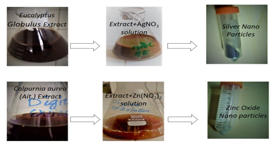
Figure 1. Image of the Synthesised Nanoparticles After Optimization

3.2. Characterization of Ag and ZnO NPs

a) UV-vis spectroscopy: The UV-vis spectrum of the synthesized AgNPs and ZnONPs is shown in Fig 2. Synthesis of the AgNPs and ZnONPs was confirmed by recording the absorption spectrum at a wavelength range of 200-800 nm [39]. The characteristic surface plasmon absorption band (SPAB) was observed at 420 nm for AgNPs. There is a report that SPAB of AgNPs from leaf extract was in range of 436 to 446 nm [29]. Other study has also reported that SPAB of AgNPs was observed at 440 nm [16]. Broadening of peak of silver nano particles formed in the reaction indicated that the particles were poly dispersed. It was similar with the study conducted on AgNPs [30]. For ZnONPs, the characteristic surface plasmon absorption band was observed at 300 to 400 nm (Fig 2). It was reported that ZnONPs biosynthesized from different plants have showed characteristic surface plasmon absorption around 400 nm [31]. Another report has also revealed that the sharp band of zinc colloids was observed at 361 nm [32]. There is also reported result that the SPAB of ZnNPs was in range of 299 to 311 nm [17]. In addition to these, other report shows that synthesized zinc oxide nano particles were confirmed by the UV-vis absorption spectra at the wavelength range of 380 to 386 nm [31], which is the characteristic wavelength range of zinc oxide nano particles. The minor difference between these reports and the current result may be raised from plant source difference.
The broadening of peaks of ZnONPs from Calpurnia aurea (Ait.) Benth extract was greater than that of AgNPs from Eucalyptus extract which indicates that ZnONPs were smaller. Broadening of peak of zinc oxide nano particles formed in the reaction indicated that the particles are polydispersed. There is study proved that the produced ZnONPs was poly-dispersed which means not aggregated in its nature [17]. It was similar with finding of [33] that conducted on ZnONPs.
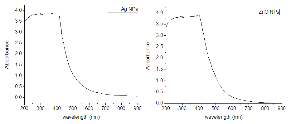
Figure 2. UV-Vis Spectrum of Ag and ZnONPs at 200-800nm Wavelength
b) X-Ray diffractometer (XRD): The powdered form of silver and ZnONPs were analyzed to confirm to estimate their cristalite size. XRD analysis of sample was conducted in 10 to 80 degree range of 2θ. The peaks at all 2θ were noted to confirm the presence of the nano particles [33].
The analysis of AgNPs shows that seven peaks at 2θ values of 27.72, 32.24, 46.34, 54.94, 57.5, 67.66, and 76.74 degrees were observed with miller indices (hkl) of (111), (200), (220), (311), (222), (400) and (420), respectively. There was a report [33] that the 2θ values of AgNPs were observed at 31.002, 46.761, 56.700, which is very similar with current result. Other researcher [34] has reported that AgNPs has five peaks at 38.1, 44.5, 64.3, 75.9, and 81.7 with miller indices (hkl) of (111), (200), (220), (311), and (222), respectively.
The broadening of Bragg’s peaks around their bases indicates the formation of small sized silver nano particles. The broadening of peaks shows the small size formation of nano particles [35].
The average crystallite size estimated from sherrer equation was 84.07 nm. The average sizes calculated from the intense peaks at 2θ (27.72, 32.24, 46.34) were 80.17, 82.55 and 89.48, respectively. Somewhat similar results were reported by different researchers which ranges from 28 to 61 nm [36] and 80-100 nm [36].
For ZnONPs, major peaks at 2θ values of 28.88, 32.03, 34.69 36.54, 47.77, and 40.50 and 56.78 degrees were observed with miller indices (hkl) of (101), (202), (003), (301), (200), (201) and (112), respectively. The report by [31] has shown that peaks were formed at 2θ for ZnONPs at 31.8, 34.5, 36.3 47.6, 56.6, 62.9, 66.4, 68.0, 72.6, 77.0, 81.4, 89.7 that correspond to indexed peaks (hkl) of (100), (002), (101), (102), (110), (103), (200), (112), (004), (202), (104) and (203), respectively. Also [37] has reported that peak position (2θ) of ZnNPs are 31.91, 36.48, 56.71, 66.57, 68.09, and 69.36 with corresponding miller indexes (hkl) of (100), (101), m (110), (200), (112), and (201). These reports are very similar to miller indices obtained currently confirming that ZnONPs were synthesised.
The crystallite size of ZnONPs was calculated from positions of the intense peaks. The average sizes obtained from the intense peaks at 2θ of 28.88, 32.03, 34.69, 36.54, and 56.78 were 70.40, 76.62, 70.40, 63.04, and 40.79 nm, respectively, whereas the average crystallite size of peaks was 64.25 nm. One study shows that ZnONPs with size of 20-65 nm [38] whereas another resesarchers [39] have found ZnONPs with about 19 nm. Also, it was reported that ZnONPs having 30-41 nm in size were synthesised [40]. In addition, another study [31] found ZnONPs with about 48.0-65.4 nm. The ZnONPs biosynthesized in the present study has smaller size when compared with the previous study, which is preferable to achieve better antimicrobial activity.
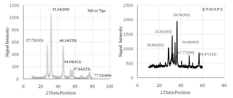
Figure 3. XRD Spectra of AgNPs and ZnONPs from Eucalyptus Calpurnia aurea (Ait.) Benth, Extract
c) Scanning Electron Microscope (SEM) Image
The SEM image of Ag and ZnO nano particles synthesized from Eucalyptus glubulus and Calpurnia aurea (Ait.) Benth leaves extract, respectively is shown in Fig. 4. AgNPs has rod shape (mostly) and spherical shape. The report by [41] has revealed that biosynthesized AgNPs had both spherical and rod shape. There is also another report which shows that the shape of biosynthesized AgNPs was an irregular and spherical [34].
These reports were in agreement with current morphological appearance of biosynthesized AgNPs. The shape of AgNPs was found to be spherical in shape by different researchers [42,43]. The SEM image disclosed a number of discrete and other larger groups that shows poly-dispersion of the crystalline AgNPs. This image indicates that the ZnNPs are not aggregated. The SEM image of ZnONPs also revealed that rod (mostly) and an irregular shaped nano particles were obtained.
These images demonstrated that produced zinc oxide nano particles were rectangular and rod shaped (mostly). The report by [43] has revealed that the shape of biosynthesized ZnONPs was both spherical and rod shape. In addition, [39] have found rod shaped ZnONPs, which also mostly observed shape (Fig 6) in current study. This study is in agreement with present study, which is mostly rod shaped ZnONPs. The report by [38] has revealed that biosynthesized ZnONPs have spherical and hexagonal shape. The difference may come from plant type used for reduction of the metal (Zn2+ to Zn°).
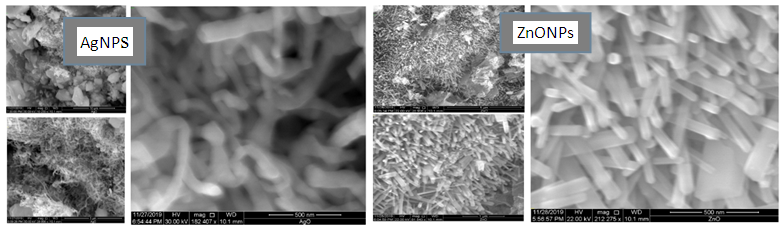
Figure 4. SEM Image of Biosynthesized AgNPs and ZnONps

3.3. Antimicrobial Activity of Ag and ZnONPs

The results on the antimicrobial test of both Ag and ZnONPs at different percentages are shown in Fig 5. It shows the patterns of antimicrobial capacity of Ag and ZnONPs which was determined by applying them against Aspegillus Niger, Penicillum spp and Rhizopus fungal types.
Both nano particles (AgNPs and ZnONPs) had significantly (p<0.05) affected the growth of all fungus. The diameter of inhibited zone was increased as the concentration level of Ag and ZnONPs increased. There was a numerical difference between both types of treatments.
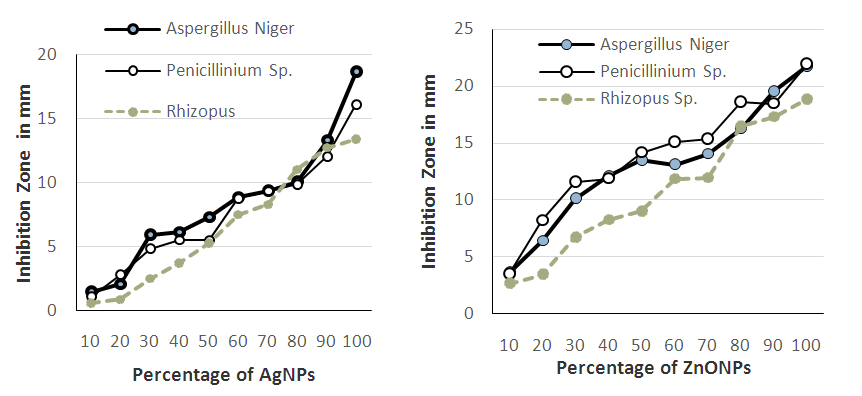
Figure 5. Antimicrobial Activity of Both Ag and ZnONPs
The inhibition zone of ZnONPs against Aspergillus niger, Penicillinium sp. and Rhizopus sp. was higher than that of AgNPs at the same concentrations. For instant, the inhibition zones of 100% ZnONPs against Aspergillus, Penicillinum and Rhizopus were 18.69, 16.13, and 13.44 mm whereas those of 100% AgNPs were 21.75, 21.72, and 18.87 mm, respectively. In addition, the inhibition zone of ZnONPs and AgNPs at 10% of concentration level against Aspergillus, Penicillinum and Rhizopus were (1.47, 1.08, 0.57) and (3.61, 3.49, 2.65 mm), respectively. Both treatments were not significantly different from the control sample at 100% concentration. For penicillinium sp., 90% of both treatments were also not significantly different from the control sample (Mancozeb). This effect may come from attachment of the NPs to the cells of the fungi, which destroy their cellular integrity. This destruction of their fungal cells may also leads to inability to multiply to form more mycelia, which appeared physically on Injera during storage. This growth inhibition may also be the results of enzyme inhibition through attachment of these small sized crystallite nano particles to their cells that affects way of their metabolization.
Silver ion is highly toxic to most microorganisms [44] and at least one mode of antimicrobial action of nano particles is through a slow release of silver ions via oxidation within or outside the cell. Silver nano particles are known to affect the permeability of membranes of microbial cells [45].
The antimicrobial activity of ZnONPs falls into two major pathways. These are interfering with the cell membrane by damaging the membrane integrity and permeability and increasing oxidative stress through reactive oxygen species (ROS) generation. At low concentrations, the antimicrobial effect is induced by nano materials themselves through physical absorption or electronic interaction–induced membrane damage [46] whereas excessive ROS production has been noted when Zn2+ concentrations reach 50 mg/L. ZnONPs possess positive charges in water, whereas bacterial membranes composed of acidic phospholipids and lipopolysaccharides present negative charges. This provides the basis for electrostatic interaction between ZnONPs and bacterial cells [47]. The ZnONPs first bind to the hydrophilic site on the lipopolysaccharide and then deform the outer membrane. This deformed membrane may not be able to multiply and continue the normal growth.

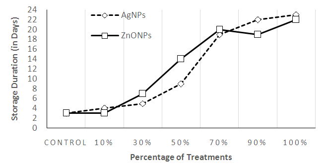
3.4. Storage Duration (Shelf Life) of Injera

The storage duration of Injera without visible mold growth was determined by taking the time from day of baking to the moment in which the samples of a batch show any visible signs of molding. The samples were stored at average temperature of 25.6 ± 2°C and average relative humidity of 50.3 ± 5%.
The visible mold sign-free storage period of Injera under unpreserved condition is 3-4 days [3]. Similarly, the current study shows that the shelf life of Injera packed with conventional package was 3 days. The plastic packages coated with Ag and ZnONPs had a better visible mold sign-free storage period (>18 days) at above 70% concentrations. The present results showed that treatments with Ag and ZnO nano particles have a better effect on elongation of Injera shelf life.
When comparing Ag and ZnONPs, 10% of ZnONPs has recorded similar result to the control sample at 4th day. However, at same concentration, the shelf life of Injera packed with plastic package coated with AgNPs at 10% was upto 5 days.
Also, the result (23 days) from AgNPs at 90% was greater than the result of ZnONPs at 90% (19 days). However, in other treatments (30, 50, 70%), ZnONPs has resulted in higher shelf life than AgNPs. As shown in Fig. 6, the shelf life of Injera samples packaged in plastics with ZnONPs were 8, 15, and 21 days at 30, 50, and 70% concentrations whereas those of AgNPs at same concentrations were 5, 9 and 19th days, respectively.
Figure 6. The Shelf Life of Injera Stored in plastics bag treated with Ag and ZnONPs

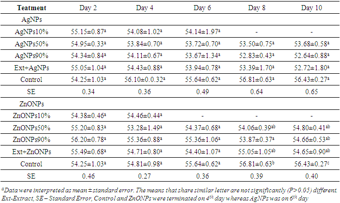
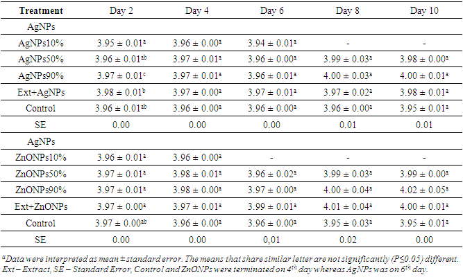
3.5. Moisture Content and pH of the Stored Injera

3.5.1. Moisture Content
Results of this investigation showed that there was a significant difference (p<0.05) on the moisture content of Injera samples with different treatments.
As showed in Table 1, the moisture content of Injera samples were ranging from 52.6 - 56.7%. The moisture content of Injera samples were decreased. However, after maximum shelf life reached, it starts to be increased that may be due to deterioration caused by overgrowth of fungus.
Table 1. Moisture Contents of Injera stored at different days
     
The significant difference was observed between the control and other treatments starting from day 8 of storage. The difference between the previous report and the present result may come from storage temperature and relative humidity difference. Thickness of the Injera that is only manually controlled during baking may also be a factor for the difference. However, the report by [49] has shown that moisture content of Injera was from 45.2 to 56.2%. This report is in agreement with current result of Injera moisture content.
Results of moisture content showed better maintenance of moisture content for breads stored by active coatings compared to the control one [50]. In contradiction to this report, there is no difference between the control and the treatment samples of current study when compared in moisture contents.
Moisture is an important parameter in baked foods that significantly affects shelf life and growth of microbial contaminants [51]. The report by [52] has shown that the overall mean moisture content of different kinds of bread made from wheat range from 37 - 47% during storage period. However, Injera had higher moisture content that make it more perishable than most bread [3]. High Moisture content is a serious problem in many bakery products that can result in high microbial growth and leads to microbiological spoilage of foods [3]. However, using of active food packaging provides interaction between food and packaging material, inhibiting microbial growth more than other factors considered in storage of foods [53].
3.5.2. pH of Injera
As it can be seen from Table 2 that pH of Injera was not significantly (p>0.05) different from the control sample. However, as storage days increases the pH of Injera treated with AgNPs and ZnONPs at different percentages increased, whereas the result from the control (untreated plastic zipper bag) decreased numerically (acidity increased). This increment may be raised from reaction between metal nano particles and other compounds in the Injera samples.
Table 2. The pH of Injera stored for 10 days
     
When seen by storage day difference, at day 2, only the pH of Injera samples stored in plastic bag treated with solution of Ext +AgNPs was significantly (p≤0.05) different from all other treatments. This difference may came from Injera thickness difference which is not fully controlled during baking. At day 6, Injera stored in only plastic packages treated with 10% AgNPs was significantly different from Injera samples stored in plastic packages coated with 90% ZnONPs. This may be resulted from Injera thickness difference, there is a numerical increase at day 8 and 10 as compared with other previous days, which may be due to reaction with these metals coated on the surfaces of the packages.
The report by [3] has shown that the pH values of the Injera samples were found to be between 3.31 and 4.41. This report showed that Injera samples under different treatments were not significantly (p>0.05) affected the pH value of the samples and this result is in agreement with the current result.
The pH results of Injera samples obtained in current study were comparable with the results reported in other related studies. The result reported by [3] also showed that the pH of the control (sample without preservatives) was 3.40. These reported results were closer to the results obtained in current study, which were from 3.94 to 4.02. The pH values obtained was quite low (more acidic) which makes Injera favorable for yeast and mold growth in addition to its high moisture content. To overcome this problem, the current study is promising to use biosynthesized Ag and ZnONPs on packaging of Injera in a future.

3.6. Total Mold and Yeast Count

In current study, it was also observed that the control sample (uncoated plastic package) had recorded high number of colony forming unit per gram of Injera whereas sample treated with AgNPs or ZnONPs had a low colony-forming unit at each storage day based on their concentration. Even though Injera has only 3-4 days shelf life when packed in conventional packages, it was possible to increase its shelf life to above 20 days using packaging plastic bag treated with Ag and ZnONPs (see Fig 7).
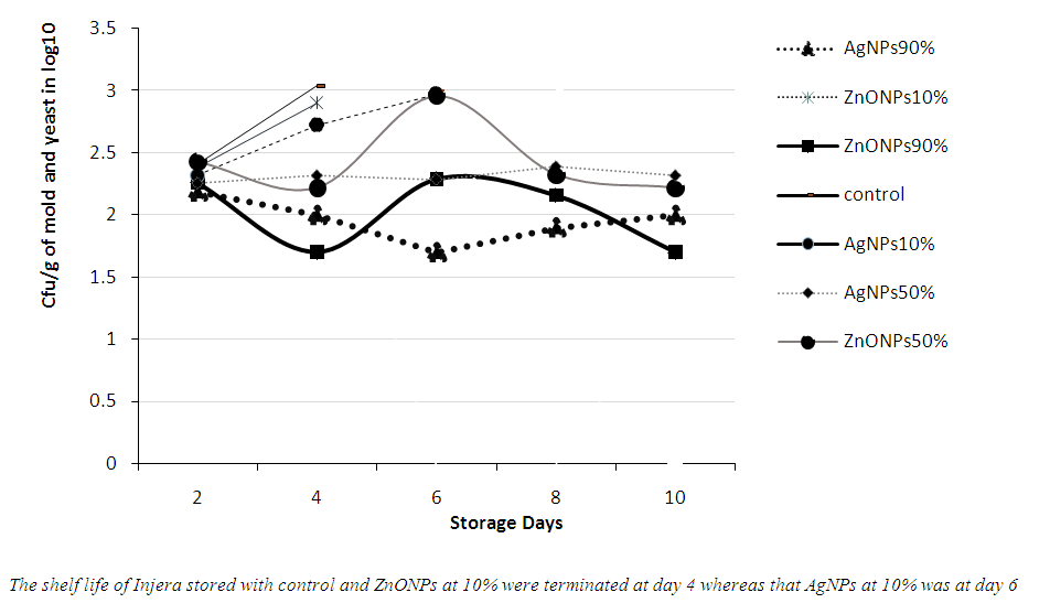
Figure 7. Colony Count of Mold and Yeast in Stored Injera using plastic coated with Ag and ZnO nano particles
At day 2, the colony forming units (cfu/g) of each Injera were not significantly (p>0.05) different. However, at day 4, the cfu/g of yeast and fungus for the control sample (3.0 log10) was highly significantly different from all the treatment samples. The results from the control and treatments at 10% were significantly increased with storage days. The shelf life of Injera stored in treated plastic bag were increased. This shows that treating of plastics with these biosynthesized nano particles can improves shelf life of Injera. According to [54], standard maximum permissible limits of baked products for yeast and mold is <4 in log10. Since there are no available previous findings that deal with storage of Injera with nano packaging, the results of this study were compared with findings on related products. The report by [50] showed that the shelf life of sliced wheat bread increased from 3 to 35 days for package incorporated with 2% ZnONPs. The limit between acceptable and marginal quality in terms of molds and yeasts is 2 x 10³ (3.30 log10) Cfu/g and the limit separating marginal quality from unacceptable quality is 5 x 10⁴ (4.5 log10) cfu/g [55].
High moisture level promotes the growth of yeasts and molds in bakery products [56]. Generally, molds are tolerant of acid conditions and favor an acidic pH (3.5-5.5). Therefore, foods with pH value <4.5 are not usually spoiled by bacteria but are more susceptible to mold spoilage [57]. Even though shelf life Injera is affected more by moulds, using of Ag and ZnONPs were proved to reduce its growth significantly in present study.

3.7. Migration of Ag and ZnONPs

The migration level of the control and AgNPs (10%) were 0 and 0.02 mg/Kg, respectively (Fig 8) whereas other treatments’ results were higher i.e. 0.45, and 0.59 mg/Kg for 50% AgNPs and 90% AgNPs, respectively. The result of the control samples at different days shows similarity showing absence of Ag in tef Injera. At day 2 of storage, 10% AgNPs (0.02 mg/Kg) records lower amount of migration followed by Ext +AgNPs (0.22 mg/kg). There was significant (P ≤ 0.05) difference between each treatments and the control sample except 10% AgNPs. The control sample was significantly different from all other treatments at day 6 and 8 storage days. The results of 10% AgNPs and Ext + AgNPs were similar at both days. But, the results of 50% AgNPs and 90% AgNPs were highly significantly different from other treatments. At day 10, all treatments were statistically different from the control sample. For ZnONPs (Fig 8), there was less migration at day 2. Each treatment was significantly (P≤0.05) different from each other at same storage day. The control sample had about 23.6 Zn (mg/Kg) content whereas treatments at 10, 50 and 90 % had 135.4, 285.6, 348.3 mg/Kg, respectively. Similarly, Ext + ZnONPs has recorded higher migration (511.3 mg/kg) as compared to other treatments. At day 4, the result from the control sample was similar with results at day 2. However, all the other treatments had recorded a higher migration results with their respective order of increased concentration.
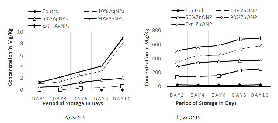
Figure 8. Migration of Ag+ and Zn2+ to Injera sample stored in plastics coated with Ag and ZnONPs
This shows that migration has increased with increasing of storage duration. For instant, the results of 10% ZnONPs and 90% ZnONPs had increased from 135.4 and 285.6 mg/Kg at day two to 253.0 and 583.4 mg/Kg at day ten, respectively. Similarly, the results of Ext + ZnONPs had also increased with increasing of storage duration According to [58], the amount of migrated silver increased with time and temperature. This study was in agreement with present finding since migration of AgNPs increased with time. The reports of [59] and [60] have shown that white tef (used in this study) had 9.58 mg/Kg and 40.2 mg/Kg of Zinc, respectively. Currently, the amount of Zn found in treatment samples were above that of the control smples confirming the presence of migration.
Using of nano particles in the food industry might pose risks that do not occur in macro size materials [61]. The report by [62] has mentioned some specific problems, such as certain nano-composites migrating from packaging to the food. European Food Safety Authority [12] recommended upper limits of Ag migration from packaging to be less than 0.05 mg/kg in food. However, in the current study, the amount of migration results of AgNPs is above this limit that needs further study to use it for Injera packaging. Eventhough ZnO is considered “generally recognized as safe (GRAS) [63], characteristics of its nano form may be different due to its increased surface area.
Different studies [64,65] have proved that nano particles incorporated into polymeric matrices had potential antimicrobial in increasing shelf life of food. Also, [27] has concluded that the incorporation of AgNPs into polymers can be used in food contact materials to decrease migration. Based on these previous studies, it could be expected that migration problem can be solved by incorporation of them as ingredients of plastic polymers.

4. Conclusions

Injera is Ethiopian daily stable foods, which is very perishable due to its high moisture content and its acidic nature that favors fast mould and yeast growth. Traditional preservation methods are not convenient for Injera storage. Nano technology is becoming a promising option in elongation of shelf life of food, which can also perform for Injera. In this study, biosynthesized Ag and ZnONPs had better antimicrobial activity against all tested fungus. Aspergillus sp. is more inhibited than Penicillium sp. and Rhizopus sp., respectively. Both NPs increased shelf life of Injera to more than 10 days at concentrations above 50% when packed aseptically. However, the amount of migration of Ag and ZnONPs was relatively high that may be due to direct contact between Injera and dip coated plastic package. The difficulty in using of this dip coated plastics for Injera storage is migration wich is accerated with Injera moisture and transferred to Injera easily. To reduce this migration, it would be advisable to find improved way of using biosynthesized nano particles in packing of Injera which may be inclusion of these nano particles as ingredients of packaging materials during processing.

Conflicts of Interest

The authors have declared that there is no conflicts of interest in any case.

AKNOWLEDGEMENTS

The authors thankfully acknowledged Ethiopian Biotechnology Institute, Addis Ababa University School of Food Science and Nutrition and Ethiopian Institute of Agricultural Research for their funding and equipment assistance that helped us to successfully complete this study.

References

[1]  Kyzlink, and Int. V., 2001. Principles of food preservation. Elsevier Publ., Amsterdam, 247-370 international, Journal of Food Microbiology, 101: 161-168.
[2]  Smith, J.P., 1993. Bakery products. In: Principles and Application of Modified Atmosphere Packaging of Food (ed. R. T. Parry). Blackie Academic and Professional, Glasgow, UK. pp. 134-169.
[3]  Ashagrie, Z., and Abate, D., 2012. Improvement of Injera Shelf Life Through the Use of Chemical Preservatives. African Journal of Food, Agriculture, Nutrition and development, 12(5), 6409-6423.
[4]  Ball, L. J., Sands, D. C., and Newman, R. D., 1996. A biotechnological approach to world hunger:the use of lysine excreting microbial mutants as inoculum in Ethiopia bread. Journal of the American Dietetic Association, 96: 9(1) A55.
[5]  Carbone, M., Sabbatella, G.S., AntonaroliRemita, H., Orlando, V., and Biagioni, S., 2015. .Exogenous control over intracellular acidification: enhancement via proton-caged compounds coupled to gold nano particles. Biochimica et Biophysica Acta - General Subjects., 1850, 2304-2307.
[6]  Espitia, P.J.P., Soares, N.D., dos Reis Coimbra, J.S., de Andrade, N.J., Cruz, R.S., and Medeiros, E.A.A., 2012. Zinc oxide nano particles: synthesis, antimi-crobial activity and food packaging applications. Food and Bioprocess Technology, 5 (5), 1447-1464.
[7]  Toker, R.D., Kayaman-Apohan,N., and Kahraman M.V., 2013. UV-curable nano-silver containing polyurethane based organic–inorganic hybrid coatings. Progress in Organic Coatings., 76, 1243-1250.
[8]  Martinez-Abad, A., Lagaron, J.M., and Ocio, M.J., 2012. Development and characterization of silver-based antimicrobial ethylene-vinyl alcohol copolymer (EVOH) films for foodpackaging applications. Journal of Agricultuaral and Food Chemistry, 60, 5350-5359.
[9]  Vermeiren, L., Devlieghere, F., and Debevere, J., 2002. Effectiveness of some recent antimicrobial packaging concepts. Food Addit. Contam, 1, 163-171.
[10]  Malhotra, B., Keshawn, A., and Kharkwal, H., 2015. Antimicrobial food packaging: potential and pitfalls Frontiers in Microbiology., 6, 1-9.
[11]  Metak, A.M., 2015. Effects of nanocomposites based nano-silver and nano-titanium dioxideon food packaging materials. Int. J. Appl. Sci.Technol., 5, 26-40.
[12]  European Food Safety Authority [EFSA], 2011. Guidance on the risk assessment of the application of nanoscience and nanotechnology in the food and feed chain.., 9(5), 2140.
[13]  Echegoyen,Y., and Nerín, C., 2013. Nanoparticle release from nano-silver antimicrobial food containers. Food and Chemical Toxicology, 62, 16-22.
[14]  Cushen, M., Kerry, J., Morris, M., Cruz-Romero, M., and Cummins, E., 2014. Evaluation and simulation of silver and copper nanoparticle migration from polyethylene nanocomposites to food and an associated exposure assessment. Journal of Agricultral and Food Chemistry. 62, 14031411.10.1021/jf404038y.
[15]  Agency (2005). Mancozeb Facts. EPA 738-F-05-XX
[16]  Rose, L., and Priya, F.J., 2017. Phytochemical screening and Green Synthesis of Silver Nano particles Using Aqueous Extract of Catharanthus roseus Stem Bark. International Journal of Pharma Sciences and Research, 8(5), ISSN 0975-9492.
[17]  Manokari, M., Ravindran, C.P., and Shekhawat, M.S., 2016. Production of zinc oxide nano particles using aqueous extracts of a medicinal plant micrococca mercurialis (l.) benth. World Scientific News, 30, 117-128.
[18]  Banerjee, P., and Nath, D., 2015. A Phytochemical Approach to Synthesize Silver Nano particles for Non-Toxic Biomedical Application and Study on their Antibacterial Efficacy. Nanoscience Technology, 2(1), 1-14.
[19]  Thomas R., Janardhanan, A., Varghese, RT., E.V., Soniya, E.V., Mathew, J., and Radhakrishnan E.K., 2014. Antibacterial properties of silver nano particles synthesized by marine Ochrobactrum sp.. Brazilian Journal of Microbiology, 45(4), 1221-1227.
[20]  Meruvu, H., Vangalapati, M., Chippada S.Ch., and Bammidi S.R., 2011. Synthesis and Characterization of Zinc Oxide Nano particles and Its Antimicrobial Activity against Bacillus Subtilis and Escherichia Coli., Rasayan J., Chemistry.4( 1), 217-222.
[21]  Abdallaha, Y., Ogunyemia, S.O., Zhanga, M., Fouadc, D.H., Honga, X., Ibrahima, E., Masuma, Md.I., Hossaina, A., Moc, J., and Lia B. 2019. Green Synthesis of Zinc Oxide Nano particles Using Different Plant Extracts and Their Antibacterial Activity against Xanthomonas Oryzae Pv. Oryzae. Artificial Cells, Nanomedicine, and Biotechnology, 47(1), 341–352.
[22]  Ravimannan, N., 2016. Study on microbial profile of bread during storage: International Journal of Advanced Reseach. Biological sciences., 3(9): 60-63.
[23]  Clara, C., Matasyoh, J. C., Wagara I. N., and Nakuvuma, J., 2013. Antifungal activity of Monathotaxis littoralis essential oil against mycotoxigenic fungi isolated from maize. International Journal of Microbiology Research and Reviews, 2(6), 103109.
[24]  Al-Zubaidi, Sh., Al-Ayafi, A., and Abdelkader, H., 2019. Biosynthesis, Characterization and Antifungal Activity of Silver Nano particles by Aspergillus Niger Isolate. Nanotechnology Research., 1 (1), 023-036.
[25]  AOAC., 2000. Official Methods of Analysis (17th ed., Vol. 2) Washington D.C: Association of Official Analytical chemistry.
[26]  Katsinis, G., Rigas, F., and Doulia, D., 2008. Synergistic effect of chemical preservatives with ethanol on the micriobial shelf life of bread by factorial design. International Journal of Food Science an Technology, 43, 208-215.
[27]  Azlin-Hasim, S., Cruz-Romero, MC., Morris, MA., Enda Cummins, E., and Kerry, KJ., 2015. Effects of a combination of antimicrobial silver low density polyethylene nanocomposite films and modified atmosphere packaging on the shelf life of chicken breast fillets. Food Packaging and Shelf Life, http://dx.doi.org/10.1016/j.fpsl.2015.03.003014-9271-8.
[28]  Tournas, V., Stack, M.E., Philip, B. Mislivec,Ph.B., Koch, H.A., and Ruth Bandler R., 2001. Bacteriological Analytical Manual, 8th Edition, Revision A, 1998. Chapter 18.
[29]  Ahmed, S., Ullah S., Ahmad, M., Swami, B., and Ikram, S., 2015. Green synthesis of silver nano particles using Azadirachta indica aqueous leaf extract. Journal of Radiation Research and Applied Sciences, http://dx.doi.org/10.1016/j.jrras.2015.06.006.
[30]  Omara, ST., Zawrah, MF., and Samy, AA., 2017. Minimum bactericidal concentration of chemically synthesized silver nano particles against pathogenic Salmonella and Shigella strains isolated from layer poultry farms. Journal of Applied Pharmaceutical Science, 7(08), 214-221.
[31]  Abraham, J., Singh, N., Saha, P., and Rajkumar K., 2014. Biosynthesis of Silver and Selenium Nano particles By Bacillus Sp. Japsk And Evaluation of Antimicrobial Activity. Der Pharmacia Lettre, 6 (1), 175-181.
[32]  Ogunyemi, S.O., Abdallah,Y., Zhang, M., Fouad, H., Hong, X., Ibrahim, E., Masum, M.I., Hossain, A., Mo J., and Li, B. 2019. Green synthesis of zinc oxide nano particles using different plant extracts and their antibacterial activity against Xanthomonasoryzaepv.oryzae, Artificial Cells. Nanomedicine, and Biotechnology, 47(1), 341-352.
[33]  Maity, J., Pal, S., Mondal, S., and Mukherjee, R., 2018. Synthesis and Characterization of ZnO Nano particles using Moringa Oleifera Leaf Extract: Investigation of Photocatalytic and Antibacterial Activity. International journal of Nanoscience and Nanotechnololgy, 14(2), 111-119.
[34]  Ahmed, J., Hiremath, N., and Jacob, H., 2016. Antimicrobial efficacies of essential oils/nano particles incorporated polylactide films against L. monocytogenes and S. typhimurium on contaminated cheese. International Journal of of Food Properties.
[35]  Velavan S, and Amargeetha A., 2018. X-ray Diffraction (XRD) and Energy Dispersive Spectroscopy (EDS) Analysis of Silver Page 3 of 5 Nano particles Synthesized from Erythrina Indica Flowers. Nanoscience and Technoogyl, 5(1), 1-5.
[36]  Govindarajan, M., Rajeswary, M., Muthukumaran, U., Hoti, SL., Khater, H., and Benelli, G., 2016. Single-step biosynthesis and characterization of silver nano particles using Zornia diphylla leaves: a potent eco-friendly tool against malaria and arbovirus vectors. Journal of Photochemistry and Photobiology B: Biology, 161:482–489.
[37]  Kanchana, R., and Zantye, P., 2016. Plant-mediated synthesis of silver nano particles with diverse applications. Asian Journal of Pharmaceutical and Clinical Research, 9, 124128.
[38]  Pal, Sh., Pal, K., Mukherjee, S., Bera, D., Karmakar, P., and Sukhen, D., 2020. Green cardamom mediated phytosynthesis of ZnONPs and validation of its antibacterial and anticancerous potential, Materials. Research. Express, 7 015068.
[39]  Banumathi, B., Malaikozhundan, B., and Vaseeharan, B., 2016. Invitro acaricidal activity of ethnoveterinary plants and green synthesis of zinc oxide nano particles against Rhipicephalus (Boophilus) microplus. Veternary Parasitology, 216: 93-100.
[40]  Hassan, SM., Abdel-Shafy, HI., and Mansour, MSM., 2016. Removal of pharmaceutical compounds from urine via chemical coagulation by green synthesized ZnO-nano particles followed by microfiltration for safe reuse. Arab J Chem. https://doi.org/10.1016/j.arabjc.2016.04.009 70.
[41]  Malaikozhundan, B., Vaseeharan, B., Vijayakumar, S., Pandiselvi, K., Kalanjiam, MAR., Murugan, K., and Benelli, G., 2017. Biological therapeutics of Pongamia Pinnata coated zinc oxide nano particles against clinically important pathogenic bacteria, fungi and MCF-7 breast cancer cells. Microbial Pathogenesis, 104, 268–277.
[42]  Nagababu, P., and Rao V.U., 2017. Pharmacological Assessment, Green synthesis, and Characterization of Silver Nano particles of Sonneratia apetala Buch.-Ham. Leaves. Journal of Applied Pharmaceutical Science, 7 (08), 175-182.
[43]  Vishwakarma,V., Samal,S., and Manoharan, N., 2010. Safety and risk associated with nano particles—a review. Journal of Minerals & Materials Characterization & Engineering., 9, 455-459.
[44]  Veerasamy, R., Xin, T. Z., Gunasagaran, S., Xiang, T. F. W., Yang, E. F. C., Jeyakumar, N., Dhanaraj S.A., . 2011. Biosynthesis of silver nano particles using mangosteen leaf extract and evaluation of their antimicrobial activities. Journal of Saudi Chemical Society, 15, 113e120. 36.
[45]  Jung, WK., Koo, HC., Kim, KW., Shin, S, Kim, SH., and Park, YH., 2008. Antibacterial activity and mechanism of action of the silver ion in Staphylococcus aureus and Escherichia coli. Applied. And Environmental Microbialogy, 74, 21718.
[46]  Li, WR. Xie, XB. Shi, QS. Zeng, HY, Ou-Yang, YS., and Chen, YB., 2010. Antibacterial activity and mechanism of silver nano particles on Escherichia coli. Applied Microbiology and. Biotechnology, 85(4), 111522.
[47]  Yong, L.Q., Cheng Teng, Hande, M.P., Ong, Ch.N.,Yu, L.E., Bay, B.H., and Baeg, G.H., 2017. Zinc oxide nano particles exhibit cytotoxicity and genotoxicity through oxidative stress responses in human lung fibroblasts and Drosophila melanogaster. International journal of Nanomedicine, 12: 1621–1637.
[48]  Sarwar, S., Chakraborti, S., Bera, S., Sheikh, I.A., Hoque, K.M., and Chakrabarti, P., 2016. “The antimicrobial activity of ZnO nano particles against Vibrio cholerae: variation in response depends on biotype,” Nanomedicine: Nanotechnology, Biology and Medicine, 12( 6), 1499–1509.
[49]  Abinet T., 2019. Improvement of Injera Shelf Life and Staling Through Vacuum and Non-Vacuum Polyethylene Packaging: Their Synergistic Effect With Chemical Preservative. MSc Thesis, Addis Ababa University, College of Natural nad Computational Sciences, Addis Ababa, Ethiopia.
[50]  Godebo, D., Dessalegn, E., and Niguse, G., 2019. Nutritional Composition, Microbial Load and Sensory Properties of Fenugreek (Trigonella foenum-graecum L.) Flour Substituted Injera. Journal of Food Processing Technology, 10, 799.
[51]  Noshirvania, N., Ghanbarzadeha, B., Mokarrama, R.R., and Hashemib, M., 2017. Novel active packaging based on carboxymethyl cellulose-chitosan-ZnO NPs nanocomposite for increasing the shelf life of bread. Food Packaging and Shelf Life, 11, 106-114.
[52]  Teshome, E., Tola, Y. B., and Mohammed, A., 2017. Optimization of baking temperature, time, and thickness for production of gluten free biscuits from Keyetena Teff (Eragrostis tef) variety. Journal of Food Processing and Technology, 8(5).
[53]  Ayub, M., Wahab, S., and Durrani, Y., 2003. Effect of water activity (aw), moisture content and total microbial count on the overall quality of bread. Intrenational Journal of Agriculture and Biology, 274-278.
[54]  De Azeredo, and H. M.C., 2012. Antimicrobial activity of nano materials for food packaging applications. Ch, 13. dioxideon food packaging materials. International journal of Applied. Science Technology., 5, 26-40.
[55]  Saddozai, AA., and Samina, K., 2009. Microbiological Status of bakery products available in Islamabad. Pakistan Journal of Agricultural Research 22(1/2): 93-96.
[56]  Rayman, M.K., 1981. Microbiological quality of pasta products sold in Canada. Journal of Food Protection, 44(10), 746-749.
[57]  Jay, M. J., 2005. Modern food microbiology (4th ed., p. 187). New York:Chapman and Hall.
[58]  Khan, Sh., Hashmi, s., Saleem, Q., 2013. Microbial Spoilage of Bakery Products and Its Control by Preservatives. Shodhankan, 2(3), 169-177
[59]  Huang, Y., Chen S., X., Bing, X., Gao, C., Wang T., and Yuan, B., 2011. Nano silver migrated into food-simulating solutions from commercially available food fresh containers. Packaging Technology and Science, 24, 291-297.
[60]  Dame, Z.T., 2018. Analysis of major and trace elements in tef (Eragrostis tef). A Short Communication: Journal of King Saud University Science, https://doi.org/10.1016/j.jksus.2018.03.020.
[61]  Abebe, Y., Alemtsehay, B., Hambidge, K.M.; Stoecker, B.J., Bailey, K., amd Gibson, R. S., 2007. Phytate, zinc, iron and calcium content of selected raw and prepared foods consumed in rural Sidama, Southern Ethiopia, and implications for bioavailability. Journal of Food Composition and Analaysis., 20 (3): 161-168.
[62]  Sozer, N., and Kokini, J. L., 2009. Nanotechnology and its applications in the sector. Illinois Agricultural Experiment Station, 82-89.
[63]  Wei, H., YanJun, Y., NingTao, L., and LiBing, W., 2011. Application and safety assessment for nano-composite materials in food packaging. Journal of Materials Science, 56(12), 1216–1225.
[64]  Rasmussen, JW. Martinez E., Louka P., and Wingett, DG., 2010. Zinc Oxide Nano particles for Selective Destruction of Tumor cells and Potential for Drug delivery Applications. Expert Opinion on Drug Delivery, 7(9), 1063–1077.
[65]  Radusin, T., Ristić, I.S., Branka, M., Pilić, and Novakovi, A.R., 2016. Antimicrobial nano materials for food packaging applications, Food and Feed Research, 43 (2), 119-126.
[66]  Eslami, M., Bayat, M., Nejad, A.S.M., Sabokbar, A., and Anvar, A.A. (2016). Effect of polymer/nanosilver composite packaging on long-term microbiological status of Iranian saffron (Crocus sativus L.). Saudi J. Biological Sciences, 23 (3), 341-347.