-
Paper Information
- Paper Submission
-
Journal Information
- About This Journal
- Editorial Board
- Current Issue
- Archive
- Author Guidelines
- Contact Us
International Journal of Food Science and Nutrition Engineering
p-ISSN: 2166-5168 e-ISSN: 2166-5192
2018; 8(6): 142-150
doi:10.5923/j.food.20180806.02

Effects of Dietary Supplemental Ascorbic Acid and Cholecalciferol on Bone Characteristics of Hens at the Late Laying Stage
 Abstract
 Reference
 Full-Text PDF
 Full-text HTML
Ogunwole O. A.1, Adedeji B. S.1, Olumide M. D.2, Mosuro A. O.1, I. E. Adewemimo1
1Agricultural Biochemistry and Nutrition Unit, Department of Animal Science, University of Ibadan, Nigeria
2Department of Animal Science, School of Agriculture and Industrial Technology, Babcock University, Ilishan Remo, Ogun State, Nigeria
Correspondence to: Ogunwole O. A., Agricultural Biochemistry and Nutrition Unit, Department of Animal Science, University of Ibadan, Nigeria.
| Email: |  |
Copyright © 2018 The Author(s). Published by Scientific & Academic Publishing.
This work is licensed under the Creative Commons Attribution International License (CC BY).
http://creativecommons.org/licenses/by/4.0/

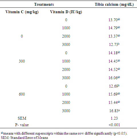
Effect of dietary inclusion of vitamin C and D3 supplementation on bone characteristics of hens aged 70 weeks at the late laying stage. Lohman Brown (n=180) were randomly allotted to 12 dietary treatments in triplicates of 5 birds each. The formulated basal diet was supplemented with three levels of vitamin C (0, 300 and 600 mg/kg) and four levels of vitamin D3 (0, 1000, 2000 and 3000 IU/kg). The experiment was a 3x4 factorial arrangement in a completely randomised design. Two hens per replicate were selected, sacrificed and bone physico-chemical attributes assessed. Tibia length of hens on 2000 IU D3 (115.60) was significantly higher (P < 0.05) than those on 0 (114.76), 1000 (114.47) and 3000 IU D3 (113.66) supplementation. Bone robusticity index of hens on supplemental vitamin C were higher (P<0.05) at 0 mg (30.36) than at 300 (29.10) and 600 mg (28.60) while inclusion of D3 at 1000 (29.20), 2000 (29.32) and 3000 IU (28.86) were similar (P>0.05) but lower than when supplemented at 0 IU (30.04). Bone calcium, phosphorus, magnesium, potassium and ash differed significantly (P<0.05) at various inclusion levels of vitamin C and D3 while the effect of interaction of supplemental vitamin C and D3 on tibia bones of the hens was only significant (P<0.05) for tibia calcium. Tibia calcium ranged between 12.69 to 16.83% in hens on combined dietary supplement of 0 IU D3+ 600 mg vitamin C and 3000 IU D3+600 mg vitamin C, respectively. In conclusion, dietary supplements of vitamin C and D3 improved the physical and chemical characteristics of spent laying hens’ bone.
Keywords: Robusticity index, Tibia bone composition, Supplemental vitamin, Lohmann Brown hens, Laying hens bone attributes
Cite this paper: Ogunwole O. A., Adedeji B. S., Olumide M. D., Mosuro A. O., I. E. Adewemimo, Effects of Dietary Supplemental Ascorbic Acid and Cholecalciferol on Bone Characteristics of Hens at the Late Laying Stage, International Journal of Food Science and Nutrition Engineering, Vol. 8 No. 6, 2018, pp. 142-150. doi: 10.5923/j.food.20180806.02.
Article Outline
1. Introduction
- Deficiency and loss in bone mineral structure is a function of skeletal problem (osteoporosis) in modern highly productive laying chickens [1]. Bone fracture is one of the most serious skeletal problems in modern laying hens [2]. Randall and Duff [3] reported osteoporosis as the major reason for bone loss and subsequent fractures in laying hens. The role of nutrition in the maintenance of bone integrity cannot be undermined. Birds that have laid consistently over a long time are usually susceptible to bone brittleness and fracture. Adequate supplemental vitamin C or D3 may help reduce the incidence of skeletal abnormality often observed in spent birds. Diets of hens well fortified with the required nutrients for bone health will impact the skeletal integrity of hens at the late laying stage.Available reports on the roles of vitamins on skeletal integrity in laying hens were inconsistent. Earlier report by Newman and Leeson [4] revealed that dietary vitamin C had little effects on bone breaking strength of laying leghorn hens. Improved bone parameters were reported in broiler chickens [5] while [6] did not observe any influence of dietary vitamin C on tibia bone physical parameters. The influence of dietary vitamin D on tibia and femur bones of chickens has been similarly reported [7-10]. Increased bone strength was only achieved in broiler chickens when dietary vitamin C supplementation was concomitant with vitamin D [11]. Dietary ascorbic acid affects bone resorption marker enzymes [11] and thus its concentration may affect the integrity of bone matrix. Fritts and Waldroup [10] reported that vitamin C influences the conversion of vitamin D into its metabolic active form calcitriol which is essential for calcium and phosphorous regulation and calcification processes. However, information on the combined effect of dietary vitamin C and D3 on bone health is scanty in literature. Therefore, this study was aimed at assessing the effects of supplemental vitamin C and D3 on bone characteristics of spent laying hens.
2. Materials and Methods
2.1. Experimental Site
- The experiment was carried out at the Poultry Unit, Teaching and Research Farm, University of Ibadan, Ibadan, Nigeria. The university is located in Ibadan, in the tropical rain forest zone of Nigeria within latitude 7° 26 N and longitude 3° 54 E, with a mean altitude of 277 meters above sea level.
2.2. Experimental Animal and Diets
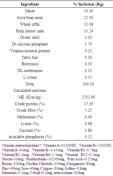
- A total of 180 Bovan brown hens aged 12 weeks were randomly allocated into 12 treatments of three replicates of five hens per replicate in a 3x4 factorial arrangement of a completely randomized design. The basal diet for the study was supplemented with three levels of vitamin C (0, 300 and 600 mg/kg) and four levels of vitamin D (0, 1000, 2000 and 3000 IU/kg) and their combination to produce 12 treatments. The basal experimental diet composition is shown in Table 1. The experiment lasted 60 weeks.
|
2.3. Data Collection
- At week 70, two hens per replicate were selected and sacrificed. They were carefully dissected into primal cuts. The left and right tibia of each bird was removed with the drumsticks and flesh intact. The drumsticks were labeled and immersed in boiling water (100°C) for 10 minutes. After cooling to room temperature, the drumsticks were defleshed manually and then air dried for 24 hours at room temperature. Bone physical parameters measured were: bone weight, bone length and robusticity index while bone chemical composition measured included calcium, phosphorus, magnesium and potassium. The bone weight/length index was obtained by dividing the tibia weight by its length. The tibiotarsal and robusticity indexes were determined according to Adebiyi et al. [12]. The percentage ash was determined relative to dry weight of tibia while calcium, phosphorus, magnesium and potassium composition were analysed according to AOAC [13].
2.4. Statistical Analysis
- Data were subjected to polynomial regression, descriptive statistics and analysis of variance (SAS, 2003) while means were separated using Tukey’s HSD option of the same package at α0.05.
3. Results
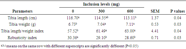
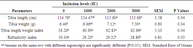
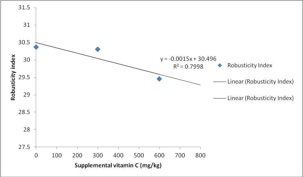
- The main effect of dietary supplementation of vitamin C on bone physical characteristics of laying chickens is shown in Table 2. Tibia length, tibia weight, tibia length/weight index and robusticity index differed significantly (P<0.05) across the treatments. Tibia length of hens fed supplemental vitamin C at 0 (116.70) and 300 mg (114.35) were similar (P>0.05). They were however higher (P<0.05) than 113.11 cm in hens fed 600 mg vitamin C. Tibia weight increased significantly (P<0.05) with supplemental vitamin C from 6.73g in hens fed 0 mg vitamin C to 7.04 and 7.11g in hens fed 300 and 600 mg vitamin C, respectively. Tibia length/weight index in hens on 0 mg dietary vitamin C (57.52) was significantly lower (P<0.05) than those on 300 (61.49) and 600 mg (63.00) supplemental vitamin C. Robusticity index was significantly higher (P<0.05) in hens on 0 mg dietary vitamin C (30.36) compared with 29.10 and 28.60 in hens fed 300 and 600 mg vitamin C, respectively. Robusticity index of bone in hens fed 300 mg (29.10) and 600 mg (28.60) dietary vitamin C were similar (P<0.05) but were lower (P<0.05) than those on 0 mg dietary vitamin C (30.36). The main effect of inclusion of dietary vitamin D3 supplementation on bone physical characteristics of laying chickens is shown in Table 3. Tibia length, tibia weight, tibia length/tibia weight, and robusticity index differed significantly (P<0.05) with dietary supplement of vitamin D3. Tibia length of hens on 0 IU (114.76) was not significantly different (P>0.05) from those on 1000 (114.47) and 3000 IU (113.66) vitamin D but were lower (P<0.05) than those on 2000 IU D3 (115.60). Tibia length of hens on 1000 and 2000 IU however were not significantly different (P>0.05). Tibia weight of hens on 0 IU (6.46) was similar (P>0.05) to those of hens on 1000 IU (6.96) but lower than 7.12 and 7.30 in hens on 2000 and 3000 IU, respectively. Tibia length/weight index of hens on 0 IU (56.26) was significantly lower compared with hens fed 1000 (60.90), 2000 (62.83) and 3000 IU D3 (62.69). Significantly higher (P<0.05) bone robusticity index was observed in hens fed 0 IU D3 (30.04) compared with 29.20, 29.32 and 28.86 in hens on 1000, 2000 and 3000 IU D3, respectively. The relationship between increased supplemental vitamin C in the diet of hens and the bone robusticity index was negatively correlated and is shown in Figure 1 and further explained by equation 1.
 | (1) |
 | (2) |
|
|
 | Figure 1. Relationship between supplemental vitamin C and bone robusticity index of laying hens |
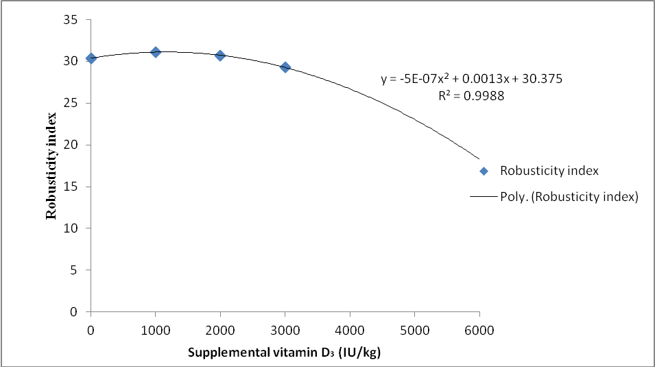 | Figure 2. Relationship between supplemental vitamin D3 and bone robusticity index of laying hens |
|
 | (3) |
 | (4) |
 | (5) |
 | (6) |
 | (7) |
 | (8) |
 | (9) |
 | (10) |
|
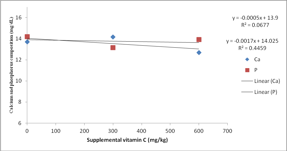 | Figure 3. Relationship between supplemental vitamin C and laying hen tibia calcium and phosphorus |
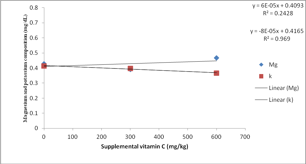 | Figure 4. Relationship between supplemental vitamin C and laying hen tibia magnesium and potassium |
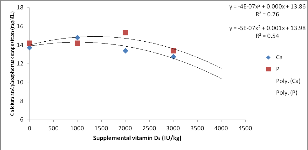 | Figure 5. Relationship between supplemental vitamin D3 and laying hens tibia calcium and phosphorus |
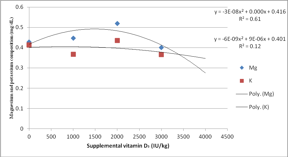 | Figure 6. Relationship between supplemental vitamin C and laying hens tibia magnesium and potassium |
|
4. Discussion
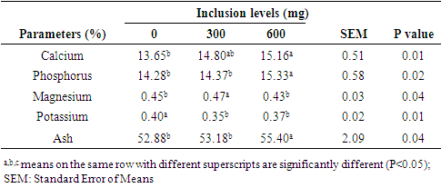
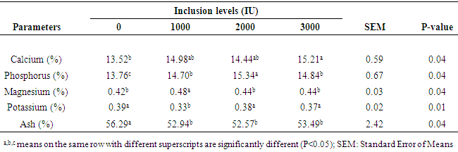
- Bone mineralisation has been studied with respect to processes (chemical and physical) that improves bone matrix as mineralisation affects bone strength with an implication on the ability of the skeleton to withstand gravity and additional loading. Biochemical parameters are direct indicators of bone quality which include bone density [15] and bone flexibility and strength [16]. Rao et al. [17] reported that different measurements can be used to determine bone mineralisation, structure and health in poultry. The improvements observed with respect to the bone parameters monitored in this study could be linked to the documented roles of both vitamins in bone mineralisation. Leeson and Summer [18] emphasized the crucial roles of vitamin C in the hydroxylation of proline residues necessary for synthesis of procollagen a precursor of bone formation. It has been recognized to play a role as a cofactor to several key enzymes involved in procollagen synthesis [19] and bioconversion of D3 to its metabolic active form [20]. Similarly, the importance of vitamin D3 in the absorption and utilization of calcium for bone development and strength have been reported [21, 22] reported improved bone resistance with vitamin C supplementation in chicks while Ogunwole [6] observed no beneficial effect of vitamin C on tibia bone physical parameters measured in broiler chickens. Conversely, Bruno et al. [7] and [5] observed decreased tibia and femur parameters with vitamin C supplementation in broiler chickens reared under heat stress. Fritts and Waldroup [10] reported increased bone weight while improvement in bone parameters was reported by Matilla et al. [23] with vitamin D supplementation in chickens. Sahin et al. [24] however observed no influence of vitamin D3 on bone improvement in laying quails. Higher bone weight/length index observed in this study with supplementation of vitamin C and D3 corroborated earlier report [25] that increased bone weight/length index indicated more bone density and increased bone strength. Similar observation was reported for the effect of vitamin C and vitamin D on robusticity index of the birds as supplementation increased. Lower robusticity index has been associated with increased bone strength [26] thus emphasising that vitamin C and D supplementation in this study favoured formation of stronger bone. Calcitriol the metabolically active form of vitamin D3, is a secosteroid hormone involved in Ca and P metabolism [27] while vitamin C is a key player in the bioconversion of vitamin D to this active form [20]. This role of vitamin C and D could be attributed to the increased bone Ca and P observed in this study. High rate of bone resorption during lay resulted in bone weakness at the end of their production cycle but with the inclusion of vitamin D3 there would be more calcium in the bone which reduced osteoporosis [28]. Although, there was increased calcium and phosphorus deposition in the tibia with the supplementation of either of the vitamins, magnesium and potassium content in the bone did not follow a particular trend thus emphasizing the importance of Ca and P as the key players in bone mineralization. The importance of vitamin D was further strengthened by Pereira [29] that vitamin D3 deficiency could impair bone formation and as such ascribed vitamin D, calcium and phosphorus as extremely important for bone mobilization and increased bone strength. Tibia ash was reportedly increased with supplemental vitamin D3 [10]. The effect of interaction of dietary vitamin C and D3 supplementation on bone chemical characteristics of laying chickens was only significant for bone calcium. This further suggests the synergistic role of vitamin C and D3 in improving calcium absorption and utilization. The involvement of vitamin C in the bioconversion of vitamin D had earlier been highlighted [20].The influence of vitamin C and D3 observed on increased tibia calcium deposition may be attributed to the several metabolic functions of calcium in poultry [30]. The high regression coefficients in Figures 2 and 3 suggest that robusticity index of the tibia was strongly dependent on supplemental vitamins C and D. This implied that 79.9 and 99.8% of the observed improvement in robusticity index were adduced to supplemental vitamin C and D3, respectively. As shown in Figure 3, it seems vitamin C supplementation was more favourable to phosphorus deposition in the tibia compared to calcium as 44.5% of the deposition of P in the bone was ascribed to supplemental vitamin C. A very strong relationship between vitamin C supplementation and K deposition (R2=0.96) compared with 0.24 for Mg deposition in the tibia suggests that increasing supplemental vitamin C would result in improved bone K deposition.Supplemental vitamin D3 on the other hand was more favourable to Ca deposition. Optimal tibia Ca and P were attained at 1400 IU supplementation of vitamin D3. Tibia magnesium and potassium improved with supplemental vitamin D3 as optimal tibia magnesium and potassium were attained at 1600 and 1000 IU of supplemental vitamin D3, respectively. This implied that supplementation of vitamin D3 beyond these levels would reduce deposition of these minerals in the bone of laying hens. Though tibia calcium in hens on the control was similar (P>0.05) to those of hens from other treatment combinations, hens on combination of 600 mg ascorbic acid and 3000 IU D3 had higher tibia calcium than those on sole supplementation of 600 mg ascorbic acid and 3000 IU cholecalciferol, respectively. This observation suggests a synergy between these vitamins for tibia calcium deposition when combined at higher levels in the diets of hens.
5. Conclusions
- In conclusion, supplemental vitamin C and D3 improved physical and chemical characteristics of tibia bone in laying hens while the interaction of both vitamins in the diet of laying hens increased tibia calcium deposition.
ACKNOWLEDGEMENTS
- Authors appreciate the Tertiary Education Trust Fund (TETFUND) for their provision of research materials for this study.