International Journal of Diabetes Research

p-ISSN: 2163-1638    e-ISSN: 2163-1646

2016;  5(4): 75-85

doi:10.5923/j.diabetes.20160504.03


Role of Adipokines in Controlling Insulin Signaling Pathways in Type-2 Diabetes and Obesity. Current and Future Perspectives

Tasneem Bouzid 1, Frederick G. Hamel 2, 3, Jung Yul Lim 1, 4

1Department of Mechanical and Materials Engineering, University of Nebraska-Lincoln, Lincoln, NE, USA

2Departments of Internal Medicine and Pharmacology and Experimental Neuroscience, University of Nebraska Medical Center (UNMC), Omaha, NE, USA

3Research Service (151), Veterans Affairs Nebraska-Western Iowa Health Care System, Omaha, NE, USA

4The Graduate School of Dentistry, Kyung Hee University, Seoul, South Korea

Correspondence to: Jung Yul Lim , Department of Mechanical and Materials Engineering, University of Nebraska-Lincoln, Lincoln, NE, USA.

Email:

Copyright © 2016 Scientific & Academic Publishing. All Rights Reserved.

This work is licensed under the Creative Commons Attribution International License (CC BY).
http://creativecommons.org/licenses/by/4.0/

Abstract

Adipose cells produce several types of cytokines, known as adipokines, which influence a variety of endocrine signaling pathways. Specifically, several adipokines have been proposed to play a vital role in regulating insulin signaling pathways within various tissues in the body and may thus provide a molecular link between obesity and type 2 diabetes mellitus (type-2 DM). Adiponectin and leptin adipokines have been demonstrated to improve insulin sensitivity, while other adipokines such as resistin, tumor necrosis factor-α (TNF-α), and interleukin-6 (IL-6) have been evidenced to induce insulin resistance. The low-grade chronic inflammation and greater adipose deposition accompanied by obesity may lead to dysregulation in the production and secretion of adipokines, thus introducing metabolic complications. In this mini review, we will highlight the findings on the role of adipokines in insulin sensitivity/resistance in an effort to better understand the mechanism of type-2 DM. Then, we will provide a perspective on the potential control of adipokine secretion by extracellular mechanical loading cue. This is based on the reported correlation between the whole body exercise and circulating adipokine level. The understanding of cellular level mechanism of the mechanical loading control of adipokine biology may provide a new and improved insight into the missing link among exercise, obesity, and type-2 DM.

Keywords: Adipocytes, Adipokine, Type 2 diabetes, Insulin sensitivity, Insulin resistance, Mechanical loading

Cite this paper: Tasneem Bouzid , Frederick G. Hamel , Jung Yul Lim , Role of Adipokines in Controlling Insulin Signaling Pathways in Type-2 Diabetes and Obesity. Current and Future Perspectives, International Journal of Diabetes Research, Vol. 5 No. 4, 2016, pp. 75-85. doi: 10.5923/j.diabetes.20160504.03.

1. Introduction

Obesity greatly increases the risk for associated metabolic diseases, including type 2 diabetes mellitus (type-2 DM). Although adipose tissue was thought to serve mainly to store energy in the form of triacylglycerides, numerous studies spanning the last 20 years have provided evidence that adipose also behaves as an endocrine organ by secreting bioactive cytokines known as adipokines. Adipokines have been implicated in mediating insulin signaling pathways in various tissues within the body [1]. The chronic low-grade inflammation and corresponding increase in macrophage infiltration, the characteristics of obese white adipose tissue (WAT) [2], correlate with increased adipocyte secretion of pro-inflammatory molecules such as resistin, tumor necrosis factor-α (TNF-α), and interleukin-6 (IL-6) [1]. Such molecules impair the ability of tissues to correctly sense and respond to insulin (i.e., causing insulin resistance) via various mechanisms that intervene in glucose transport and lipid metabolism. Conversely, adipocytes are also capable of secreting insulin-sensitizing molecules such as adiponectin and leptin, which improve the response of tissues to insulin and glucose [3].
Insulin instigates its metabolic effects by first binding to the insulin receptor (IR) in the plasma membrane, which initiates a phosphorylation cascade. IR is a tetrameric membrane protein consisting of two αβ dimers linked by disulfide bonds and belongs to a family of tyrosine kinases which includes insulin-like growth factor (IGF)-I receptor and the insulin receptor-related receptor (IRR) (see reviews [4-6]). Alternative exon splicing leads to tissue-specific synthesis of two different IR isoforms, IRa and IRb, which have different binding affinities to insulin [5]. IGF-I and IRR are also capable of forming functional complexes with insulin upon binding [4].
Insulin binds to IR at the extracellular α-subunit, which then derepresses the kinase activity of intracellular β-subunit [4-5]. Conformational change and transphosphorylation of specific tyrosine residues on the β-subunit further increases kinase activity, leading to the recruitment of intracellular docking proteins such as insulin-receptor-substrate-1 (IRS-1), -2 (IRS-2), Src-homology/collagen (Shc), and growth factor receptor-bound protein 2 (Grb2) [4-6]. The major substrate for IR in adipose and muscle tissues is IRS-1, which once phosphorylated mediates the association with and activation of phosphatidylinositol-3-kinase (PI3K) [4-7]. Serine phosphorylation caused by cytokines and fatty acid metabolites can inhibit IRS-1 signaling [7]. PI3K can catalyze the formation of phosphatidylinositol4,5biphosphate and phosphatidylinositol3,4,5triphosphate (PIP3), and the latter then recruits protein kinase-B (PKB, or Akt) by binding to its SHT domain [4, 6, 8]. Akt activation results in the translocation of glucose transporters to the membrane, leading to increased glucose uptake by the adipocyte cell. Glucose transporters (GLUT) are facilitative membrane transporter proteins that employ the diffusion gradient of glucose and other sugar molecules across the plasma membrane [9]. GLUT1 is primarily expressed in the brain, with moderate levels of expression in muscle, adipose tissue, and liver [9]. GLUT2, which is important in the sensing of glucose levels, is predominantly expressed in pancreatic β-cells, liver, and kidneys [9]. GLUT3 is largely found in the brain, and displays a strong affinity to for glucose [9]. The insulin-responsive GLUT4 is found in the heart, skeletal muscle, and adipose tissue [9]. The insulin-Akt pathway leads to the translocation of GLUT-4-containing vesicles to the plasma membrane, causing an immediate 10 to 20-fold increase in glucose uptake [9]. Other metabolic downstream effects of the IRS in adipocytes include reduced fatty acid secretion and regulation of the expression of genes important in adipocyte differentiation and lipogenesis [1, 4]. The mitogenic response to insulin is transduced via the mitogen-activated protein kinase (MAPK) cascade, in which Grb2 interacts with IRS and associates with the guanine nucleotide exchange factor son-of-sevenless (SOS) [6, 8]. However, Grb can activate the MAPK pathway independently from IRS by associating with activated Shc [6]. Insulin resistance associated with type-2 DM is a result of various disturbances in the intracellular signaling cascade mentioned above. These include reductions in receptor concentration, kinase activity, IRS phosphorylation, and GLUT transportation [4].
Numerous extracellular factors can influence obesity and related adipocyte metabolism and macrophage infiltration. Interestingly, recent research provides increasing evidence of the importance of mechanical loading cue in regulating adipocyte function, particularly in cellular adipogenesis and lipid accumulation and potentially on adipose tissue metabolism and inflammation (see other reviews [10, 11]). In this mini review, we will first briefly highlight the roles that adipokines (adiponectin, leptin, resistin, TNF-α, and IL-6) have been shown to have in regulating insulin sensitivity and resistance. Then, we will explore recent literatures that provide evidence of the mechanical loading control of metabolic regulation in adipocytes. From this review, we expect to suggest a new perspective that extracellular mechanical loading environments of adipocytes may regulate insulin sensitivity/resistance and glucose metabolism via affecting adipokine secretion and its signaling activity.

2. Role of Adipokines in Insulin Signaling

2.1. Adiponectin

Adiponectin is an adipocyte-specific hormone known to be involved in a variety of metabolic, anti-inflammatory, and vasoprotective activities [12]. Originally termed Acrp30, adiponectin is also referred to as AdipoQ, apM1 (adipose most abundant gene transcript1), or GBP28 (gelatin-binding protein 28) [13]. Adiponectin, the result of apM1 gene, is highly expressed in adipose tissue with nominal expression in muscle and liver. Adiponectin occurs in circulation in the form of three oligomeric complexes for different target tissues and with varying physiological effects, e.g., trimers (low molecular weight, LMW), hexamers (middle molecular weight, MMW), and the oligomeric complexes composed of 18 protomers or more (high molecular weight, HMW) [12]. Adiponectin has four structural domains, N-terminal peptide, variable region, collagenous domain, and globular domain that binds to adiponectin receptors [14]. Upon cleavage by leukocyte elastase, the globular domain is liberated [15]. Then, adiponectin receptors 1 (AdipoR1) and 2 (AdipoR2) serve as receptors for full-length adiponectin and globular adiponectin, though the affinity may vary, e.g., AdipoR1 showing high affinity for globular adiponectin but very low affinity for full-length adiponectin [14].
The role of adiponectin in hepatic insulin resistance has been extensively tested, results from which have proposed adiponectin as a potential therapeutic option for patients with type-2 DM [16]. Studies have reported that adiponectin could suppress glucose production and increase insulin sensitivity [13, 14, 16-19]. As in Figure 1, adipocytes manipulated to overexpress adiponectin or incubated in adiponectin-conditioned media displayed increased insulin-stimulated glucose uptake [18]. In unfed mice, adiponectin could sensitize hepatic reactivity to insulin by reducing hepatic glucose production and thus serum glucose levels [13]. The insulin-sensitizing effect of thiazolidinedione (TZD), one of the anti-diabetic agents that promotes glucose homeostasis without increasing insulin release, was diminished in diabetic obese mice by a targeted mutation in the adiponectin gene, thus indicating that adiponectin may play a vital role in peroxisome proliferator-activated receptor γ (PPARγ)-mediated insulin sensitization [3]. Ligand-activated PPARγ heterodimerizes with the retinoid X receptor and regulates transcription by binding to specific PPARγ-responsive elements within the promoters of target genes via regulatory mechanisms that involve 5’-AMP-activated protein kinase (AMPK). This process is thought to eventually result in increased fatty acid oxidation, which may in turn divert the flux of free fatty acids from the muscle and liver to the adipose tissue [20, 21].
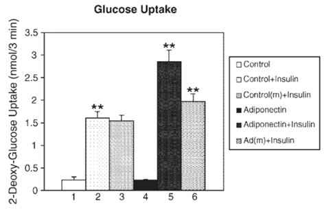
Figure 1. Mature adipocytes overexpressing adiponectin or incubated in adiponectin-conditioned medium (m) enhances their rate of glucose uptake in response to insulin. Means ± SEM from three separate experiments are shown. **: p < 0.01 for Adiponectin+Insulin vs. Control+Insulin and for Ad(m)+Insulin vs. Control(m)+Insulin. Reprinted with permission from American Society for Biochemistry and Molecular Biology [18]
Moreover, TZDs were shown in animal models to be effective inducers of adiponectin expression in muscle and adipose tissue of mice [22, 23] and in clinical patients with type-2 DM [24]. Studies have specifically implicated HMW adiponectin oligomers in reducing hepatic insulin resistance. For instance, in the presence of an insulin stimulator, pioglitazone, HMW adiponectin expression was significantly increased, which could be correlated with improved hepatic insulin activity [25]. Together, adiponectin can improve insulin sensitivity and has been proposed to be an effective target for developing anti-diabetic drugs. However, further research is required to determine the entire mechanism by which adiponectin regulates insulin signaling in combination with other diabetes controlling factors.

2.2. Leptin

The 16 kDa obese (ob) gene product, leptin protein, was discovered to be a critical regulator of body weight. Named from the Greek root leptόs meaning thin, leptin increased energy expenditure and reduced food intake in obese mice resulting in a 30% reduction in body weight over 2 weeks [26]. Leptin secreted from adipocytes have been suggested to have an important role in the regulation of a variety of body functions such as appetite, satiety, food intake, reproductive function, fertility, puberty, activity, energy expenditure, and atherogenesis (see the review [27]).
It was shown that the level of circulating leptin increased under increased caloric intake, and leptin resistance was produced after overfeeding [28]. The infusion of recombinant leptin in voluntarily overfed rats did not alter the contribution of gluconeogenesis to total glucose output, whereas the infusion nearly doubled it in control and pair-fed rats (Figure 2) [28]. Among obese humans, the leptin level strongly correlated with body mass index (BMI) [29]. Moreover, the correlation between leptin and insulin has been relatively well established to be dependent on obesity [30]. On the other hand, some studies reported a correlation less dependent on measures of obesity. For instance, it was demonstrated for lean rats that hyperinsulinemia can upregulate obese gene expression and hypoinsulinemia can downregulate it; while in obese rats, obese gene expression can only be upregulated by hyperinsulinemia [31]. Additionally, treating obese mice with leptin improved overall glucose clearance even prior to the weight loss [32].
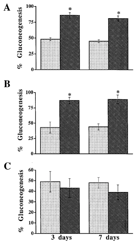
Figure 2. In control (A) and pair-fed (B) rats, moderate increases in circulating leptin levels caused a marked increase in gluconeogenesis relative to total glucose output. In overfed rats (C), increased leptin levels failed to change the contribution of gluconeogenesis to the total glucose output. , Lep-; , Lep+. Means ± SEM. *: p < 0.01 between Lep-and Lep+. Reprinted with permission from American Diabetes Association [28]
The mechanisms involved in hepatic insulin sensitization by leptin are not fully understood, but the effect may be mediated by the hypothalamus. A leptin receptor gene therapy in the arcuate nucleus (ARC) area of the hypothalamus in leptin receptor-deficient rats was shown to improve insulin sensitivity via suppressing hepatic glucose production rather than increasing glucose uptake [33]. In a study that investigated the expression and signaling of different isoforms of the leptin receptor (or obese receptor, OB-R) expressed in the hypothalamus, homozygous diabetes (db/db) mice showed a dramatic reduction in expression of the long isoform, while the short isoform was unable to activate signal transducer and activator of transcription (STAT) protein which is an important component in the ras/PI3K pathways [34]. The subcutaneous infusion of physiological level leptin enhanced insulin sensitivity in lipodystrophic mice, and this effect may be attributed to the diversion of fatty acids into adipose tissue for storage [35]. Interestingly, it was also demonstrated that PPARγ activators reduce obese gene expression both in vivo and in vitro, leading to the unwanted and counterproductive side effect caused by TZDs [36, 37, 38]. In summary, leptin is a multifunctional adipokine that primarily regulates body weight and food intake, and has been shown to reverse insulin desensitization. Additional efforts need to be made to fully understand the mechanisms by which leptin operates to regulate insulin signaling both in vitro and in vivo.

2.3. Resistin

Resistin is a member of cysteine-rich proteins known as found-in-inflammatory zone (FIZZ) or resistin-like molecules (RELM) which include RELM-α (FIZZ-1), RELM-β (FIZZ-2), and RELM-γ (FIZZ-3). Named for its ability to induce insulin resistance, resistin was discovered to be circulating in mouse serum with increased levels in both genetically- and diet-induced obese mice [39]. Interestingly, resistin has become a focus of several recent studies due to its potential to provide an insight into the possible mechanisms involved in insulin signaling [40].
In mice, resistin is secreted from adipose tissue and acts on skeletal muscle myocytes, hepatocytes, and adipocytes. Exposure to higher glucose concentrations significantly increased resistin expression in 3T3-L1 adipocytes, while treatment with insulin reduced it [41]. Resistin was found to adjust flux through G6Pase by regulating AMPK phosphorylation in the liver, indicating a possible overlap in the mechanism of adipose-derived hormones such as adiponectin (described above) [14, 40]. For example, resistin caused a decrease in AMPK phosphorylation and thus the rate of gluconeogenesis in mice [40]. Moreover, significant decreases in the phosphorylation of Akt and glycogen synthase kinase3 (GSK3), key effectors of the insulin signaling pathway, in human liver cells were induced with resistin in a dose-dependent manner [42].
Although the insulin desensitizing effects of resistin in rodents is well documented, studies investigating the link between resistin and diabetes in humans are conflicting. Both clinically and experimentally, researchers found little or no relationship between resistin and insulin resistance. Resistin was expressed in the adipose tissue from obese patients [43], while other researchers showed that isolated adipocytes express very low resistin mRNA levels [44, 45]. The inconsistency may be due to the differences in the genomic organizations of mouse and human resistin in that human resistin is only 59% identical to its mouse counterpart at the amino acid level [46]. On the other hand, resistin was revealed to be relatively more abundant in peripheral mononuclear cells [45]. Thus, the higher levels of resistin found in the adipose tissue may originate from a greater proportion of mononuclear cells obtained from patients with severe obesity rather from the adipose cell itself [1, 2, 45]. Together, resistin affects insulin sensitivity by regulating the activity of Akt and GSK3, key intermediates of the insulin signaling. However, results from both in vivo and in vitro human studies showed inconsistency, potentially due to differences in the genetic sequence code. More studies to clarify such contradiction would be helpful for a potential clinical application of resistin.

2.4. TNF-α

In 1975, it was discovered that endotoxins induced the host to release a substance, which was called tumor necrosis factor, TNF [47]. TNF-α-converting enzyme (TACE) cleaves the 26-kDa precursor into the secreted 17-kDa form under exposure to stimuli that triggers inflammatory reaction such as oxidative stress [48]. In addition to inflammatory cells such as macrophages and lymphocytes, non-inflammatory cells including adipocytes have also been evidenced to express TNF-α mRNA [49]. Thus, as a primary pro-inflammatory cytokine, TNF-α has been of interest for its possible effects on adipogenesis, lipid metabolism, and insulin sensitivity [50, 51].
Numerous studies have established for TNF-α a role to induce insulin resistance in both animals and humans [49-57]. Endogenous adipose expression of TNF-α was observed higher in obese humans in comparison with controls [49, 52], having a positive correlation with BMI [58]. In a study that transplanted TNF-α-deficient (KO) mice maintained on a high-fat diet with either TNF-α-sufficient (TNF-α+/+) or TNF-α-deficient (TNF-α-/-) bone marrow, KO TNF-α-/- mice exhibited increased insulin sensitivity (Figure 3) as well as decreased PPARγ mRNA levels [51]. Recently, patients with non-diabetic rheumatoid arthritis showed improved insulin sensitivity in response to anti-TNF-α therapy [53]. Moreover, TNF-α receptor (TNFR)-immunoglobulin G (IgG) treated mice required substantially more glucose to maintain normal glucose level [49], and the administration of TNF-α reversed the insulin effects on glycemic levels [54].
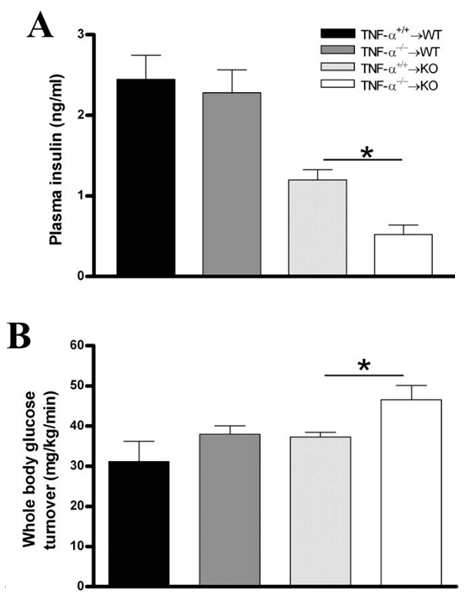
Figure 3. Wild-type and knock-out mice underwent bone marrow transplantation and received either or bone marrow from donor mice. The mice were put on a high-fat diet (HFD) for 26 weeks. Measured plasma insulin level (*: p = 0.002; A) and whole body glucose turnover (*: p = 0.02; B) indicate that mice lacking TNF-α had the highest insulin sensitivity. Reprinted with permission from the American Physiological Society [51]
Whereas the exact mechanism of TNF-α control of the insulin signaling is still yet to be fully understood, studies have pointed to the phosphorylation of IRS-1. TNF-α levels were detected as higher in obesity, and TNF-α could act as a potent activator of JNK which phosphorylates IRS-1 at serine residues, effectively inactivating it [55]. As mentioned previously, the binding of insulin to IR causes auto-phosphorylation by an intrinsic tyrosine kinase, and IR then phosphorylates and activates IRS-1 [56]. The obese TNF-α-/- mice displayed a significant increase in the auto-phosphorylation of IR, suggesting IR mediation in the TNF-α control of insulin signaling [57]. Together, TNF-α, the prime pro-inflammatory cytokine, has consistently been shown to trigger insulin resistance both in vitro and in vivo. Further research on the regulatory mechanism may be needed for better understanding of the involvement of TNF-α in insulin sensitivity/resistance.

2.5. IL-6

IL-6 is a multifunctional cytokine that plays numerous roles in addition to controlling immune cell function, such as behaving as a hepatocyte-stimulating factor and a growth factor for human myeloma and promoting the transition of hematopoietic stem cells from the G0 to G1 phase [59]. The binding of IL-6 to its low affinity receptor, IL-6 receptor-α (IL-6Rα), forms a high affinity hexameric complex with two gp130 proteins to activate the JAK/Tyk tyrosine kinase and STAT pathways [60]. IL-6Rα is abundantly detected on the surface of certain cells including macrophages, neutrophils, myocytes, and hepatocytes [61].
There has been an interest in clarifying the potential role of IL-6 in type-2 DM and insulin sensitivity. Although patients with type-2 DM have shown elevated circulating IL-6 [62], results from studies examining the correlation between IL-6 and insulin resistance have not been fully consistent. IL-6 has been shown to activate SOCS-1 and -3 proteins in the liver, thus accompanying insulin resistance [63, 64]. However, another study reported that while IL-6 increased SOCS-3 expression in murine myotubes, glucose uptake was increased rather than decreased [65]. It was shown that prolonged IL-6 exposure caused insulin resistance in both differentiating and fully differentiated adipocytes accompanied by decreased expression of PPARγ [66]. As in Figure 4, the treatment of differentiating preadipocytes with IL-6 significantly reduced both lipogenesis and glucose transport. Moreover, IL-6 could also downregulate the expression of adiponectin, the insulin signaling sensitizer as described above, in 3T3-L1 adipocytes [67]. On the other hand, a recent study showed that global deletion of IL-6 in mice induced maturity-onset obesity, liver inflammation, and whole-body insulin resistance [68].
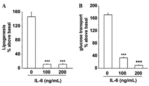
Figure 4. Administration of IL-6 (100 and 200 ng/mL) during differentiation of 3T3-F442A preadipocytes greatly reduces (A) lipogenesis and (B) glucose transport at maturity. Each bar represents mean ± SEM of 3-4 experiments performed in duplicate. All values are expressed as percentages above basal (control) measurements. ***: p < 0.001. Reprinted with permission from Elsevier [66]
Data implicating IL-6 in obesity-associated insulin resistance may be explained by its established function in inflammation and immunity, as obesity can be described as systemic low-grade inflammation. In regard to mechanisms, IL-6 was shown to enhance fatty acid oxidation in both healthy and type-2 DM humans, mice, L6 myotubes, and 3T3-L1 adipocytes via AMPK [65, 69]. However, such results were obtained from in vitro studies at supra-physiological IL-6 concentrations and studies with mice that have IL-6 displaying only ~42% sequence identity with human IL-6. Interestingly, some studies reported no significant changes in glucose homeostasis in response to an acute administration of IL-6 [70]. Such a contradictory nature of IL-6 can be attributed partly to its complex signaling mechanism. IL-6 is mediated by trans-signaling, in which IL-6R is cleaved by metalloproteinases resulting in a soluble form of IL-6R, sIL-6R. This may allow IL-6 to signal in cells that express gp130 but not the receptor itself in the plasma membrane [70]. To summarize, the role of IL-6 in the development of insulin resistance is still not clear. Evidences suggest a positive, negative, or neutral effect on insulin sensitivity for various tissues and for rodent or human models. The effect also appears to depend on whether IL-6 is administered acutely or chronically, where chronic exposure leads to insulin resistance.

3. Perspective on the Potential Role of Mechanical Environment in Adipokine Secretion and Insulin Sensitivity

Physical exercise has been suggested as a powerful tool not only to control body weight but also to improve systemic insulin sensitivity. The anabolic control of insulin signaling activity through dynamic physical exercise can be achieved through enhancing glucose transport, regulating hepatic glucose output, and repairing lipid metabolism. Studies have tested the effects of human exercise on the expression of adipokines (see detailed reviews in [71, 72]). These include testing of both moderate aerobic exercise and resistance training protocols incorporating cycling, running, and ramp exercises [73-75]. These studies have reported that regular exercise training, sometimes even after a single bout of exercise, may cause a reduction of pro-inflammatory adipokine expression while inducing insulin sensitivity supporting adipokine secretion. Obese young women, after the exercise program, showed significantly increased level of circulating adiponectin while displaying decreased circulating TNF-α (Figure 5) [73]. On the other hand, the level of leptin, another insulin sensitizing adipokine, decreased after exercise. While it is not fully established, it may be hypothesized that mechanical loading applied to the adipose tissue during exercise may play a role in the exercise control of insulin sensitivity/resistance via affecting the level of insulin signaling regulatory adipokine secretion.
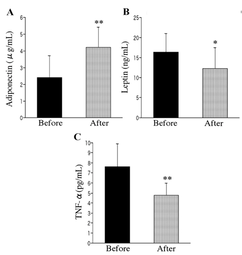
Figure 5. Levels of circulating adiponectin (A), leptin (B), and TNF-α (C) in obese young women before and after a single bout of moderate intensity exercise. The results indicate that the exercise increases plasma adiponectin while decreasing plasma leptin and TNF-α. Each bar represents mean ± SEM. *: p < 0.05 and **: p < 0.01. Reprinted with permission from the Japan Endocrine Society [73]
Regardless of these investigations on the correlation between exercise and adipokine secretion and circulation at the whole body level, surprisingly, there is very little research focusing on the cellular level connection. Studies applying mechanical stimulations, e.g., stretch, compression, and fluid shear, to adipocytes or their precursor cells have only examined the effects of mechanical stimuli on their adipocytic commitment and differentiation (see the review [76]). Since the circulating adipokine level is reported to be altered by the whole body exercise as described above, the functional activity of adipocytes to produce various adipokines is expected to be affected by extracellular mechanical loading environments. Altered adipokine secretion, in both repertoire and amount, by the adipocytes in response to external mechanical loading may then play a role in affecting insulin sensitivity/resistance. Such a mechanism based on what may be called ‘adipokine mechanobiology’ may provide a new and advanced insight into the adipokine control of insulin signaling.
Cellular mechanotransduction, or the translation of external mechanical stimuli into cellular biomechanical signals, involves mechanical force-based conformational and functional changes in mechanosensitive proteins, membrane, cytosol, and nucleus. Mechanotransduction plays a key role in the homeostasis and regeneration of many cell types and tissues (see our review [77]). In case cellular mechanotransduction activities malfunction, the physiology of many known mechanically functional cells is disrupted, often leading to various diseases and even cancer [78]. The mechanotransduction research has mostly focused on established “mechanically functional” cells such as muscle, bone, cartilage, and blood vessel cells or as a tool to achieve multipotent stem cell differentiation toward these known mechanosensitive cell types [77-79].
The concept of mechanotransduction has recently been applied to adipocytes but only focusing on commitment and differentiation [76], as noted above. Under the static bodyweight load and weight-bearing or from the whole body movement/exercise, cells in the adipose tissue are to be physiologically exposed to compound external mechanical stimulations including tension, compression, pressure, shearing, and their combinations [76]. It would thus be natural to suppose that adipocyte function and fate would also be affected by external mechanical loading similar to the cases of other known mechanically functional cells. To introduce the key data of adipocytic mechanotransduction, studies demonstrated that adipogenesis encounters an inhibitory effect from cyclic stretching. It was shown that cyclic mechanical cell stretching downregulated the terminal adipogenesis of 3T3-L1 preadipocytes [80, 81]. Umbilical cord perivascular cells also displayed suppressed adipogenesis when exposed to cyclic stretch [82]. For mesenchymal stem cells, it was evidenced that cyclic stretch may direct their fate by enhancing osteogenesis at the expense of decreasing adipogenesis [83]. The molecular mechanism of the cyclic stretch inhibition of adipogenesis may be accomplished via the mechanosensitive upregulation in extracellular signal-regulated kinase (ERK) [80, 83], cyclooxygenase-2 (Cox-2) [81], TGFβ1/Smad signaling [82], and β-catenin signaling [84]. In our recent study [85], bone morphogenetic protein 4 (BMP4) induction of mesenchymal stem cell adipocytic commitment and differentiation could be suppressed by cyclic cell stretching and this was observed to be mediated by ERK activation (Figure 6) but not through the downregulation of BMP4 signaling through Smad or p38 cascade.
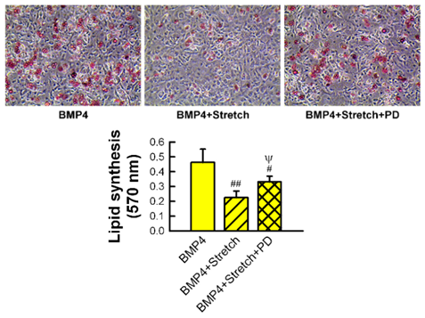
Figure 6. Bone morphogenetic protein 4 (BMP4)-induced lipid synthesis in murine MSCs during adipogenic differentiation is impeded by cyclic stretching. The inhibitory effect of cyclic stretch on adipocytic differentiation is reversed by the addition of ERK inhibitor, PD98059 (PD). Oil red O stained images and lipid quantification by spectrophotometer. Comparison with BMP4 (#, ##) and BMP4 plus stretch (ψ) shown at p < 0.05 (#, ψ) and p < 0.01 (##). Reprinted with permission from Elsevier [85]
Interestingly, it was proposed for 3T3-L1 preadipocytes that static stretching, i.e., elongation of the cells to a certain strain then keeping the strain for prolonged time, might provide an opposite effect contrast to the cyclic stretching. When 3T3-L1 cells were exposed to static stretching, they actually showed promoted adipogenesis [86, 87]. This was attributed to the upregulation of RhoA and Rho-kinase (ROCK) signaling under static stretching [86]. The combined data may lead to an intriguing question on the role of cyclic vs. static stretching in the adipose mechanobiology, which may respectively function as an inhibitor vs. promoter of adipocytic commitment and differentiation (as in our recent perspective article [88]). See Figure 7 for potential mechanistic signaling pathways reported so far to govern the effects of cyclic vs. static stretching on adipogenic differentiation [76].
These mechanical loading data in adipocytic commitment and differentiation may be further tested in terms of adipokine mechanobiology. Based on the fact thatadipocytes are mechanically responsive as just described, how mechanical loading will affect adipocytes in the adipokine secretion may be investigated as a missing link among exercise, obesity, and insulin sensitivity. Such a question has the same context with the reported correlation between whole body exercise and adipokine biology as described above, but may provide unprecedented data at the cellular level. Knowing the repertoire and secretion level of various adipokines by adipocytes under external mechanical loading will significantly improve the understanding of how exercise will affect the level of circulating adipokines and then insulin signaling. Recalling that the mode of mechanical loading, e.g., static vs. cyclic stretch, is a critical factor to differentially control cellular adipogenesis, exploring the potential differences in adipokine secretion profile under various loading regimens (static vs. cyclic, strain, frequency, duration, rest period, etc.) would be required. The data can be further correlated with the effect of exercise mode in influencing circulating adipokines and insulin signaling. Taken together, the study of adipocyte level cascades of the mechanical loading control of adipokine biology in regulating insulin signaling may suggest a new research topic, i.e., adipokine mechanobiology, and this approach may solve the missing link between obesity and insulin-mediated metabolic syndrome, type-2 DM.
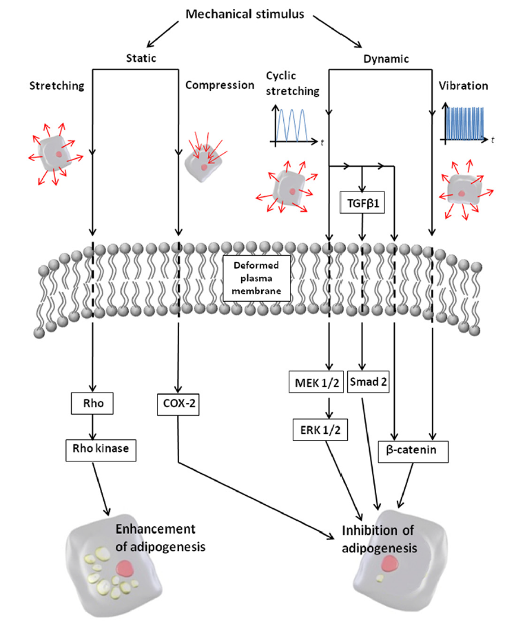
Figure 7. Potential mechanotransduction pathways involved in cellular adipogenesis. Static stretching activates the Rho and Rho Kinase (ROCK) signaling pathway, leading to enhanced adipogenesis. Cyclic stretching activates ERK, TGFβ1/Smad, and β-catenin pathways, leading to reduced adipogenesis. Reprinted with permission from Elsevier [76]

4. Conclusions

Researches provide strong evidence that adipose tissue plays important roles outside of energy storage. Adipose tissue has been shown to behave as an endocrine organ capable of regulating many physiological processes by the secretion of protein factors known as adipokines. Of great clinical relevance to type-2 DM in particular are adiponectin, leptin, resistin, TNF-α, and IL-6. Adiponectin has long been of great interest to researchers as an insulin-sensitizer and a potential target of anti-diabetic drugs, and leptin is a multifunctional protein that has been shown to reverse insulin resistance. Resistin, on the other hand, has been shown to target key nodes of the insulin signaling pathway to induce insulin desensitization. TNF-α is a pro-inflammatory cytokine thought to be increased in obese individuals due to the chronic low-grade inflammation associated with obesity, and has been shown to also induce insulin resistance. While further research is still required, IL-6, a multifunctional cytokine, has been shown to alter insulin sensitivity both in vivo and in vitro studies. Recent advances in research indicate that physical exercise may reduce pro-inflammatory adipokine expression while improving insulin sensitivity by causing an increase in the secretion of insulin regulatory adipokines. Furthermore, research is providing increasing evidence that adipose tissue is mechanically responsive, much like muscle and bone. Thus, it may be of interest to investigate the cellular level effect that mechanical loading environment has on the adipokine secretory response of adipocytes in an effort to further illuminate the role of adipokines in insulin signaling. The broader implications of such research include a more advanced understanding of type-2 DM and revealing potential novel targets for anti-diabetic drugs.

ACKNOWLEDGEMENTS

This literature review was supported by NSF Graduate Research Fellowship 1610400 (awarded to Bouzid), Nebraska Tobacco Biomedical Seed Grant (awarded to Hamel and Lim), NSF CAREER Award 1351570, Nebraska Research Initiative, and UNL Layman New Directions Awards (all awarded to Lim). We also thank the support from the Department of Veterans Affairs (for Hamel).

References

[1]  Jung UJ, Choi M (2014) Obesity and its metabolic complications: The role of adipokines and the relationship between obesity, inflammation, insulin resistance, dyslipidemia and nonalcoholic fatty liver disease. 15: 6184-6223.
[2]  Xu H, Barnes GT, Yang Q, Tan G, Yang D, et al. (2003) Chronic inflammation in fat plays a crucial role in the development of obesity-related insulin resistance. J Clin Invest 112: 1821-1830.
[3]  Blüher M, Mantzoros CS (2015) From leptin to other adipokines in health and disease: Facts and expectations at the beginning of the 21st century. Metab Clin Exp 64: 131-145.
[4]  Saltiel AR, Kahn CR (2001) Insulin signalling and the regulation of glucose and lipid metabolism. Nature 414: 799-806.
[5]  White MF (2003) Insulin signaling in health and disease. Science 302:1710-11.
[6]  Iliya IA, Mohammed B, Akuyam SA, Nok AJ, Bauchi ZM, et al. (2016) Molecular mechanisms of insulin signaling. Sub-Saharan Afr J of Med 3:3.
[7]  Hansen BC, Bray GA (2008) Chap. Insulin signaling in adipocytes and the role of inflammation, in “The Metabolic Syndrome: Epidemiology, Clinical Treatment, and Underlying Mechanisms”. Springer Science & Business Media.
[8]  Copps KD, White MF (2012) Regulation of insulin sensitivity by serine/threonine phosphorylation of insulin receptor substrate proteins IRS1 and IRS2. Diabetologia 55:2565-82.
[9]  Wood IS, Trayhurn P (2003) Glucose transporters (GLUT and SGLT): expanded families of sugar transport proteins. Br J Nutr 89:3-9.
[10]  Franz T (2014) Does cell mechanics in adipogenesis offer new keys for the prevention and management of obesity? Biophys J 106: 1231-1232.
[11]  Lee K, Kuo CK (2015) Chap. Extracellular matrix remodeling and mechanical stresses as modulators of adipose tissue metabolism and inflammation, in “The mechanobiology of obesity and related diseases”. Edited by Gefen A and Benayahu D, Springer International Publishing.
[12]  Simpson F, Whitehead JP (2010) Adiponectin-It’s all about the modifications. Int J Biochem Cell Biol 42: 785-788.
[13]  Berg AH, Combs TP, Du X, Brownlee M, Scherer PE (2001) The adipocyte-secreted protein Acrp30 enhances hepatic insulin action. Nat Med 7: 947-953.
[14]  Yamauchi T, Kamon J, Ito Y, Tsuchida A, Yokomizo T, et al. (2003) Cloning of adiponectin receptors that mediate antidiabetic metabolic effects. Nature 423: 762-769.
[15]  Waki H, Yamauchi T, Kamon J, Kita S, Ito Y, et al. (2005) Generation of globular fragment of adiponectin by leukocyte elastase secreted by monocytic cell line THP-1. Endocrinology 146: 790-796.
[16]  Yamauchi T, Kamon J, Waki H, Terauchi Y, Kubota N, et al. (2001) The fat-derived hormone adiponectin reverses insulin resistance associated with both lipoatrophy and obesity. Nat Med 7: 941-946.
[17]  Vrachnis N, Belitsos P, Sifakis S, Dafopoulos K, Siristatidis C, et al. (2012) Role of adipokines and other inflammatory mediators in gestational diabetes mellitus and previous gestational diabetes mellitus.
[18]  Fu Y, Luo N, Klein RL, Garvey WT (2005) Adiponectin promotes adipocyte differentiation, insulin sensitivity, and lipid accumulation. J Lipid Res 46: 1369-1379.
[19]  Baranova A, Gowder SJ, Schlauch K, Elariny H, Collantes R, et al. (2006) Gene expression of leptin, resistin, and adiponectin in the white adipose tissue of obese patients with non-alcoholic fatty liver disease and insulin resistance. Obes Surg 16: 1118-1125.
[20]  Nawrocki AR, Rajala MW, Tomas E, Pajvani UB, Saha AK, et al. (2006) Mice lacking adiponectin show decreased hepatic insulin sensitivity and reduced responsiveness to peroxisome proliferator-activated receptor gamma agonists. J Biol Chem 281: 2654-2660.
[21]  Hardie DG, Carling D, Carlson M (1998) The AMP-activated/SNF1 protein kinase subfamily: Metabolic sensors of the eukaryotic cell? Annu Rev Biochem 67: 821-855.
[22]  Tsuchida A, Yamauchi T, Takekawa S, Hada Y, Ito Y, et al. (2005) Peroxisome proliferator-activated receptor (PPAR) alpha activation increases adiponectin receptors and reduces obesity-related inflammation in adipose tissue: Comparison of activation of PPARalpha, PPARgamma, and their combination. Diabetes 54: 3358-3370.
[23]  Liu Y, Chewchuk S, Lavigne C, Brule S, Pilon G, et al. (2009) Functional significance of skeletal muscle adiponectin production, changes in animal models of obesity and diabetes, and regulation by rosiglitazone treatment. Am J Physiol Endocrinol Metab 297: E657-64.
[24]  Bajaj M, Suraamornkul S, Piper P, Hardies LJ, Glass L, et al. (2004) Decreased plasma adiponectin concentrations are closely related to hepatic fat content and hepatic insulin resistance in pioglitazone-treated type 2 diabetic patients. J Clin Endocrinol Metab 89: 200-206.
[25]  Li Y, Ding L, Hassan W, Abdelkader D, Shang J (2013) Adipokines and hepatic insulin resistance. J Diabetes Res 2013: 170532.
[26]  Halaas JL, Gajiwala KS, Maffei M, Cohen SL, Chait BT, et al. (1995) Weight-reducing effects of the plasma protein encoded by the obese gene. Science 269: 543-546.
[27]  Fasshauer M, Bluher M (2015) Adipokines in health and disease. Trends Pharmacol Sci 36: 461-470.
[28]  Wang J, Obici S, Morgan K, Barzilai N, Feng Z, et al. (2001) Overfeeding rapidly induces leptin and insulin resistance. Diabetes 50: 2786-2791.
[29]  Zimmet P, Hodge A, Nicolson M, Staten M, de Courten M, et al. (1996) Serum leptin concentration, obesity, and insulin resistance in western samoans: Cross sectional study. BMJ 313: 965-969.
[30]  Dagogo-Jack S, Fanelli C, Paramore D, Brothers J, Landt M (1996) Plasma leptin and insulin relationships in obese and nonobese humans. Diabetes 45: 695-698.
[31]  Cusin I, Sainsbury A, Doyle P, Rohner-Jeanrenaud F, Jeanrenaud B (1995) The ob gene and insulin. A relationship leading to clues to the understanding of obesity. Diabetes 44: 1467-1470.
[32]  Harris RB, Zhou J, Redmann SM,Jr, Smagin GN, Smith SR, et al. (1998) A leptin dose-response study in obese (ob/ob) and lean (+/?) mice. Endocrinology 139: 8-19.
[33]  German J, Kim F, Schwartz GJ, Havel PJ, Rhodes CJ, et al. (2009) Hypothalamic leptin signaling regulates hepatic insulin sensitivity via a neurocircuit involving the vagus nerve. Endocrinology 150: 4502-4511.
[34]  Ghilardi N, Ziegler S, Wiestner A, Stoffel R, Heim MH, et al. (1996) Defective STAT signaling by the leptin receptor in diabetic mice. Proc Natl Acad Sci U S A 93: 6231-6235.
[35]  Shimomura I, Hammer RE, Ikemoto S, Brown MS, Goldstein JL (1999) Leptin reverses insulin resistance and diabetes mellitus in mice with congenital lipodystrophy. Nature 401: 73-76.
[36]  De Vos P, Lefebvre AM, Miller SG, Guerre-Millo M, Wong K, et al. (1996) Thiazolidinediones repress ob gene expression in rodents via activation of peroxisome proliferator-activated receptor gamma. J Clin Invest 98: 1004-1009.
[37]  Kallen CB, Lazar MA (1996) Antidiabetic thiazolidinediones inhibit leptin (ob) gene expression in 3T3-L1 adipocytes. Proc Natl Acad Sci U S A 93: 5793-5796.
[38]  Larsen TM, Toubro S, Astrup A (2003) PPARgamma agonists in the treatment of type II diabetes: is increased fatness commensurate with long-term efficacy? Int J Obes Relat Metab Disord 27: 147-161.
[39]  Steppan CM, Bailey ST, Bhat S, Brown EJ, Banerjee RR, et al. (2001) The hormone resistin links obesity to diabetes. Nature 409: 307-312.
[40]  Muse ED, Obici S, Bhanot S, Monia BP, McKay RA, et al. (2004) Role of resistin in diet-induced hepatic insulin resistance. J Clin Invest 114: 232-239.
[41]  Shojima N, Sakoda H, Ogihara T, Fujishiro M, Katagiri H, et al. (2002) Humoral regulation of resistin expression in 3T3-L1 and mouse adipose cells. Diabetes 51: 1737-1744.
[42]  Zhou L, Sell H, Eckardt K, Yang Z, Eckel J (2007) Conditioned medium obtained from in vitro differentiated adipocytes and resistin induce insulin resistance in human hepatocytes. FEBS Lett 581: 4303-4308.
[43]  McTernan PG, McTernan CL, Chetty R, Jenner K, Fisher FM, et al. (2002) Increased resistin gene and protein expression in human abdominal adipose tissue. J Clin Endocrinol Metab 87: 2407.
[44]  Janke J, Engeli S, Gorzelniak K, Luft FC, Sharma AM (2002) Resistin gene expression in human adipocytes is not related to insulin resistance. Obes Res 10: 1-5.
[45]  Nagaev I, Smith U (2001) Insulin resistance and type 2 diabetes are not related to resistin expression in human fat cells or skeletal muscle. Biochem Biophys Res Commun 285: 561-564.
[46]  Ghosh S, Singh AK, Aruna B, Mukhopadhyay S, Ehtesham NZ (2003) The genomic organization of mouse resistin reveals major differences from the human resistin: Functional implications. Gene 305: 27-34.
[47]  Carswell EA, Old LJ, Kassel RL, Green S, Fiore N, et al. (1975) An endotoxin-induced serum factor that causes necrosis of tumors. Proc Natl Acad Sci U S A 72: 3666-3670.
[48]  Togashi N, Ura N, Higashiura K, Murakami H, Shimamoto K (2002) Effect of TNF-alpha--converting enzyme inhibitor on insulin resistance in fructose-fed rats. Hypertension 39: 578-580.
[49]  Hotamisligil GS, Shargill NS, Spiegelman BM (1993) Adipose expression of tumor necrosis factor-alpha: Direct role in obesity-linked insulin resistance. Science 259: 87-91.
[50]  Araujo EP, De Souza CT, Ueno M, Cintra DE, Bertolo MB, et al. (2007) Infliximab restores glucose homeostasis in an animal model of diet-induced obesity and diabetes. Endocrinology 148: 5991-5997.
[51]  De Taeye BM, Novitskaya T, McGuinness OP, Gleaves L, Medda M, et al. (2007) Macrophage TNF-alpha contributes to insulin resistance and hepatic steatosis in diet-induced obesity. Am J Physiol Endocrinol Metab 293: E713-E725.
[52]  Rask-Madsen C, Dominguez H, Ihlemann N, Hermann T, Kober L, et al. (2003) Tumor necrosis factor-alpha inhibits insulin’s stimulating effect on glucose uptake and endothelium-dependent vasodilation in humans. Circulation 108: 1815-1821.
[53]  Huvers FC, Popa C, Netea MG, van den Hoogen FHJ, Tack CJ (2007) Improved insulin sensitivity by anti-TNFα antibody treatment in patients with rheumatic diseases. Ann Rheum Dis 66: 558-559.
[54]  Ofei F, Hurel S, Newkirk J, Sopwith M, Taylor R (1996) Effects of an engineered human anti-TNF-alpha antibody (CDP571) on insulin sensitivity and glycemic control in patients with NIDDM. Diabetes 45: 881-885.
[55]  Hirosumi J, Tuncman G, Chang L, Gorgun CZ, Uysal KT, et al. (2002) A central role for JNK in obesity and insulin resistance. Nature 420: 333-336.
[56]  Hotamisligil GS, Peraldi P, Budavari A, Ellis R, White MF, et al. (1996) IRS-1-mediated inhibition of insulin receptor tyrosine kinase activity in TNF-alpha- and obesity-induced insulin resistance. Science 271: 665-668.
[57]  Uysal KT, Wiesbrock SM, Marino MW, Hotamisligil GS (1997) Protection from obesity-induced insulin resistance in mice lacking TNF-alpha function. Nature 389: 610-614.
[58]  Hotamisligil GS, Arner P, Caro JF, Atkinson RL, Spiegelman BM (1995) Increased adipose tissue expression of tumor necrosis factor-alpha in human obesity and insulin resistance. J Clin Invest 95: 2409-2415.
[59]  Kishimoto T, Akira S, Taga T (1992) Interleukin-6 and its receptor: A paradigm for cytokines. Science 258: 593-597.
[60]  Boulanger MJ, Chow DC, Brevnova EE, Garcia KC (2003) Hexameric structure and assembly of the interleukin-6/IL-6 alpha-receptor/gp130 complex. Science 300: 2101-2104.
[61]  Fried SK, Bunkin DA, Greenberg AS (1998) Omental and subcutaneous adipose tissues of obese subjects release interleukin-6: Depot difference and regulation by glucocorticoid. J Clin Endocrinol Metab 83: 847-850.
[62]  Vozarova B, Weyer C, Hanson K, Tataranni PA, Bogardus C, et al. (2001) Circulating interleukin-6 in relation to adiposity, insulin action, and insulin secretion. Obes Res 9: 414-417.
[63]  Starr R, Willson TA, Viney EM, Murray LJ, Rayner JR, et al. (1997) A family of cytokine-inducible inhibitors of signalling. Nature 387: 917-921
[64]  Senn JJ, Klover PJ, Nowak IA, Zimmers TA, Koniaris LG, et al. (2003) Suppressor of cytokine signaling-3 (SOCS-3), a potential mediator of interleukin-6-dependent insulin resistance in hepatocytes. J Biol Chem 278: 13740-13746.
[65]  Carey AL, Steinberg GR, Macaulay SL, Thomas WG, Holmes AG, et al. (2006) Interleukin-6 increases insulin-stimulated glucose disposal in humans and glucose uptake and fatty acid oxidation in vitro via AMP-activated protein kinase. Diabetes 55: 2688-2697.
[66]  Lagathu C, Bastard JP, Auclair M, Maachi M, Capeau J, et al. (2003) Chronic interleukin-6 (IL-6) treatment increased IL-6 secretion and induced insulin resistance in adipocyte: Prevention by rosiglitazone. Biochem Biophys Res Commun 311: 372-379.
[67]  Fasshauer M, Kralisch S, Klier M, Lossner U, Bluher M, et al. (2003) Adiponectin gene expression and secretion is inhibited by interleukin-6 in 3T3-L1 adipocytes. Biochem Biophys Res Commun 301: 1045-1050.
[68]  Matthews VB, Allen TL, Risis S, Chan MH, Henstridge DC, et al. (2010) Interleukin-6-deficient mice develop hepatic inflammation and systemic insulin resistance. Diabetologia 53: 2431-2441.
[69]  Van Snick J, Cayphas S, Szikora JP, Renauld JC, Van Roost E, et al. (1988) cDNA cloning of murine interleukin‐HP1: homology with human interleukin 6. Eur J Immunol 18: 193-197.
[70]  Kraakman MJ, Kammoun HL, Allen TL, Deswaerte V, Henstridge DC, et al. (2015) Blocking IL-6 trans-signaling prevents high-fat diet-induced adipose tissue macrophage recruitment but does not improve insulin resistance. Cell Metab 21: 403-416.
[71]  Borghouts LB, Keizer HA (2000) Exercise and insulin sensitivity: A review. Int J Sports Med 21: 1-12.
[72]  Gorgens SW, Eckardt K, Jensen J, Drevon CA, Eckel J (2015) Exercise and regulation of adipokine and myokine production. Prog Mol Biol Transl Sci 135: 313-336.
[73]  Kondo T, Kobayashi I, Murakami M (2006) Effect of exercise on circulating adipokine levels in obese young women. Endocr J 53: 189-195.
[74]  Polak J, Klimcakova E, Moro C, Viguerie N, Berlan M, et al. (2006) Effect of aerobic training on plasma levels and subcutaneous abdominal adipose tissue gene expression of adiponectin, leptin, interleukin 6, and tumor necrosis factor alpha in obese women. Metabolism 55: 1375-1381.
[75]  Baturcam E, Abubaker J, Tiss A, Abu-Farha M, Khadir A, et al. (2014) Physical exercise reduces the expression of RANTES and its CCR5 receptor in the adipose tissue of obese humans. Mediators Inflamm 2014: 627150.
[76]  Shoham N, Gefen A (2012) Mechanotransduction in adipocytes. J Biomech 45: 1-8.
[77]  Riehl BD, Park JH, Kwon IK, Lim JY (2012) Mechanical stretching for tissue engineering: Two-dimensional and three-dimensional constructs. Tissue Eng B 18: 288-300.
[78]  Jaalouk DE, Lammerding J (2009) Mechanotransduction gone awry. Nat Rev Mol Cell Biol 10: 63-73.
[79]  Haasper C, Jagodzinski M, Drescher M, Meller R, Wehmeier M, et al. (2008) Cyclic strain induces FosB and initiates osteogenic differentiation of mesenchymal cells. Exp Toxicol Pathol 59: 355-363.
[80]  Tanabe Y, Koga M, Saito M, Matsunaga Y, Nakayama K (2004) Inhibition of adipocyte differentiation by mechanical stretching through ERK-mediated downregulation of PPARgamma2. J Cell Sci 117: 3605-3614.
[81]  Tanabe Y, Matsunaga Y, Saito M, Nakayama K (2008) Involvement of cyclooxygenase-2 in synergistic effect of cyclic stretching and eicosapentaenoic acid on adipocyte differentiation. J Pharmacol Sci 106: 478-484.
[82]  Turner NJ, Jones HS, Davies JE, Canfield AE (2008) Cyclic stretch-induced TGFβ1/Smad signaling inhibits adipogenesis in umbilical cord progenitor cells. Biochem Biophys Res Commun 377: 1147-1151.
[83]  David V, Martin A, Lafage-Proust MH, Malaval L, Peyroche S, et al. (2007) Mechanical loading down-regulates peroxisome proliferator-activated receptor gamma in bone marrow stromal cells and favors osteoblastogenesis at the expense of adipogenesis. Endocrinology 148: 2553-2562.
[84]  Sen B, Xie Z, Case N, Ma M, Rubin C, et al. (2008) Mechanical strain inhibits adipogenesis in mesenchymal stem cells by stimulating a durable β-catenin signal. Endo crinology 149: 6065-6075.
[85]  Lee JS, Ha L, Park J, Lim JY (2012) Mechanical stretch suppresses BMP4 induction of stem cell adipogenesis via upregulating ERK but not through downregulating smad or p38. Biochem Biophys Res Commun 418: 278-283.
[86]  Hara Y, Wakino S, Tanabe Y, Saito M, Tokuyama H, et al. (2011) Rho and rho-kinase activity in adipocytes contributes to a vicious cycle in obesity that may involve mechanical stretch. Sci Signal 4: ra3.
[87]  Levy A, Enzer S, Shoham N, Zaretsky U, Gefen A (2012) Large, but not small sustained tensile strains stimulate adipogenesis in culture. Ann Biomed Eng 40: 1052-1060.
[88]  Stoll H, Hamel FG, Lee JS, Ha L, Lim JY (2015) Mechanical control of mesenchymal stem cell adipogenesis. Endocrinol Metab Synd 4: 152.