International Journal of Diabetes Research

p-ISSN: 2163-1638    e-ISSN: 2163-1646

2013;  2(3): 39-44

doi:10.5923/j.diabetes.20130203.01

The Effect of Some Biochemical Parameters in Brain Tissue of Rats Pine Oil Streptozotocin with Experimental Diabetes in Rats

Ersin Demir1, Okkes Yilmaz1, Ayse Dilek Ozsahin2

1FiratUniversity, Faculty of Science, Biology Department, Elazig, 23169, Turkey

2Bitlis Eren University, Faculty of Science, Biology Department, Bitlis, 13000, Turkey

Correspondence to: Ayse Dilek Ozsahin, Bitlis Eren University, Faculty of Science, Biology Department, Bitlis, 13000, Turkey.

Email:

Copyright © 2012 Scientific & Academic Publishing. All Rights Reserved.

Abstract

The aim of this study pine oil Type-2 diabetes created the rats in the brain tissue of some biochemical parameters on the investigation of the effect of the purposes. To create diabetes in male rats,a 40mg/kg STZ (streptozotocin)intraperitoneal injection was given. These rats (n=20) were divided into two groups. The diabetes control group was given a 1mg/kg dose of intraperitoneal injection DMSO (dimethyl sulfoxide) two days a week. The diabetes treatment group was given pine oil DMSO with a 1/1 dilutionratio and this mixture was given to the diabetes+pine oil group two days a week, and also 0.5 ml of pure pine oil was added to 500 ml of water and given to the rats.This practice lasted for 8 weeks.Diabetes control group, compared to the diabetes+pine oil group of MDA (malondialdehyde) and total protein levels significantly decreased, the GSH (glutathione) level significant level was found to increase.Diabetes group, compared to the diabetes+pine oil group applied to the pine oil in the brain tissue, fatty acid composition, cholesterol, stigmasterol and β-sitosterol with vitamin K outside the lipophilic vitamin at the level of the resulting change to prevent were determined.Experimental diabetes is created in rats’ brain tissue, examined, the parameters obtained positive, according to the data of the pine oil diabetes during brain tissue occurring metabolic abnormalities correction would be useful to us suggests.

Keywords: Diabetes, Pine Oil, Brain, Lipid Peroxidation, Fatty Acid, Cholesterol

Cite this paper: Ersin Demir, Okkes Yilmaz, Ayse Dilek Ozsahin, The Effect of Some Biochemical Parameters in Brain Tissue of Rats Pine Oil Streptozotocin with Experimental Diabetes in Rats, International Journal of Diabetes Research, Vol. 2 No. 3, 2013, pp. 39-44. doi: 10.5923/j.diabetes.20130203.01.

1. Introduction

Diabetes mellitus (DM) is the most significant chronic diseaseand cause of death in modern society. Diabetes is a metabolic disorder characterized by hyperglycemia resulting from defective insulin secretion, resistance to insulin action or both. DM involves high level ofblood glucose, which contributes to an increase in free radical production[1,2]. It is a global healthcare problem that affects 5–7% of the world population[3].
This disease is divided into two broad categories, including type 1 and type 2. Type1 diabetes or insulin- dependent diabetes mellitus occurs due to immunological destruction of pancreatic β cells and consequent insulin deficiency. Type 2 diabetesornon-insulin-dependentdiabetes mellitus is characterized by impaired insulin secretionor insulin resistance[4].
The neurological consequences of diabetes mellitus in the Central Nervous System (CNS) are now receiving greater attention. Cognitive deficits, along with morphological and neurochemical alterations illustrate that the neurological complications of diabetes are not limited to peripheral neuropathies[5]. The central complications ofhyperglycemia also include the potentiation of neuronal damage observed following hypoxic/ischemic events, aswell as stroke[6,7]. Experimental diabetes hyperglycemia revealed significant changes in neuronal and glial cells; damage is caused by causing transient ischemia[8]. Glucose utilization is decreased in the brain during diabetes[2,5], providing a potential mechanism for increased vulnerability to acute pathological events.An increased level of lipid peroxidation in diabetes[9] similar to the result of experimentally induced hypoglycemia was reported with an increase in oxidative damage in the brain tissue of rats[10].
Streptozotocin (STZ) is preferred by many researchers in the creation of experimental diabetes. The induction of experimental diabetes in the rat using chemicals which selectively destroy pancreatic B cells is very convenient and simple to use. The most usual substances to induce diabetes in the rat isstreptozotocin. Streptozotocin action in B cells is accompanied by characteristic alterations in blood insulin and glucose concentrations[11].
Ethnopharmacological surveys indicate that more than 1200 plants are used in traditional medicine for their alleged hypoglycemic activity[12-14]. Literature has shown specific chemical constituents of these plants, such as phytochemicals to be the active hypoglycemic and hypolipidemic principle in many medicinal plants with blood glucose and lipid-lowering attributes[15]. Since antiquity, diabetes mellitus has been treated with plant medicines. Recent scientific investigation has confirmed the efficacy of many of these preparations, some of which are remarkably effective[15]. The aim of this study pine oil Type-2 diabetes created the rats in the brain tissue of some biochemical parameters on the investigation of the effect of the purposes.

2. Materials and Methods

Animals
Animals, experimental design the experimental protocols were approved by the local Animal Use Committees of Firat University (Elazig, Turkey). Animal care and experimental protocols complied with the NIH Guide for the Care and Use of Laboratory Animals (NIH publication no. 05.05.2011/81). Thirty healthy adult male Wistar albino rats, aged 8–10 weeks were obtained and maintained from Firat University Experimental Research Centre (Elazig, Turkey). The animals were housed in polycarbonate cages in a room with a 12 h day-night cycle, temperature of 24 ± 3℃, and humidity of 45% to 65%. During the whole experimental period, animals were fed with a balanced commercial diet (Elazig Food Company, Elazig, Turkey) ad libitum and fresh distilled drinking water was given ad libitum.
Experimental Design
The animals were randomly divided into three groups with ten animals in each group. The first group was taken as:
1. Control group: Normal control received 10% DMSO (pure DMSO) intraperitoneallyonly two days a week for a period of 2 months.
2. Diabetes group (D): Streptozotocin (STZ) was dissolved in sodium citrate buffer (pH 4.5) and injected two days a week for a period of 2 months at a dose of 40mg/kg body weight. Blood glucose levels were determined 3 days after STZ injection. Rats with a blood glucose concentration above 140-200mg/dl were declared diabetic[16-17].
3. Diabetes+Pine Oil group (D+PO): Rats received pine oil dissolved in DMSO (1:1 v/v) intraperitoneally for two days a week for a period of 2 months at a dose of 1mg/kg body weight. Also the addition of 0.5 ml of pure pine oil added to 500 ml drinking water was given to the rats.
All protocols described were reviewed and approved by the local institutional committee for the ethical use of animals. Eight weeks STZ diabetic rats and age-matched controls were killed by decapitation. The brain tissues were removed and samples were used fresh or kept at –70°C.
Homogenate Preparation
Tissue samples were homogenized in Tris-HCl buffer (pH 7.5) and centrifuged at 9000xgfor 20 min at 4°C. Supernatants were collected, aliquoted, and stored at –70°C until use. The supernatant obtained from the TBARS, reduced glutathione and total protein analysis, the pellets ADEK vitamins, cholesterol, and fatty acid analysis was performed.
Biochemical Determinations
Determination of MDA-TBA level
Lipid peroxides (TBARS) in tissue homogenate were estimated using thiobarbituric acid reactive substances by the method of Okhawa et al.[18]. To 1,0 ml tissue homogenate, 0,5 ml of 8,1% SDS, 1,0 ml of (20% acetic acid/NaOH pH 3,5), 1,0 ml of 10% TCA,50 µl of 2% BHT and 1,0 ml of 0,8% TBA were added. The mixture was heated in a water bath at 95℃ for 60 min. After cooling, 4 ml of n-butanol / pyridine mixture were added and shaken vigorously. After centrifugation at 4250 rpm for 15 min, the organic layer was taken and its absorbance at 532 nm was measured. 1.1.3.3-tetramethoxypropane was used as standard. The resulting nmol MDA/g tissue was calculated.
Determination of GSH level in tissue samples
Reduced glutathione (GSH)was determined by the method of Ellman[19]. Briefly, 1 mL tissue homogenate was treated with 1 mL of 5 trichloroacetic acid (% 10) (Sigma, St. Louis, MO), the mixtures were centrifuged at 5000 rpm and the supernatant was taken. After deproteinization, the supernatant was allowed to react with 1 mL of Ellman’sreagent (30 mM 5, 5’-dithiobisnitro benzoic acid in 100 mL of 0.1% sodium citrate). The absorbance of the yellow product was read at 412 nm in spectrophotometer. Pure GSH was used as standard for establishing the calibration curve[20].
Lipid extraction
Lipid extraction of tissue samples were extracted with hexane-isopropanol (3:2 v/v) by the method of Hara and Radin[21]. A tissue sample measuring 1 g was homogenized with 10 mL hexane-isopropanol mixture. Fatty acids in the lipid extracts were converted into methyl esters including 2% sulphuric acid (v/v) in methanol[22]. The fatty acidmethyl esters were extracted with 5 mL n-hexane. Analysis of fatty acid methyl ester was performed in a Shimadzu GC-17A instrument gas chromatograph equipped with a flame ionization detector (FID) and a 25m, 0.25 mm i.d.permabond fused-silica capillary column (Macherey- Nagel, Germany). The oven temperature was programmed between 145–215℃, 4℃ / min. Injector and FID temperatures were 240 and 280℃, respectively. The rate of nitrogen carrier gas was at 1 mL / min. The methyl esters of fatty acids were identified by comparison with authentic external standard mixtures analyzed under the same conditions. Class GC 10 software version 2.01 was used to process the data. The results were expressed as (%) tissue.
Saponification and extraction
Alpha-tocopherol and cholesterol were extracted from the lipid extracts by the method of Sanchez-Machado et al.[23] with minor modifications. Five milliliters n-hexane / isopropyl alcohol mixture was treated with 5 mL of KOH solution (0.5 M in methanol), which was immediately vortexedfor 20 s. The tubes were placed in a water bath at 80℃ for 15 min. Then after cooling in iced water, 1 mL of distilled water and 5 mL of hexane was added, and the mixture was rapidly vortexed for 1 min, then centrifuged for 5 min at 5000 rpm. The supernatant phase was transferred to another test tube and dried under nitrogen. The residue was re-dissolved in 1 mL of the HPLC mobile phase (68:28:4 (v/v/v) methanol:acetonitrile:water). Finally, an aliquot of 20 μL was injected into the HPLC column. Before injection, the extracts were maintained at −20℃ away from light.
Total Protein Assay
Total protein contents of brain tissue were determined as Lowry’s method described. The procedure for measuring protein was followed according to Lowry et al.[24]using BSA (Bovin serum albumin) as standard. The absorbance was read at 750nm using aspectrophotometer.
Statistical analysis
Values were given as means±S.D for ten rats in each group. Data were analyzed by one way analysis of variance (ANOVA) followed by Duncan's Multiple Range Test (DMRT) using SPSS-15. The limit of statistical significance was set at p<0.05.

3. Results and Discussion
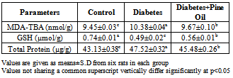
Lipid Peroxidation and Glutathione
The levels of TBARS, GSH and total protein in the brain of control and diabetic rats are presented in the Tables 1. The TBARS and total protein levels was found to be significantly high in the diabetes group (p<0.001) when compared to the control group.The GSH level in the diabetes groups was found to decrease significantly compared to the control group (p<0.001).
When the TBARS and total protein amounts was compared with the diabetes group, significant decreases were observed in diabetes+pineoil group (p<0.001) and GSH levels was determined to increase significantly.
Table 1. Pine oil in experimentally induced type 2 diabetes in the brain tissue MDA-TBA, GSH and total protein levels
     
The brain exhibits numerous morphological and functional alterations during diabetes. The chronic hyperglycemias cause an imbalance in the oxidative status of the nervous tissue, and the resulting free radicals damage the brain through a peroxidative mechanism. In diabetes, brain tissue is more vulnerable to oxidative stress[25]. Oxidative stress that leads to an increased production of reactive oxygen species (ROS) and finally cellular lipid peroxidationhas been found to play an important role in the development of diabetes mellitus LPO is one of the cellular features of chronic diabetes[26]. Extensive evidence has demonstrated that the increase of lipid peroxidation plays an important role in the progression of diabetes by altering the transbilayer fluidity gradient which could hamper the activities of membrane-bound enzymes and receptors[27]. In our study there was a significant increase in LPO levels in the diabetic group whereas the level of LPO in the diabetic rats significantly decreased as a result of the pine oil and was found to approximate the values of the control group. Lipids are particularly vulnerable to oxidation because the brain is rich in polyunsaturated fatty acids[28]. The resulting decrease in the level of LPO is very important in this respect. Lipid peroxide-mediated tissue damage has been observed in the development of both type 1 and 2 diabetes mellitus[29]. It has been reported that oxidative stress increases neuronal death that, in turn, contributes to the neuropathology associated with diabetes[30]. In agreement with the previous studies, herein we found that LPO level was elevated in brain tissues of STZ treated rats while the amount of GSH decreased[31,32].
GSH functions as free radical scavenger and in the repair of free radical caused biological damage[33]. In the present study, GSH levels decreased in the diabetic group; pine oilapplied to diabetic rats caused an increase in GSH levels in brain tissue. Increase in LPO levels in diabetic rats, decreased GSH level which has been suggested to be one of the most important indicators in the formation of the complications of diabetes. Pine oil’s positive impact on brain tissue is important in this regard.
Fatty Acids
The change of fatty acids in the brain of control and diabetic rats are presented in the Table 2. The palmitic, stearic, linoleic, arachidonic and docosahexaenoic acid levels were found to be significantly high in the diabetes group (p<0.001) when compared to the control group. The oleic acid level in the diabetes groups was found to decrease significantly compared to the control group (p<0.001).
When the palmitic, linoleic, arachidonic and docosahexaenoic acid amounts were compared with the diabetes group, significant increases were observed in the diabetes+pineoil group (p<0.001) and oleic acid level was determined to decreased significantly (p<0.001).Oleic, linoleic and arachidonic acid values were determined to approximate the values of the control group. There was not statistically significant in the level of stearic acid (p>0.05).
Table 2. Pine oil in experimentally induced type 2 diabetes in the brain tissue fatty acid composition
     
Lipid profile abnormalities form 40% of cases of diabetes and diabetic complications and are one of the most common.Fatty acid is required for both the structure and function of every cell in the body and forms an important component of cell membranes. Fatty acid composition is changed in humans[34] and animals with diabetes[35]. Diabetes group in brain tissue, palmitic and stearic acids have been shown to increase.Expression of this increase may be due to a lack of insulin. Because the palmitic and stearic acid biosynthesis, acetyl-CoA carboxylase and fatty acid synthase enzyme activity occurs. Pine oil is applied to the diabetic rats by affecting the activity of these enzymes according to the group of Diabetes, D + PO group were increased biosynthesis of palmitic and stearic acids. Brain tissue of diabetic rats, increased levels of palmitic and stearic acids was determined to be in compliancewith previous studies[36]. Pine oil in the brain tissue of diabetic rats decreased the levels of palmitic and stearic acid. Oleic acid is in monounsaturated fatty acids and stearic acid is substrate of oleic acid. Oleic acid is synthesized with activity of stearoyl CoA desaturase enzyme. This enzyme activity is dependent on insulin. Oleic acid levels decreased in thediabetes group compared to the control group, diabetic rats were determined to prevent the changes that emerged from pine oil. The resulting change in stearoyl CoA desaturase enzyme activity of pine oil may have been as a result of affected.
An increase in the level of linoleic acid in in diabetes group compared to the control group was observed; this increase in brain tissue of diabetic rats was determined to decrease the pine oil. This data we have obtained is in accordance with previous studies.Diabetes inhibitsdelta-6-desaturase[37], which converts linoleic acid into gamma linolenic acid, the precursor of arachidonic acid, and ultimately, several vasoactive prostanoids. In experimental and clinical diabetes, gamma linolenicacid production is reduced[34]. Consequently, the levels of dihomo gamma linolenic acid, which is a product of gamma linolenic acid elongation, and arachidonic acid also are reduced.
Experimental studies of diabetes found diabetic rats had decreased levels of arachidonic acid in brain tissue, but in our study we found increased levels of arachidonic acid in brain tissue when pine oil is applied.By reducing the level of arachidonic acid approximately the values of the control group were detected. Delta 6 desaturase enzyme is the major enzyme responsible for changes in fatty acid composition in diabetes.
The conditionally essential PUFA arachidonic acid[AA,4 20:4(n-6)] and docosahexaenoic acid[DHA, 22:6(n-3)] make up; 20% of fatty acids in the mammalian brain[38]. Both fatty acids and the functioning of brain function have a significant impact on learning[39]. Docosahexaenoic acid levels increased in the diabetic group compared to the control group, applied to pine oil prevent this change has been detected.
Vitamins and Cholesterol Levels
The difference of A, D, E, K vitamins, cholesterol and sterol in the brain of control and diabetic rats are presented in the Table 3. The vitamin K1, vitamin K2, retinol and β-sitosterol amounts were found to be significantly increased in the diabetes group (p<0.001) when compared to the control group. The α-tocopherol, cholesterol and stigmasterol levels in the diabetes groups was found to decrease significantly compared to the control group (p<0.001). There was not statistically significant in the level of δ-tokopherol(p>0.05).
When the vitamin K1, vitamin K2, α-tocopherol, cholesterol and stigmasterolamounts were compared with the diabetes group, significantly high levels were observed in the diabetes+pineoil group (p<0.001) and retinol level was determined to decreased significantly (p<0.001). There was no statistical significance in the level of δ-tocopherol (p>0.05).
In the diabetic group compared with the control group, lipophilic vitamins, cholesterol and sterol level were found to be significantly changed. Lipophilic vitamins are necessary to maintain the physiological functions of the organism, as well as supporting immune and growth activity, undertake an important role in stimulating digestion and synthesis. In addition, several studies tend to concur that with some diseases vitamin deficiency has been suggested to increase[40]. Other antioxidants such as vitamin E can protect cells from oxidative damage by cleaning. In our study the level of α-tocopherolin the brain tissue of diabetic rats decreased compared to the control group;we found the pine oil in the brain tissue of diabetic rats increased levels of α-tocopherol.
Table 3. Pine oil in experimentally induced type 2 diabetes in the brain tissue A,D,E,K vitamins, cholesterol and sterol change (µg/g)
     
Brain tissue is rich in cholesterol. In our study the level of cholesterol in the brain tissue of diabetic rats decreased compared to the control group; the pine oil in the brain tissue of diabetic rats, we found, increased levels of cholesterol. Earlier studies showed that diabetes produces a global suppression of the enzymes of cholesterol synthesis and their master transcriptional regulator SREBP-2 in the brain.The pine oil application may have inhibitive or stimulative effects on the activities of the enzymes responsible for the transcription of cholesterol which result in differences in cholesterol quantities.

4. Conclusions

In conclusion, experimental diabetes created in rats brain tissue, examined, the parameters obtained positive, according to the data of the pine oil diabetes during brain tissue occurring metabolic abnormalities correction would be useful to us suggests.

ACKNOWLEDGEMENTS

This work has been supported by the Firat University Research Fund (FÜBAP Project number 1652 and 1670)

References

[1]  Saravanan G, Ponmurugan P. Ameliorative potential of S-allyl cysteine on oxidative stress in STZ induceddiabetic rats. Chemico-Biological Interactions2011, 189: 100-106.
[2]  Perry IJ. Commentary: smoking and diabetes – accumulating evidence of a causal link. International Journal of Epidemiology 2001, 30:554-555.
[3]  Sundaram R, Naresh R, Shanthi P, Sachdanandam P. Ameliorative effect of 20-OH ecdysone on streptozotocin induced oxidative stress and cell damage in experimental hyperglycemic rats. Process Biochemistry 2012, 47: 2072-2080.
[4]  Arya A, Looi CY,Cheah SC, Mustafa MR, Mohd MA. Anti-diabeticeffectsof Centratherumanthelminticum seeds methanolic fraction onpancreaticcells, b-TC6 and its alleviating role in type2 diabetic rats.Journal of Ethnopharmacology 2012, 144: 22-32.
[5]  Pari L, Latha M. Effect of Scopariadulcis (Sweet Broom weed) plant extract on plasma antioxidants in streptozotocin- induced experimental diabetes in male albino Wistar rats. Pharmazie 2004, 59: 557-560.
[6]  Gipsen WH, Biessels GJ. Cognition and synaptic plasticity in diabetes mellitus.Trends in Neurosciences2000, 23: 542-549.
[7]  McCall AL. The impact of diabetes on the CNS. Diabetes 1992, 41: 557-570.
[8]  Li PA, Gisselsson J, Keuker J, Vogel ML, Kuschinsk SW. Hyperglycemia-exaggerated ischemic brain damage following 30 min of middle cerebral artery occlusion is not due to capillary obstruction.Brain Research1998, 804:36-44.
[9]  Wolff SP. Diabetes mellitus and free radicals: Free radicals, transition metals and oxidative stress in the aetiology of diabetes mellitus and complications. British Medical Bulletin1993, 49 (3): 642-652.
[10]  Aragno M, Brignardello E, Tamagno O, Boccuzzi G. Dehydroepiandrosterone administration prevents the oxidative damage induced by acute hyperglycemia in rats. Journal of Epidemiology 1997, 155:233-240.
[11]  Szkudelski T. The mechanism of alloxan and streptozotocinaction in B Cells of the rat pancreas. Physiological Research 2001, 50: 536-546.
[12]  Zheng XK, Zhang L, WangWW, WuYY, Zhang QB. Anti-diabetic activity and potential mechanism of total flavonoids of Selaginellatamariscina (Beauv.) Spring in rats induced by high fat diet and low dose STZ. Journal of Ethnopharmacology 2011, 137: 662-668.
[13]  Jouad H, Haloui M, Rhiouani H, El Hilaly J, Eddouks M. Ethnobotanical survey of medicinal plants used for the treatment of diabetes, cardiac and renal diseases in the North centre region of Morocco (Fez-Boulemane). Journal of Ethnopharmacology 2001, 77: 175-182.
[14]  Grover JK, Yadav S, Vats V. Medicinal plants of India with anti-diabetic potential. Journal of Ethnopharmacology2002, 81: 81-100.
[15]  Oladele SB, Ayo JO, Adaudi AO. Medicinal and physiological properties of flavonoids, coumarin derivatives and anthraquinones of plant origin.West African Journal of Pharmacology and Drug Research1995, 11: 134-144.
[16]  Ramesh B, PugalendiKV.Protective effect of Umbelliferone on membranous fatty acid composition instreptozotocin-induced diabetic rats.Life Sciences2006, 79: 306-310.
[17]  Masiello P, Broca C, Gross R, Roye M, Manteghetti M. Development of a new model in adult rats administered streptozotocin and nicotinamide. Diabetes1998, 47(2): 224-229.
[18]  Ohkawa H,Ohishi N, Yagi K. Assay for lipid peroxides in animal tissues by thiobarbituric acid reaction. Analytical Biochemistry 1979, 95(2):351-358.
[19]  Ellman GL. Tissue sulfhydryl groups. Archives of Biochemistyr and Biophysics 1959, 82: 70-77.
[20]  Akerboom TP, SiesH.Assay of glutathione, glutathione disulfide, and glutathione mixed disulfides in biological samples. Methods in Enzymology 1981, 77: 373-82.
[21]  Hara A, Radin NS. Lipid extraction of tissues with a lowtoxicity solvent. Analytical Biochemistry 1978, 90: 420-426.
[22]  Christie WW. Gas chromatography and lipids. The Oil 1999, Glasgow.
[23]  Sanchez-Machado DI, Lopez-Hernandez J, Paseiro-Losada P, Lopez-CervantesJ. An HPLC method for the quantification of sterols in edible seaweeds. Biomedical Chromatography 2004, 18: 183-90.
[24]  Lowry OH,Rosebrough NJ, Farr AL, Randall RJ.Protein measurement with the Folin phenol reagent. The Journal of Biological Chemistry 1951, 193(1):265-275.
[25]  Nazaroğlu NK,Dincel AS,Altan N.The effects of sulfonylurea glyburide on superoxide dismutase, catalase, and glutathione peroxidase activities in the brain tissue ofstreptozotocin-induced diabetic rat.Journal of Diabetes and Its Complications2009, 23: 209-213.
[26]  Sarkhail P,Rahmanipour S, Fadyevatan S,Mohammadirad A, Dehghan G, Amin G, Shafiee A, Abdollahi M. Antidiabetic effect of Phlomisanisodonta: effects on hepatic cells lipid peroxidation and antioxidant enzymes in experimental diabetes. Pharmacological Research2007, 56: 261–266.
[27]  Dmitriev LF, Titov VN.Lipid peroxidation in relation to ageing and the role of endogenous aldehydes in diabetes and other age-related diseases.Ageing Research Reviews2010, 9:200–210.
[28]  Reed TT. Lipid peroxidation and neurodegenerative disease. Free Radical Biology & Medicine2011, 51: 1302-1319.
[29]  Baydas G, Dönder E, Kiliboz M, Sonkaya E,Tuzcu M, Yasar A, Nedzvetskii VS. Neuroprotection by alpha-lipoic acid in streptozotocin-induced diabetes.Biochemistry (Mosc)2004, 69(9): 1001-1005.
[30]  Greene DA, Stevens MJ, Obrosova I,Feldman EL. Glucose-induced oxidative stress and programmed cell death in diabetic neuropathy. Europen Journal of Pharmacology 1999, 375: 217-223.
[31]  Raza H, Ahmed I, John A, Sharma AK.Modulation of xenobiotic metabolism and oxidative stress in chronic streptozotocin-induced diabetic rats fed with Momordicacharantia fruit extract. Journal of Biochemical and Molecular Toxicology2000, 14:131-139.
[32]  Celik S,Baydas G,Yilmaz O. Influence of vitamin E on the levels of fatty acids in MDA in some tissues of diabetic rats. Cell Biochemistry andFunction2002, 20:67-71.
[33]  Prabakaran D, Ashokkumar N.Protective effect of esculetin on hyperglycemia-mediated oxidative damage in the hepatic and renal tissues of experimental diabetic rats. Biochimie 2012,1-8.
[34]  Ruiz-Gutierrez V, Stiefel P, Villar J, Garcia-Donas MA, Acosta D, Carneado J. Cell membrane fatty acid composition in type 1 (insulindependent) diabetic patients: Relationship with sodium transport abnormalities and metabolic control. Diabetologia1993, 36: 850-856.
[35]  Holman RT, Johnson SB, Gerrard JM, Mauer SM,Kupcho-Sandberg S, Brown DM. Arachidonic acid deficiency in streptozotocin-induced diabetes. Proceedings of the National Academy of Sciences of the United States of America1983, 80:2375-2379.
[36]  Kulkarni CR, Joglekar MM,Patil SB, Arvindekar AU. Antihyperglycemic and antihyperlipidemic effect of Santalum album in streptozotocin induced diabetic rats. Pharmaceutial Biology2012, 50(3):360-365.
[37]  Murugan P, Pari L.Protective role of tetrahydrocurcumin on changes in the fatty acid composition instreptozotocin-nicotinamide induced type 2 diabetic rats. Journal of Applied Biomedicine2007, 5: 31-38.
[38]  Friedmann N, Gellhorn A, Benjamin W. Synthesis of Arachidonic Acid from Linoleic Acid in vivo in Diabetic Rats. Israel Journal of Medical Sciences1966, 2:677-682.
[39]  Rapoport SI.Arachidonicacid and the brain1–3.American Society for Nutrition2008, 138: 2515-2520.
[40]  Horrocks LA, Yeo YK.Health benefits of docosahexaenoic acid (DHA). Pharmacological Research1999, 40(3):211-225.
[41]  XueX, You J, He P.Simultaneous determination of five fat-soluble vitamins in feed by high-performance liquid chromatography following solid-phase extraction. Journal of Chromatographic Science2008, 46:345-350.
[42]  Suzuki R, Lee K, Jing E, Biddinger SB, McDonald JG, Montine TJ, Craft S, Kahn CR. Diabetes and insulin in regulation of brain cholesterol metabolism. Cell Metabolism 2010, 12(6):567-579.