Clinical Practice

p-ISSN: 2326-1463    e-ISSN: 2326-1471

2016;  5(1): 1-5

doi:10.5923/j.cp.20160501.01


Comparison of Surgical Techniques in T1 and T2 Thymomas: The Possibility of VATS Thymectomy Compared with Open Resection

Madiyorov B. T. 1, Krotov N. F. 2, Rasulov A. E. 1, Chernyshova T. V. 1, Sabirov D. R. 1, 3

1National Cancer Research Center, Ministry of Healthcare of the Republic of Uzbekistan, Uzbekistan

2FGBI "Oncology Institute named by N.N. Petrov" of Russian Ministry of Health, Uzbekistan

3Head of the Military Medical Faculty at the Tashkent Medical Academy, Tashkent, Uzbekistan

Correspondence to: Madiyorov B. T. , National Cancer Research Center, Ministry of Healthcare of the Republic of Uzbekistan, Uzbekistan.

Email:

Copyright © 2016 Scientific & Academic Publishing. All Rights Reserved.

This work is licensed under the Creative Commons Attribution International License (CC BY).
http://creativecommons.org/licenses/by/4.0/

Abstract

Background: The objective of this study was to compare the results of VATS thymectomy, with standard operations (thoracotomy, sternotomy), and assessment of overall survival in the long term. Patients and Methods: A retrospective review of 27 patients who underwent thymectomy, in last 13 years. The key point of the study was overall survival in the two groups. Results: Of the 27 patients, 11 patients have undergone VATS thymectomy in stage I and II; in 16 patients thymectomy with standard access in stages I and II was performed. Postoperative mortality was 0%. Stage of the tumor and the number of patients undergoing radio and chemotherapy, were comparable in both surgical groups. The average length of stay in the hospital for the main group was 7 days, in the control group 16 days. Essential differences in index of 5-10 year survival have not been identified between the 2 groups. Conclusions: VATS thymectomy is an effective and less traumatic treatment for thymomas. There were no significant differences in patient survival depending on the used technique.

Keywords: Mediastinum, Videothoracoscopy, Thymoma, Thymectomy

Cite this paper: Madiyorov B. T. , Krotov N. F. , Rasulov A. E. , Chernyshova T. V. , Sabirov D. R. , Comparison of Surgical Techniques in T1 and T2 Thymomas: The Possibility of VATS Thymectomy Compared with Open Resection, Clinical Practice, Vol. 5 No. 1, 2016, pp. 1-5. doi: 10.5923/j.cp.20160501.01.

1. Background

Thymus tumors according to different data make up from 10% to 60% of all tumors of the mediastinum, which constitute about 3-7% of all oncologic pathology and 10- 20% of primary mediastinal disorders. [1-10, 11, 13-15, 20, 22]
All variety of thymic tumors are divided into 2 groups: I group consists of tumor histogenesis which is associated with the epithelial component of the thymus gland (thymoma, thymic cancer); Group II - tumor histogenesis which is not associated with the epithelial component (carcinoid, germ cell tumors, lymphomas, soft tissue tumors, hyperplasia and thymic cysts) [3, 7, 16, 19, 22].
To date, histological classification of thymomas proposed by WHO experts, under the general editorship of J. Rosai was accepted in 1999 and modified in 2004: spindle cell (type A), lymphoid (type B1), mixed (type AB, B2), epithelial (type B3), a cancer of the thymus (type C). [11, 16, 19].
For staging, prognosis, and surgical treatment, thymoma classification based on the degree of infestation, the roposed Masaoka is used [5, 9]. 1st degree for this classification corresponds to the absence of macro- and microscopic invasion; 2nd degree: macroscopic invasion of the thymus tissue of the capsule; IIIa degree: no invasion of large vessels; IIIb degree: an infestation of large vessels; IVa degree: dissemination on the pericardium or pleura; IVb degree: Lymphogenous or hematogenous metastasis [2, 3, 9, 11, 13, 21].
Until now, a surgical method is the "golden standard" in treatment of patients with tumors of the thymus gland. This is true both for the surgery and for the combined treatment. Radical surgery - the most important factor in prognosis of surgical treatment of patients with benign and malignant thymoma [2, 4, 15, 17, 19, 25, 26].
Tumors of the thymus gland (thymoma), tumor-like growths, and gravis myastheniaare considered as the indications for surgical treatment. Approximately 30% of patients with thymoma manifests myasthenia gravis (MG) [1, 2, 6, 7, 12-15, 20]. Blalock, et al [1] is one of the first to describe a series of patients with myasthenia gravis (MG) for whom thymectomy was performed.
The main factor determining the clinical manifestations, course and prognosis of thymoma, is the presence and extent of invasive growth. So according to various authors, surgical treatment is the key to long-term survival of patients, 5 and 10-year survival of patients with 1 and 2-stage thymoma after thymectomy, ranges from 89% to 100% and 71 to 95%, respectively, in stage III - 74% and 46% and the 5 year survival in disease IV is less than 25% [2, 3, 7, 13, 14].
To perform radical surgery on the thymus using different approaches - neck; traspleural–right sided and transbipleural; and trasnssternal - by a total or partial median sternotomy and by oblique partial sternotomy [2, 4, 5, 7, 8, 12, 14, 17, 18, 20, 21, 26].
However, with the recent widespread introduction of thoracoscopic techniques into clinical practice, is increasingly discussed the use of video assisted thoracoscopy (VATS) thymectomy that reduce surgical trauma and associated postoperative complications [4, 7, 8, 12, 15, 21, 23, 26].
Many publications have demonstrated the safety and efficacy of VATS thymectomy in patients with myasthenia gravis. Many researchers are of the opinion that this surgery is currently the method of choice in these patients [4, 7, 8, 12, 15, 21, 23, 26].
In general, it should be recognized that the VATS interventions research data in isolated tumors of the thymus are not accompanied by myasthenia, i.e. it is underrepresented, and the question about the possibilities of VATS access in such clinical situations requires further study.
Given the above, the aim of this study was to compare the results of VATS thymectomy, versus standard operations (thoracotomy, sternotomy), and assessment of overall survival on the long term.

2. Subjects and Methods

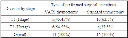
Retrospective analysis was held on 27 patients with tumors of the thymus, the first and second stages, in period from 2001 to 2014 who were treated in the thoracic surgery department. Patient characteristics: -13 males (48.15%) -14 female (51.85%), age varied: from 10 to 55 years. The mean age was 31,40 ± 16,4 years.
To evaluate the general condition of the patient, verification of tumor process carried out comprehensive investigations: general clinical research biplane standard chest X-ray, bronchoscopy, trans-thoracic puncture of the tumor (indication), and cytological and histological examination of the tumor.
For differential diagnosis used additional methods of investigation: CT scan of the chest with contrast mediastinal vessels, ultrasound examination of the mediastinum, supraclavicular area, abdomen, magnetic resonance imaging (MRI) of the chest. A number of patients used the following diagnostic procedures: supraclavicular lymph node puncture or prescalenous biopsy, thoracoscopic study.
The main source of information about the further development of the disease was hospital records, which contain information on the results of periodic inspections, information from the database of organizational and methodical department and response from the regional oncologic dispensary of the republic, which was received according to request.
Depending on the approach of surgery, patients were divided into two groups: basic - VATS thymectomy, the control - thymectomy performed through the standard approach - thoracotomy, sternotomy. Comparison between parameters of both groups by stages is summarized in Table 1.
Table 1. Parameters and comparison of open versus video thoracoscopic operations, depending on the stage (p < 0,05)
     
Depending on the histological type of thymomas, patients are divided as follows: Type A - spindle cell thymoma in 4 patients (14.8%), type AB – lymph-epithelial thymoma in 7 (25.9%), type B1- B3 in 9 (33.3%), and type C - thymus cancer in 7 (25.9%). Tumor size: in the first group of 3 to 6 cm: 5; up to 8 cm: 6 cases, on average - 4,8 ± 2,2 cm. In control group from 3 to 6 cm: 3; up to 8 cm: 13, on average 8,2 ± 3,2 cm. In 6 patients with myasthenia gravis, surgical operations were carried out, all the histological examination revealed thymoma: in two of them, operation was performed with VATS access; while in 4, by sternotomy. Patients with myasthenia gravis were divided using MGFA international classification (Myasthenia Gravis Foundation of America) [6]. 1st degree (ocular disorder); 4%, 2nd degree (mild violation of neuromuscular transmission); 38%, 3rd degree (moderate disorders of neuromuscular transmission); 58%.
In the study group, in all patients left-sided thoracoscopic access was used. This is due primarily to the topographic anatomical location of thymus. Thoracoscope with zero optics is used for visualization of the surgical field.
After surgery, 16 patients of the study and control groups with clear signs of malignant thymoma received adjuvant radiation therapy to the mediastinum from 40 to 60 Gr. These same patients underwent from 4 to 6 cycles of chemotherapy based on platinum in combination with other cytotoxic drugs (doxorubicin, cyclophosphamide, vinblastine, and etoposide).
Quantitative variables are described by the following statistics: the number of patients, the arithmetic mean value, standard deviation of the arithmetic mean, 25th and 75th percentiles, the median. Qualitative variables are presented as absolute (numbers) and relative frequencies (percentages). The significance of differences were determined by using of nonparametric Mann-Whitney U-test. If the level of error p <0,05. The survival rate after surgery was assessed on a scale of Kaplan-Meier, with a 95% confidence interval (longrang criterion), as amended by Yeats. The calculations were performed on a PC using Microsoft Excel and statistical analysis package Statistica 5.1 for Windows data ("StatInc.", USA).

3. Results and Discussion

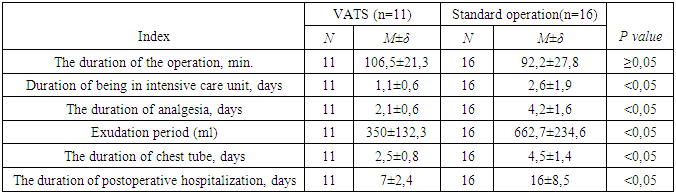
In all VATS, patients did not have dangerous complications. Conversion was performed in 1 (9.1%) patients in the early stages of the development of techniques in our clinic. The reason for the conversion in this case was the presence of pronounced adhesions in the pleural cavity.
Traumatism of surgical interventions was evaluated on the basis of exudation in the postoperative period, the timing of draining, time spent in the intensive care unit, duration of surgery, and duration of anesthesia (Table 2). In the long-term period: time to recurrence and survival after surgery, depending on the surgical approach.
Table 2. Parameters of the comparison of open and VATS operations
     
In connection with smoother postoperative period in the study group, only 70% of the patients were in the intensive care unit, whereas in the control group, all patients after thoracotomy were transferred to the intensive care unit that determined the need for prolonged mechanical ventilation. Analysis of the results established a significant reduction in the length of stay of patients in the intensive care ward after VATS operations compared to the same period in the standard: 1,1 ± 0,6 and 2,6 ± 1,9 days, respectively (p <0,05).
In Patients operated upon by open method, the use of narcotic and non-narcotic pain medication proved to be used for longer period. In the study group it was 2,1 ± 0,6 days in control - 4,2 ± 1,6 days. The average duration of use of pain medications in the study group was significantly lower than in control (p <0,05). In the group with thoracoscopy, from 2 day, pain was moderate and weak, the use of non-narcotic analgesics coped with the pain, and after removal of the drainage tubes, it was very weak or absent. In Patients in the control group, pain syndrome occurred in waves, and in the first 3 days was determined mostly severe and very severe pain requiring prescription of narcotic analgesics.
The duration of draining the pleural cavity was 1 to 4 days in the study group (median 2,5 ± 0,8 days) versus 2 to 6 days in the control group (median 4,5 ± 1,4 days). Thus, drainage of pleural cavity period was significantly lower in the study group (p<0,05). Duration of postoperative hospital stay in the thoracoscopy group ranged from 3 to 27 days (average 7±2,4 days), while in the open surgery group it was 5 to 28 days (average 16 ± 8.5), the difference was statistically significant (p<0,05).
In the surgical treatment of thymomas, complications and deaths are caused directly depending on the type of surgery performed. The most common surgical complications include bleeding and suppuration of postoperative wound. Intrapleural bleeding and festering wounds in the studied groups were not observed. Non-surgical complications were mainly pneumonia (2 patients) and various cardiovascular disorders (three patients). No deaths were observed after surgery.
Observation period of patients in the main and control groups was from 6 months up to 10 years. Currently, overall survival is the most accurate in the assessment of the results of surgical and combined treatment of patients with different histological forms of thymoma. Long-term results of surgical treatment of patients evaluated in both study and control groups. Overall survival operated VATS access at T1 and T2 illustrated in Figure 1.
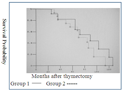
Figure 1. The survival rate after VATS thymectomy at T1 and T2: Group 1 - thymoma T1; Group 2 - thymoma in T2
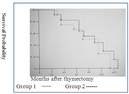
Figure 2. The survival rate after standard thymectomy at T1 and T2: Group 1 - thymoma T1; Group 2 - thymoma in T2
The analysis of our observations in groups allowed confirming absence of difference between mini-invasive and traditional thymectomy in 5-10 year survival in patients with T1N0M0 and T2N0M0 thymomas.

4. Conclusions

Thus, method of video assisted thoracoscopic surgery in thymomas decreases amount of exudation, reduces drainage time after surgery versus thoracotomy and sternotomy VATS is an effective and less traumatic treatment for thymomas. Significant differences in patient survival depending on the used technique were not established.

References

[1]  Blalock A, McGehee HA, Ford FR, et al. The treatment of myasthenia gravis by removal of the thymus gland. JAMA. 1941. Vol.117. pp.529-33.
[2]  Conrad B. Falkson, Andrea Bezjak, Gail Darling, Richard Gregg, Richard Malthaner, et al. The Management of Thymoma: A Systematic Review and Practice Guideline. J Thorac Oncol. 2009. Vol.4. N.7.pp.911–919.
[3]  Davenport E, Malthaner R. The role of surgery in the management of thymoma: A systematic review. Ann Thorac Surg. 2008. Vol.86. pp.673-84. doi:10.1016/j.athoracsur.2008.03.055.
[4]  Frank C. Detterbeck, Alden M. Parsons. Thymic Tumors. Ann Thorac Surg 2004. Vol.77.pp.1860–9.
[5]  Guimarães M., Benveniste M., Bitencourt A., Andrade V., Souza L., Gross J., Godoy M. Thymoma Originating in a Giant Thymolipoma: A Rare Intrathoracic Lesion. Ann Thorac Surg 2013. Vol.96. pp.1083–5.http://dx.doi.org/10.1016/j.athoracsur.2013.01.031.
[6]  Jaretzki A., Barohn R.J., Ernstoff R.M., et al. Myasthenia gravis: recommendations for clinical research standards. Task Force of the Medical Scientific Advisory Board of the Myasthenia Gravis Foundation of America. Neurology. 2000. № 55. Pp.16–20.
[7]  Jurado J., Javidfar J., Newmark A. et al. Minimally invasive thymectomy: outcome analysis of 263 patients. Ann. Thorac Surg. 2012. Vol.94. n.3.pp.974-981.
[8]  Koezuka S., Sato F., Hata Y., Otsuka H., Yuasa R., Kiribayashi T., Sasai D., et al. Video-Assisted Thoracoscopic Surgery for Ectopic Middle Mediastinal Thymoma in a Patient With Myasthenia Gravis. Ann Thorac Surg. 2013. Vol.95. pp.e67– 8.http://dx.doi.org/10.1016/j.athoracsur.2012.08.065.
[9]  Masaoka A., Monden Y., Nakahara K., Tanioka T. Follow-up study of thymomas with special reference to their clinical stages. Cancer. 1981.Vol. 48. pp.2485—2492.
[10]  Mineo T., Ambrogi V., Baldi A., Pompeo E., Mineo D. Recurrent intrathoracic thymomas: Potential prognostic importance of cell-cycle protein expression. J Thorac Cardiovasc Surg 2009. Vol.138. pp.40-5. doi:10.1016/j.jtcvs.2008.11.
[11]  Muller-Hermelink H., Engel P., Kuo T. et al. Tumors of thymus/Travis William D., Brambilla Elisabeth, Muller-Hermelin H., Konradk Harris Curtis C. (eds). Pathology and Genetics of Tumours of the Lung, Pleura, Thymus and Heart. Lyon: IARC-Press, 2004. pp. 148—247.
[12]  Nakamura H., Taniguchi Y., Fujioka S. et al. First experience of robotic extended thymectomy in Japan for myasthenia gravis with thymoma. Gen Thorac Cardiovasc Surg. 2012. Vol.60.n.3. pp.183-187.
[13]  Hamaji M., Allen M., Cassivi S., Nichols F., Wigle D., Deschamps C., Shen K., The Role of Surgical Management in Recurrent Thymic Tumors. Ann Thorac Surg. 2012. Vol.94. pp.247–54.
[14]  Okereke I., Kesler K., Morad M., Mi D., Rieger K., Birdas T., Badve S., et al. Prognostic Indicators After Surgery for Thymoma. Ann Thorac Surg 2010. Vol.89. pp.1071–9. doi: 10.1016/j.athoracsur.2010.01.026
[15]  Pennathur A., Qureshi I., Schuchert M., Dhupar R., Ferson P., Gooding W., Christie N., Gilbert S., Shende M., et.al. Comparison of surgical techniques for early-stage thymoma: Feasibility of minimally invasive thymectomy and comparison with open resection. J Thorac Cardiovasc Surg 2011.vol.141. pp.694-701. doi:10.1016/j.jtcvs.2010.09.003.
[16]  Rosai J. Histological Typing ofTumours of the Thymus. 2nded.World health /structures/ organization, international histological classification of tumors. Berlin: Springer, 1999.
[17]  Salma El Batti, Olaf Mercier, Adela Rohnean, Benjamin Besse, Remi Nottin, Philippe Dartevelle. Recurrence of Thymoma in the Right Atrium Arising From the Coronary Sinus. Ann Thorac Surg 2013. Vol.95. pp.e71–2. doi.org/10.1016/j.athoracsur.2012.08.114
[18]  Sivarajah M., Weksler B. Robotic-Assisted Resection of a Thymoma After Two Previous Sternotomies. Ann Thorac Surg. 2010. Vol.90. pp.668 –70.
[19]  Song Zhengbo, Jin Xiangyu, Zhang Yiping. Treatment and prognosis of type B2 thymoma. World Journal of Surgical Oncology 2014, vol.12. pp.291.http://www.wjso.com/content/12/1/291.
[20]  Stefano Margaritora, Alfredo Cesario, Giacomo Cusumano, Elisa Meacci, Rolando D’Angelillo, Stefano Bonassi, et al. Thirty-Five–Year Follow-Up Analysis of Clinical and Pathologic Outcomes of Thymoma Surgery. Ann Thorac Surg 2010. Vol.89. pp.245–52.
[21]  Takeo S., Tsukamoto S., Kawano D., Katsura M. Outcome of an Original Video-Assisted Thoracoscopic Extended Thymectomy for Thymoma. Ann Thorac Surg 2011. Vol.92. pp.2000–6. doi: 10.1016/j.athoracsur.2011.07.054.
[22]  Takeda S., Koma M., Maeda H. Huge thymoma: role of preoperative WHO histological classification. Interact. CardiovascThorac Surg. 2008. Vol.7.n.6. pp.1196-1198.
[23]  Tomulescu V., Popescu I. Unilateral extended thoracoscopic thymectomy for nontumoral myasthenia gravis: a new standard. Semin.ThoracCardiov Surg. 2012. Vol.24. n.2. pp.115-122.
[24]  Weksler B., Nason K., Mackey D., Gallagher A., Pennathur A. Thymomas and Extrathymic Cancers. Ann Thorac Surg 2012. Vol.93. pp.884 –9. doi:10.1016/j.athoracsur.2011.05.089.
[25]  Yano M., Sasaki H., Moriyama S., Kawano O., Hikosaka Yu, Masaoka A., Fujii Y. Preservation of Phrenic Nerve Involved by Stage III Thymoma. Ann Thorac Surg 2010. Vol.89. pp.1612–9. doi:10.1016/j.athoracsur.2010.01.057.
[26]  Zahid I., Sharif S., Routledge T., Scarci M. Video-assisted thoracoscopic surgery or transsternalthymectomy in the treatment of measthenia gravis? Interact. Cardiovasc. Thorac. Surg. 2011. Vol.12. n.1. pp.40-46.