-
Paper Information
- Paper Submission
-
Journal Information
- About This Journal
- Editorial Board
- Current Issue
- Archive
- Author Guidelines
- Contact Us
Clinical Practice
p-ISSN: 2326-1463 e-ISSN: 2326-1471
2013; 2(2): 10-13
doi:10.5923/j.cp.20130202.02
Medullary Nephrocalcinosis with Underlying Endocrine Abnormality: A Challenging Case to Manage
 Abstract
 Reference
 Full-Text PDF
 Full-text HTML
Azhar Amir Hamzah1, Azreen Syazril Adnan2, Mohammad Nor Gohar Rahman1, Amer Hayat Khan3
1Urology Unit, Department of Surgery, Hospital University of Malaysia, Kubang Kerian, Kelantan, Malaysia
2Chronic Kidney Disease (CKD) Resource Center, School of Medical Sciences, University Science Malaysia, Kota Bharu, Kelantan, 16150, Malaysia
3Department of Clinical Pharmacy, School of Pharmaceutical Sciences, Universiti Sains Malaysia, Penang, 11800, Malaysia
Correspondence to: Azhar Amir Hamzah, Urology Unit, Department of Surgery, Hospital University of Malaysia, Kubang Kerian, Kelantan, Malaysia.
| Email: |  |
Copyright © 2012 Scientific & Academic Publishing. All Rights Reserved.
Medullary nephrocalcinosis is an abnormality caused by the deposition of calcium salts into the renal medulla, specifically the distal convoluted tubules. The most common cause in adults is primary hyperparathyroidism, followed by renal tubular acidosis, medullary sponge kidney, and other causes of hypercalcemia (hypercalciuria). A 27 years old lady who presented with left loin pain which was later found to be bilateral medullary calcinosis. Patient experienced symptoms of colicky pain, polyuria, polydypsia, hyperthyroidism such as palpitations, tremor, sweating, loss of weight and heat intolerance. Physical examinations revealed fine tremors of hands, bilateral eyes lid lag and diffuse enlargement of the thyroid without retrosternal extension. Blood investigations raised in calcium level, 2.77 mmol/L (corrected calcium), T4 level more than 100, TSH less than 0.005 and iPTH of 0.4 (normal range 1.6-6.9). KUB radiograph was done followed by ultrasonography which showed bilateral medullary calcinosis. Present case shows the presence of hypercalcemia secondary to hyperthyroidism and was demonstrated by the plain radiography and ultrasound of the kidneys. Medullary nephrocalcinosis can be managed medically (high fluid intake and alkaline-citrate) or using Extracorporeal Shock Wave Lithotripsy (ESWL) even though the result is not so promising.
Keywords: Medullary Nephrocalcinosis, Hypercalcemia, Hyperparathyroidism
Cite this paper: Azhar Amir Hamzah, Azreen Syazril Adnan, Mohammad Nor Gohar Rahman, Amer Hayat Khan, Medullary Nephrocalcinosis with Underlying Endocrine Abnormality: A Challenging Case to Manage, Clinical Practice, Vol. 2 No. 2, 2013, pp. 10-13. doi: 10.5923/j.cp.20130202.02.
1. Introduction
- Hypercalciuria, or excessive urinary calcium excretion, occurs in about 5-10% of the population.[1] Nephrocalcinosis, is a term originally used to describe deposition of calcium salts in the renal parenchyma due to hyperparathyroidism. The term has now acquired more of a radiologic concept, and it is used to describe diffuse, fine, renal parenchymal calcification that is radiologically demonstrable. Nephrocalcinosis can be confused with renal pelvicaliceal nephrolithiasis radiographically, it is important to distinguish between these entities as they are treated differently. Nephrocalcinosisis is treated medically whereas lithiasis is generally treated by surgical intervention.[2]Nephrocalcinosis often refer to the diffuse renal parenchymal calcifications observed by ultrasonograph.[2] It may also be defined as a generalized increase in the calcium content of the kidney. It does not necessarily lead to renal calculi, but renal calculi may occur with or without the presence of nephrocalcinosis.[3] Even though these two pathologies are distinct, they are intimately related.[3] While nephrolithiasis is the condition in which renal calculi are freely mobile in the renal collecting system, nephrocalcinosis is the deposition of calcium in the renal cortex or medulla.The etiology of nephrocalcinosis is broad, arising as a consequence of distal Renal Tubular Acidosis (dRTA), or it can be induced by hypercalcemia, hyperoxaluria or certain drug therapies. In some cases nephrocalcinosis may be idiopathic.[4] Nephrocalcinosis can be subdivided into medullary and cortical nephrocalcinosis. Medullary nephrocalcinosis is the commonest pattern seen in 98% of cases, in which clusters of calcification occur around each renal pyramid.[5] This finding seems to be most commonly associated with disorder of calcium hemostasis such as in hyperparathyroidism. Other causes of medullary includes Hyperthyroidism, Medullary sponge disease, renal tuberculosis, renal tubular acidosis, renal papillary necrosis, sarcoidosis, Bartter syndrome etc.[5] Corticalnephrocalcinosis is rare involves all the renal parenchyma. The most frequent causes are chronic glomerulonephritis, acute cortical necrosis, Alport Syndrome and Oxalosis.[5] Maxime et al. Describe two forms of nephrocalcinosis, tubular and interstitial type. Tubular nephrocalcinosis is caused by the precipitation and retention of crystals in the renal tubules, whereas interstitial nephrocalcinosis is caused by the precipitation and retention of crystals in the renal interstitium. While interstitial Nephrocalcinosis is limited to the renal medullary interstitium, tubular Nephrocalcinosis can be observed in the renal cortex and medulla.[2]
2. Case Report
- A Malay lady with age of 27 years with no medical illness previously, while presented with a left loin pain from three days. Pain was colicky in nature, appeared throughout the day, with no radiation to other part of the body. Patient has on and off bilateral loin pain for the past 2 years and using analgesic. History shows that patient was defaulted previously for bilateral renal stone. Patient experienced symptoms of polyuria, polydypsia, hyperthyroidism such as palpitations, tremor, sweating, loss of weight and heat intolerance. Clinically, she was medium built with stable vital signs. Physical examinations revealed fine tremors of hands, bilateral eyes lid lag and diffuse enlargement of the thyroid without retrosternal extension. Blood investigations raised in calcium level, 2.77 mmol/L (corrected calcium), T4 level more than 100, TSH less than 0.005 and iPTH of 0.4 (normal range 1.6-6.9). Kidney, ureter and bladder (KUB) radiograph showed bilateral radiopaque calcification suggestive of calcinosis (Figure 1). Ultrasonography KUB done subsequently showed bilateral medullary calcinosis (as shown in Figure 2 and 3). Patient was referred to the endocrine for further management. Patient consent were taken prior study.
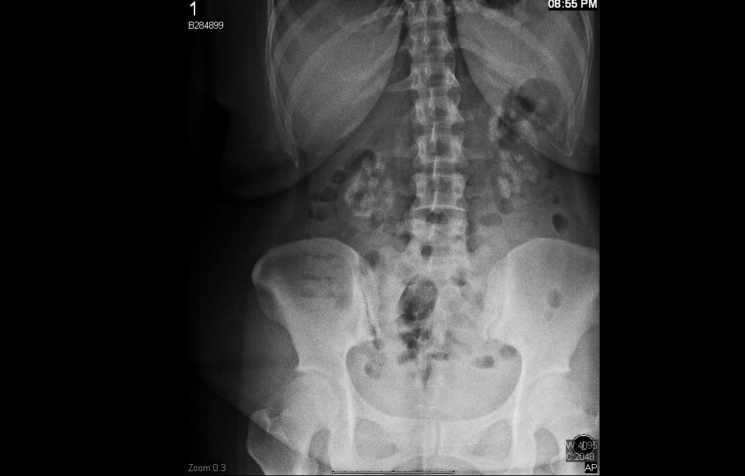 | Figure 1. Abdominal radiograph showed bilateral radiopaque calcification suggestive of calcinosis |
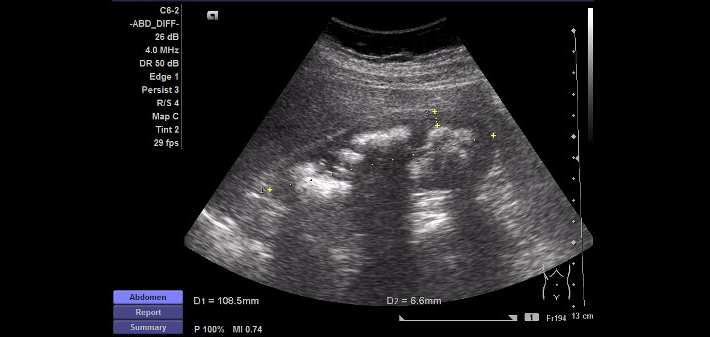 | Figure 2. Ultrasonography of right kidney showed increased in echogenicity of the medulla with posterior shadowing suggestive of calcinosis. There was no stone seen bilaterally |
 | Figure 3. Left kidney Ultrasonography KUB showed medullary calcinosis |
3. Discussion
- Nephrocalcinosis may be diagnosed with plain radiography, ultrasonography and CT scan which has the highest sensitivity to detect this disorder[4]. Ultrasound examination in experienced hand, will demonstrates the typical characteristics of papillary calcifications in the form of hyperechoic foci with or without acoustic shadowing at the tips of the pyramids.[6] Meanwhile, intravenous urography usually does not provide sufficient information to differentiate between intra-collecting systems caliceal stones lying free and those attached to or lying submucosally along the papilla. Once a diagnosis of nephrocalcinosis is confirmed, metabolic evaluation should be performed as the ultimate aimed is to initiate appropriate treatment, to delay disease progression and preserve renal function.Patients with Nephrocalcinosis do not normally present with acute symptoms, otherwise analgesic treatment has to be initiated directly and in an adequate dosage if patient presented with an acute severe pain. Prevention is the main aim and regardless of the underlying disorder, a high fluid intake is the precondition for all further treatments. Provided a stable urinary excretion rate is maintained, a high concentration level of the lithogenic substance can be excreted. Apart from high fluid intake, crystallization inhibitors, mainly citrate and magnesium, are an effective treatment option. Citrate is metabolized in the liver to bicarbonate, resulting in a higher urinary pH and therefore reduced citrate reabsorption in the renal tubule. Urinary citrate binds to calcium, forms a soluble complex reducing the precipitation of calcium with other substances, thus leading to a decreased urinary saturation index. Urinary calcium excretion can be reduced by 30% with adequate alkali citrate treatment and has been shown to decrease stone production and reduce progression of Nephrocalcinosis.[7]Typically, Nephrocalcinosis is untreated until a sufficiently large stone blocks the collecting system. Using extracorporeal shock wave lithotripsy, treating patients with nephrocalcinosis due to medullary sponge kidney. However, none of these patients became stone free as extracorporeal shock wave lithotripsy would not result in clearance of submucosal calculi.[8]Current patient presented with chronic loin pain and was controlled by adequate analgesia. She also had symptoms of hyperthyroidism, physical examination showed diffuse enlargement of thyroid gland and blood investigations showed rise in calcium and serum T4 level which is suggestive of thyrotoxicosis. Current scenario makes the diagnosis even difficult as concomitant hyperthyroidism and hyperparathyroidism is present.[5] Simultaneous occurrence of hyperthyroidism and hyperparathyroidism in the same patient is a rare combination. The clinical manifestations of hyperthyroidism may overshadow varied symptoms and signs of primary hyperparathyroidism. However, both diseases may have a profound influence on calcium metabolism and occasionally, disturbances in thyrotoxicosis may stimulate hyperparathyroidism. The diagnosis of an associated parathyroid adenoma may be missed or unnecessarily delayed because hypercalcemia is known to occur in hyperthyroidism.
4. Conclusions
- There are several causes of Nephrocalcinosis that lead to deposition of calcium in the renal parenchyma, while present case shows the presence of hypercalcemia secondary to hyperthyroidism and was demonstrated by the plain radiography and ultrasound of the kidneys. Medullary nephrocalcinosis can be managed medically (high fluid intake and alkaline-citrate) or using Extracorporeal Shock Wave Lithotripsy (ESWL) even though the result is not so promising.