Clinical Medicine and Diagnostics

p-ISSN: 2163-1433    e-ISSN: 2163-1441

2017;  7(2): 31-39

doi:10.5923/j.cmd.20170702.01


Serum Levels of High Mobility Group Box 1 (HMGB1) and Matrix Metalloprotinase 9 (MMP9) are Related to Lung Function Indices in Chronic Obstructive Pulmonary Disease

Eman Sobh 1, Asmaa Abd AL Salam Elmadbouly 2, Hend Ezzat 2, Maha Abd-Allah 3

1Chest Department, Faculty of Medicine (for girls), Al-Azhar University, Cairo, Egypt

2Clinical Pathology Department, Faculty of Medicine (for girls), Al-Azhar University, Cairo, Egypt

3Internal Medicine Department, Faculty of Medicine (for girls), Al-Azhar University, Cairo, Egypt

Correspondence to: Asmaa Abd AL Salam Elmadbouly , Clinical Pathology Department, Faculty of Medicine (for girls), Al-Azhar University, Cairo, Egypt.

Email:

Copyright © 2017 The Author(s). Published by Scientific & Academic Publishing.

This work is licensed under the Creative Commons Attribution International License (CC BY).
http://creativecommons.org/licenses/by/4.0/

Abstract

Background: High mobility group box 1 (HMGB1) and Matrix metalloproteinase 9 (MMP9) participate in inflammation and tissue remolding in various diseases. This study aimed to investigate if HMGB1 and MMP9 levels were elevated in COPD patients and if they are related to airway obstruction. Methods: Demographic and clinical characteristics, spirometry examination, and blood samples were obtained from 36 COPD patients and 39 healthy volunteers. Serum concentrations of High mobility group box 1 protein (HMGB1) and Matrix metalloproteinase-9 (MMP-9) were detected with ELISA Assay. Results: We found that serum HMGB1 and MMP9 levels were significantly increased in COPD patients compared with healthy volunteers (62.6±48.3 versus 35.4±24.9 pg/mL, p-value 0.003 and 201.8±32.8 versus 164.6±59.2 ng/ml, p-value 0.001 respectively). Also, there was asignificant positive correlation between HMGB-1 with MMR dyspnea scale and BODE index, COPD stage and negative correlation with PaO2, spirometric indices: FEV1, FVC, FEF25-75, and 6MWD. However, MMP-9 was correlated positively with MMRD score and BODE index and negatively correlated with oxygen saturation, PaO2, spirometric indices and 6MWD. There was positive correlation between HMGB-1 and MMP-9. Conclusion: Our findings first illustrate that serum HMGB1 and MMP9 levels are elevated in serum of patients with COPD, and second both markers are related to the lung function indices in these patients, suggesting that both markers may play critical roles in the pathogenesis of COPD.

Keywords: High mobility group box 1 (HMGB1), Matrix metalloproteinase 9 (MMP9), Chronic obstructive pulmonary disease (COPD), Small airway obstruction (SAO)

Cite this paper: Eman Sobh , Asmaa Abd AL Salam Elmadbouly , Hend Ezzat , Maha Abd-Allah , Serum Levels of High Mobility Group Box 1 (HMGB1) and Matrix Metalloprotinase 9 (MMP9) are Related to Lung Function Indices in Chronic Obstructive Pulmonary Disease, Clinical Medicine and Diagnostics, Vol. 7 No. 2, 2017, pp. 31-39. doi: 10.5923/j.cmd.20170702.01.

1. Introduction

Chronic obstructive pulmonary disease (COPD) is one of the leading global causes of morbidity and mortality [1, 2]. Long term inhalation of noxious gasses including cigarette smoke results in airflow obstruction and emphysema in the lungs. Persistent systemic inflammation and oxidative stress are common features of this disease [3]. A lot of studies were performed targeting this inflammatory process, its description and involved mediators [4, 5], including circulating white blood cells (WBC), C-reactive protein (CRP), interleukins 6 (IL-6) and 8 (IL-8), fibrinogen, tumor necrosis factor alpha (TNFα), transforming growth factor-β and homocysteine. [6-10]
High mobility group box 1 protein (HMGB1) is a nuclear DNA-binding protein [11] important for gene expression and proper transcriptional regulation [1]. In addition, HMGB1 works as an extracellular signaling molecule during inflammation, cell differentiation, cell migration, and tumor metastasis [12]. It exerts its actions through multiple cell-surface receptors including the receptor for advanced glycation end products (RAGE) and toll-like receptors (TLRs)(TLR2, TLR4 or TLR9) [11]. High levels of HMGB1 are found in inflammatory conditions such as sepsis, cystic fibrosis, rheumatoid arthritis and burn injury [13, 14] and in cancer development and progression [15]. High HMGB1 concentrations were found in the pulmonary epithelial lining fluid of patients with sepsis [16]. Recently, it was reported that levels of HMGB1 in the bronchoalveolar lavage (BAL) fluid were also elevated in patients with COPD [17]. In a systematic review by Gangemi et al., [18] they reported that in almost all the studies, HMGB1 levels were augmented in smokers and in patients affected by COPD.
As chronic obstructive pulmonary disease (COPD) is characterized by the progressive destruction of the extracellular matrix of the lung; Proteins belonging to the Matrix metalloproteinases (MMP) family are involved in the breakdown of extracellular matrix (ECM) in normal physiological processes such as tissue remodeling, but degradation of the ECM by MMP is also involved in the pathogenesis of a number of diseases [19]. Matrix metalloproteinase-9 (MMP9) has been implicated in numerous somatic illnesses, including cardiovascular disorders and cancer [20, 21].
Matrix metalloproteinases (MMPs) and their inhibitors play a central role in the lung remodeling in COPD [22]. MMP-9 belongs to the gelatinase subfamily of MMPs and cleaves gelatin and type 4 collagen; the main components of the basement membrane. This cleavage helps inflammatory cells reach the site of inflammation [23]. MMP-9 is also important in cytokine and protease modulation; it degrades the serine protease inhibitor alpha (1)-antitrypsin, which thus may lead to lung destruction [24]. In COPD, increased expression of MMP-9 by inflammatory cells e.g. neutrophils and macrophages are correlated with a variety of processes that cause lung damage [24]. There are numbers of human studies which have searched for relationships between COPD and MMP-9. Plasma levels of MMP-9 appear to be increased in COPD [25, 26] and were shown to correlate negatively with FEV1, carbon monoxide transfer factor and oxygen saturation, and were also able to predict both a decline in pulmonary function and numbers of exacerbations [25] MMP-9 was found to be increased in induced sputum of smokers with and without airflow obstruction [26, 27] even after smoking cessation compared to baseline [28].
The aim of the study was to evaluate the MMP-9 and HMGB1 serum concentration in COPD patientsand its relation to the degree of airway obstruction.

2. Subjects and Methods

A total of 36 patients with a diagnosis of COPD were included in the study. They were all investigated and treated at Al-Zahraa University Hospital between January 2016 andSeptember 2016. In addition to39 age-matched healthy volunteers, none of them had ever suffered frompulmonary diseases, neoplasm, or other chronic conditions. All patients and control were males to avoid bias as some studies reported gender differences in serum HMGB1 [29].
Exclusion criteria included bronchiectasis, pneumonia, pulmonary embolism, asthma, acute exacerbation of COPD and other organ infections, hemorrhagic shock, rheumatoid arthritis, acute pancreatitis, heart failure and myocardial infarction.
Clinical examination both general and local was done to all subjects.
A diagnosis of COPD was made by the pulmonologist based on history, clinical examination, and spirometry post-bronchodilator forced expiratory volume in 1st second/forced vital capacity (FEV1/FVC) ratio of <0.7 [30]. Stable COPD is defined as the absence of an exacerbation in the previous 4 weeks [31]. Symptoms of exacerbations included two or more of:a new or worsening cough, worsened dyspnoea, and increased sputum volume and/or change in its color [1].
The following data were collected: age, gender, body weight, height, smoking status, comorbidity. Body mass index (BMI) was calculated using the formula weight (Kg)/square of height (m2). Dyspnea was assessed using the Modified Medical Research Council (MMRC) scale [32], whereas exercise was assessed by the 6-min walk distance [33].
Medical Research Council (MMRC) Scale: the grade of breathlessness in COPD patients on the 5-point MMRC dyspnoea scale (for which higher scores represent more breathlessness) were recorded [32].
6-minute walk test (6MWT): was carried out according to the American Thoracic Society guidelines ATS guidelines 2002 [33] and the distance (6MWD) was recorded.
The BODEIndex (Body mass index, airflow Obstruction, Dyspnea, and Exercisecapacity) score was calculated for COPD patients according to Celli [34].
Ethical aspects
The study protocol was according to the recommendations of the local ethics committee, and informed consent was obtained from each subject before the study.
Pulmonary Function Tests:
Spirometry was performed using the MEDISOFT-HYPERAIR compact + flow meter pulmonary function testing system (Medisoft, Sorinnes, Belgium) by the pulmonologist. Spirometry was done according to European respiratory Society/American Thoracic Society guidelines for tests of pulmonary function [35]. All subjects were given verbal encouragement. Spirometric indices were calculated using the best of 3 technically satisfactory performances. Post-bronchodilator values of the forced expiratory volume in 1st second (FEV1), forced vital capacity (FVC), and the ratio FEV1/FVC were recorded. All values were expressed as percentages of the predicted normal values for age, sex, and height. Classification of severity was done according to Global Initiative for Chronic Obstructive Lung Disease guidelines. (stage I, mild COPD: FEV1 ≥ 80.0% predicted; stage II, moderate COPD: 50.0% ≤ FEV1 < 80.0% predicted; stage III, severe COPD: 30.0% ≤ FEV1 <50.0%; stage IV, very severe COPD: FEV1 < 30.0% or FEV1 < 50.0% predicted with respiratory failure) [30].
Laboratory investigations:
Complete blood count, liver function tests, kidney function tests were performed to all subjectsto exclude comorbidity (Cobas c 311 auto-analyzer system). Arterial blood gas analysis was done using a Rapid Lab 248 blood gasses analyzer (Siemens Medical Solutions, Malvern, US).
Measurement of HMGB1 and MMP9 serum levels:
Venous blood samples were collected from the median cubital vein in a serum separator tube and allow thesample to clot for two hours at room temperature, serum was separated by centrifugation at 1,000 xg for 15 minutes and stored in aliquot at -20°C until analysis. Serum levels of high mobility group protein 1 (HMGB1) and Matrix metalloproteinase-9 (MMP-9) were detected using a commercial enzyme-linked immunosorbent assay (ELISA) kit (Cusabio, China, CSB-E08223h for HMGB1 and (Affymetrix, eBioscience; BMS2016/2) for MMP9 according to the manufacturer instructions at the department of clinical pathology of Alzahraa University Hospital. The detection range of these kits is 78-5000 pg/mL for HMGB1 and 0.23 - 15.0 ng/mL for MMP9. Each sample was run in duplicate and compared with a standard curve. The mean concentration was determined for each sample.
Statistical analysis:
Statistical analysis was done using the Statistical Package for Social Science (SPSS) program version 15.0 (SPSS Inc.; Chicago, USA). Comparisons were done by the Chi-square (X2) test and correlations between different serum biomarkers and lung functions were tested using Pearson’s partial correlation test. The results were expressed as means ±SD. p-value <0.05 was considered significant (C.I 95%).

3. Results

Clinical characteristics of subjects
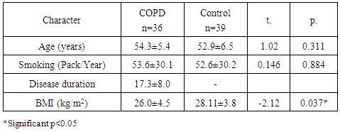
The results of this study revealed no significant statistical difference in age, smoking pack/year between both COPD and control groups, however, BMI was significantly lower in COPD group. Table (1) describes the baseline characteristics of the study groups.
Table (1). Demographic data of the studied group
     
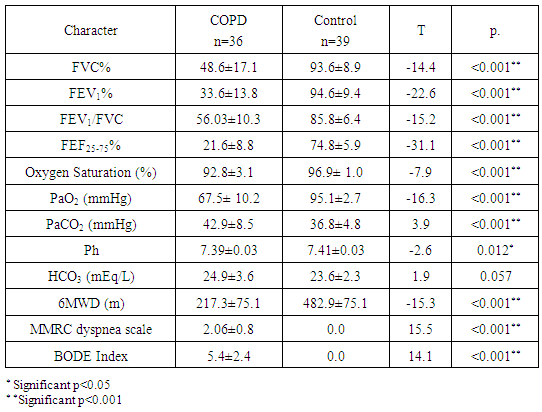
Spirometric parameters (FEV1, FVC, and FEV1/FVC), arterial blood gasses (PaO2 & PaCO2), BODE index and its parameters (6MWD, MMRD) of the COPD group were significantly lower than the control group (p<0.001) and (table 2).
Table (2). Spirometry, arterial blood gasses and BODE index parameters of the studied group
     
Serum Levels of HMGB1 and MMP9:
First, we compared HMGB1 and MMP9 serum levels between COPD patients and healthy subjects. As listed in Table 3, both levels in COPD patients were significantly increased in comparison with healthy subjects (62.6±48.3 versus 35.4±24.9 pg/mL, p-value 0.003 and 201.8±32.8 versus 164.6±59. 2 ng/ml p-value 0.001 respectively).
Table (3). Serum levels of HMGB1 and MMP9 in all groups
     
Both HMGB1 and MMP-9 were significantly higher in COPD than in control group.
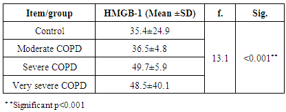
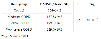
Analysis of these markers levels in each stage of COPD was done as shown in tables (4, 5).
Table (4). HMGB-1 in different stages of COPD severity
     
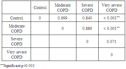
Table (5). Presentation of the results of the Tukey test for the data from Table 4
     
Serum HMGB-1 levels are increased significantly as the COPD stage worsens.
The difference in HMGB-1 value was significant in comparing control and moderate COPD groups with very severe COPD group.
Table (6). MMP-9 in different stages of COPD severity
     
MMP-9 increased significantly as the COPD stage worsens
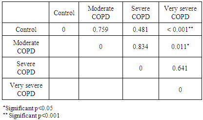
Table (7). Presentation of the results of the Tukey test for the data from Table 6
     
The difference in MMP-9 values was significant only in moderate versus very severe COPD and in control when compared to very severe COPD.
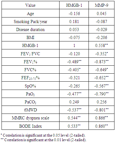
Table (8). Correlation between HMGB-1, MMP-9 and studied parameters in COPD group
     
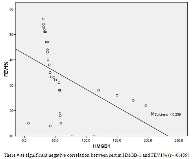
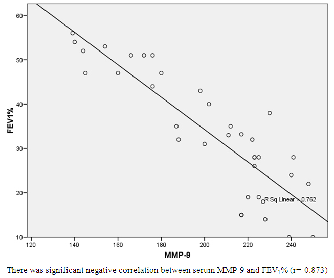
There was asignificant positive correlation between HMGB-1 with MMR dyspnea scale and BODE index, COPD stage and negative correlation with PaO2, spirometric indices FEV1, FVC, FEF25-75, and 6MWD. MMP-9 was correlated positively with MMRD score and BODE index and negatively correlated with oxygen saturation, PaO2, spirometric indices and 6MWD. There was positive correlation between HMBG-1 and MMP-9. However; both HMBG-1 and MMP-9 are not correlated with age, smoking pack/year or BMI.
Figure (1). Correlation between serum HMGB-1 and FEV1%
Figure (2). Correlation between serum MMP-9-1 and FEV1%

4. Discussion

The airflow limitation in COPD is usually progressive and associated with an abnormal inflammatory response of the lung to noxious particles or gasses. Chronic local and systemic inflammation arereasons for remodeling of lung tissue and extrapulmonary systemic change [30]. The aim of this study was to measure the serum levels of HMGB1 and MMP-9 in COPD and to study its relation with severity of airway obstruction.
In this study, we studied 36 patients with moderate to very severe COPD and 39 age and sex matched healthy controls. All patients and controls were males and all were smokers, smoking pack/years were higher in COPD than in control but the difference was non-significant (table 1). As expected, spirometric indices, arterial blood gasses and BODE index parameters were significantly different in both groups (table 2).
Previous studies searched for diagnostic tools to measure the extent of lung inflammation non-invasively [6-10, 36, 37]. However few studies examined HMBG-1 as a potential inflammatory marker in COPD. There are data showing that HMGB1 can be an inducer of inflammation [38-39]. The previous studies have shown increased sputum MMP-9 concentrations; alveolar macrophages were found to trigger larger amounts of MMP-9 with greater enzymatic activity in COPD patients [40] but few studies examined serum MMP-9 in COPD.
In the current study, we found significantly elevated serum HMGB1 in COPD group in comparison to control (table 3). The same results reported by different studies; Shang et al reported higher serum HMGB1 levels in COPD patients in comparison with control [41]. However, Zhang et al. observed the decline of serum HMGB1 levels from the acute phase to the convalescence phase of acute exacerbation of COPD [42]. Furthermore, HMGB1 levels were higher in smoker COPD patients compared with nonsmokers with COPD and exsmokers with COPD, respectively. Also Pouwels et al. studied serum and induced sputum levels of HMGB1 both during an exacerbation and during the stable phase of COPD [29]. They found no significant differences in sputum levels. however, serum levels of HMGB1 during COPD exacerbations were augmented. Cheng et al. study reported that the levels of HMGB1 in induced sputum of COPD patients were significantly higher than those of asthmatics and healthy controls [43].
On the other hand Iwamoto and his group conducted a study, of the duration of 4 years, They found no difference between plasma HMGB1 levels among the nonsmokers, smokers without COPD, and smokers with COPD [44]. In addition, there was no correlation between plasma HMGB1 levels and spirometric measurements. Ferhani et al. found that BAL levels of HMGB1 were comparable in never-smoker healthy subjects and in smokers without COPD but these levels were augmented in smokers with COPD. Bronchial biopsies revealed HMGB1-positive cells in high concentration in bronchial mucosa of smokers with COPD, more often than in healthy smokers [45].
Di Stefano et al. reported that BAL concentration of HMGB1 was significantly diminished in patients with stable COPD compared with control healthy subjects (smokers and nonsmokers). However, after matching patients for age and smoking history this difference was lost [46].
In the same context; Hou et al studied HMGB1 values both in sputum and in plasma [17]. They reported that compared with controls, HMGB1 levels in induced sputum were higher in asthmatic patients and in COPD patients. Plasma HMGB1 levels were similar to the ones obtained in sputum in COPD patients. Moreover, patients with COPD showed higher concentrations of sputum HMGB1 than patients with all severities of asthma.
Moreover; Bezerra et al. noticed higher levels HMGB-1 in alveolar macrophages from chronic cigarette smoke exposed animals [47].
Other studies involved lung tissue included Ko et al. study who enrolled twenty-eight patients with pulmonary neoplasia and found HMGB1 expression was augmented in submucosa cells, in epithelium cell, and in alveolar cells in smoker subjects with COPD but not smokers without COPD or in healthy subjects [1].
In this study on statistical analysis of the levels of HMGB1 in different stages of COPD severity, we found that the serum levels of HMGB-1 are increased significantly as the COPD stage worsens, the highest levels of HMGB1 detected in very severe COPD, the difference was significant only in comparing both control and moderate COPD with the group with versus very severe COPD (table 4, 5). Zhang et al. found HMGB1 levels were related to COPD severity [42].
Cheng et al. study reported that the levels of HMGB1 in induced sputum in COPD stage II and stage III were significantly higher than those with stage I [43].
In the current study; the levels of plasma HMGB1 negatively correlated with the post-bronchodilator FEV1%, FEF25-75 and FVC. There was no significant correlation between age and serum HMGB1 levels (table 8, fig 1). As all study subjects were smokers, so the results indicate that the elevated level of inflammatory markers may be attributed to COPD perse, however, pack/years smoking were significantly higher in COPD than in control so smoking may play anadjuvant role. Previous studies support our work Ko et al. observed a negative correlation with lung function parameters in smoker subjects with and without COPD [1]. Also, Ferhani et al. reported that the levels of HMGB1 in BAL were correlated negatively with post-bronchodilator FEV1 [45]. Kanazawa et al. demonstrated that the HMGB1 levels in the epithelial lining fluid from peripheral airway were significantly higher in COPD patients than those in non-smokers and smokers without COPD [48]. Cheng et al. identified that the sputum concentrations of HMGB1 in the COPD patients were significantly negatively correlated with FEV1% pre in all subjects [43]. Hou et al. also reported that HMGB1 levels in plasma and in induced sputum in all subjects showed a negative correlation with lung function parameters [17]. Kanazawa et al. reported no correlation between HMGB1 level in central airways and indexes of pulmonary function [48]. On the contrary, HMGB1 level in peripheral airways was related to the functional tests.
On the other hand, Iwamoto and his group didn’t find acorrelation between plasma HMGB1 levels and spirometric measurements [44].
Previous studies and the current one indicate that HMGB1 levels correlate with the degree of airflow obstruction in smokers with COPD.
In this study we found that serum levels of MMP-9 were significantly higher in COPD patients when compared to control group (table 3) and the levels were higher in severe and very severe stages (table 6, 7). This increase could result in ECM destruction in the airways and contributing to airway remodelling and the decline in lung function seen in COPD patients. Vignola et al. suggested that sputum levels of MMP-9 reflect the extent of structural changes of the airways [49].
The same results were obtained by Brajer et al. and Xin et al.who found that COPD patients had increased serum MMP-9 concentration compared with the control group [50, 51]. Beeh et al. [52] found that sputum concentration of MMP-9 was higher in COPD patients than in control subjects. Culpitt et al. observed that sputum from COPD patients contained increased levels of MMP-9 compared with non-smokers, non-symptomatic cigarette smokers, and asthmatics [26].
Mercer et al., investigated changes in sputum levels of MMP-9 and its inhibitor; the tissue inhibitors of metalloproteinase TIMP-1 during exacerbations in COPD and compared these to samples taken from the same individuals prior to exacerbation. They indicated that exacerbations are associated with an increase in MMP-9 burden in the airways. [53]
Another Egyptian study demonstrated that MMP-9 was significantly increased in both COPD patients), and healthy smokers compared with healthy non smokers [54].
In this study, MMP-9 levels are negatively correlated with FEV1%, FVC% but not correlated with neither FEF25-75% nor FEV1/FVC and negatively correlated with PaO2 but not with SpO2% or PaCO2. The levels are positively correlated with BODE Index, MMRC dyspnea scale and negatively with 6MWD but not with BMI (table 8). Similar results were obtained by Linder et al., who studied the association between serum MMP-9 and impaired lung function, assessed as FEV1, in COPD and showed that MMP-9 is related to disease severity which could indicate that MMP-9 is involved in the disease process in COPD. [55]
Some studies reported the same results; Brajer et al., found that MMP-9 levels were negatively correlated with FEV1 and FEV1/FVC [50]. Another research has shown apositive correlation between MMP-9/TIMP-1 ratio and airway obstruction assessed by FEV1 measurement [49]. Beeh et al. found that MMP-9 concentration correlated negatively with the severity of airway obstruction (FEV1%, FVC) [52]. Kang et al. revealed that lung parenchymal MMP-9 concentration correlated with the amount of cigarette smoked and inversely correlated with FEV1 [56]. Mao et al. observed that plasma MMP-9 levels were higher in patients with emphysema compared with age-matched, non-smoking control subjects [57]. Ólafsdottir et al. reported lower FEV1 values were associated with higher serum levels of MMP-9 [58]. However, Xin et al. reported that MMP9 levels did not correlate with FEV1 % values and FEV1/FVC [51]. Abd El-Fatah et al., suggested that MMP-9 serves as a biomarker for the grade and severity of COPD by assessing the mRNA levels of MMP-9 in peripheral blood samples, [59].
This study, as well as previous reports, showed that MMP-9 concentrations are associated with airflow obstruction, suggesting that MMP-9 may play a role in the pathogenesis of COPD.
In conclusion, the results of this study suggest that both HMGB1 and MMP-9 plays an important role in systemic inflammation in COPD. And higher serum concentrations are connected with higher airway obstruction and disease progression.

References

[1]  Ko HK, Hsu WH, Hsieh CC, Lien TC, Lee TS, Kou YR. High expression of high‐mobility group box 1 in the blood and lungs is associated with the development of chronic obstructive pulmonary disease in smokers. Respirology. 2014; 19(2): 253-61.
[2]  Wang H, Yang T, Li D, Wu Y, Zhang X, Pang C, Zhang J, Ying B, Wang T, Wen F. Elevated circulating PAI-1 levels are related to lung function decline, systemic inflammation, and small airway obstruction in chronic obstructive pulmonary disease. International Journal of Chronic Obstructive Pulmonary Disease. 2016;11:2369.
[3]  Gopal P, Reynaert NL, Scheijen JL, Schalkwijk CG, Franssen FM, Wouters EF, Rutten EP. Association of plasma sRAGE, but not esRAGE with lung function impairment in COPD. Respiratory research. 2014; 15(1): 24.
[4]  Barnes PJ, Shapiro SD, Pauwels RA. Chronic obstructive pulmonary disease: molecular and cellularmechanisms. European Respiratory Journal. 2003; 22(4): 672-88.
[5]  Mroz RM, Noparlik J, Chyczewska E, Braszko JJ, Holownia A. Molecular basis of chronic inflammation in lung diseases: new therapeutic approach. Journal of Physiology and Pharmacology. 2007 Nov 1; 58(5): 453-60.
[6]  Fattouh M, Alkady O. Inflammatory biomarkers in chronic obstructive pulmonary disease. Egyptian Journal of Chest Diseases and Tuberculosis. 2014; 63(4): 799-804.
[7]  Eagan TM, Ueland T, Wagner PD, Hardie JA, Mollnes TE, Damås JK, Aukrust P, Bakke PS. Systemic inflammatory markers in COPD: results from the Bergen COPD Cohort Study. European respiratory journal. 2010 Mar 1; 35(3): 540-8.
[8]  Hacievliyagil SS, Mutlu LC, Temel I. Airway inflammatory markers in chronic obstructive pulmonary disease patients and healthy smokers. Nigerian journal of clinical practice. 2013; 16(1): 76-81.
[9]  Mannino DM, Tal-Singer R, Lomas DA, Vestbo J, Barr RG, Tetzlaff K, Lowings M, Rennard SI, Snyder J, Goldman M, Martin UJ. Plasma fibrinogen as a biomarker for mortality and hospitalized exacerbations in people with COPD. Chronic obstructive pulmonary diseases (Miami, Fla.). 2015; 2(1): 23.
[10]  Seemungal TA, Lun JC, Davis G, Neblett C, Chinyepi N, Dookhan C, Drakes S, Mandeville E, Nana F, Setlhake S, King CP. Plasma homocysteine is elevated in COPD patients and is related to COPD severity. International journal of chronic obstructive pulmonary disease. 2007; 2(3): 313.
[11]  Chen Q, Guan X, Zuo X, Wang J, Yin W. The role of high mobility group box 1 (HMGB1) in the pathogenesis of kidney diseases. Acta Pharmaceutica Sinica B. 2016; 6(3): 183-8.
[12]  Tang D, Kang R, Zeh III HJ, Lotze MT. High-mobility group box 1, oxidative stress, and disease. Antioxidants & redox signaling. 2011; 14(7): 1315-35.
[13]  Fang WH, Yao YM, Shi ZG, Yu Y, Wu Y, Lu LR, Sheng ZY. The significance of changes in high mobility group-1 protein mRNA expression in rats after thermal injury. Shock. 2002; 17(4): 329-33.
[14]  Ulloa L, Batliwalla FM, Andersson U, Gregersen PK, Tracey KJ. High mobility group box chromosomal protein 1 as a nuclear protein, cytokine, and potential therapeutic target in arthritis. Arthritis & Rheumatology. 2003; 48(4): 876-81.
[15]  Sun S, Zhang W, Cui Z, Chen Q, Xie P, Zhou C, Liu B, Peng X, Zhang Y. High mobility group box-1 and its clinical value in breast cancer. OncoTargets and therapy. 2015; 8: 413.
[16]  Ueno H, Matsuda T, Hashimoto S, Amaya F, Kitamura Y, Tanaka M, Kobayashi A, Maruyama I, Yamada S, Hasegawa N, Soejima J. Contributions of high mobility group box protein in experimental and clinical acute lung injury. American journal of respiratory and critical care medicine. 2004; 170(12): 1310-6.
[17]  Hou C, Zhao H, Liu L, Li W, Zhou X, Lv Y, Shen X, Liang Z, Cai S, Zou F. High mobility group protein B1 (HMGB1) in Asthma: comparison of patients with chronic obstructive pulmonary disease and healthy controls. Molecular Medicine. 2011; 17(7): 807-15.
[18]  Gangemi S, Casciaro M, Trapani G, Quartuccio S, Navarra M, Pioggia G, Imbalzano E. Association between HMGB1 and COPD: a systematic review. Mediators of inflammation. 2015; 2015.
[19]  Feng L, Xue D, Chen E, Zhang W, Gao X, Yu J, Feng Y, Pan Z. HMGB1 promotes the secretion of multiple cytokines and potentiates the osteogenic differentiation of mesenchymal stem cells through the Ras/MAPK signaling pathway. Experimental and Therapeutic Medicine. 2016; 12(6): 3941-7.
[20]  Konjevic G, Gopčević K, V Jovic, Inic M and Jurisic V. Activity of MMP-2 and MMP-9 in sera of breast cancer patients. Stankovic S,. Pathol Res Pract. 2010 Jun 15; 206 (4): 241-7.
[21]  Radenkovic S, Konjevic G, Jurisic V, Karadzic K, M Nikitovic, Gopčević K. Values of MMP-2 and MMP-9 in the tumor tissue of basal-like breast cancer patients. Cell Biochem Biophys. 2014 Jan; 68 (1): 143-52.
[22]  Piesiak P, Brzecka A, Kosacka M, Passowicz-Muszyńska E, Dyła T, Jankowska R. Concentrations of matrix metalloproteinase-9 and tissue inhibitor of metalloproteinases-1 in serum of patients with chronic obstructive pulmonary disease. Polski merkuriusz lekarski: organ Polskiego Towarzystwa Lekarskiego. 2011; 31(185): 270-3.
[23]  Dilek F, Ozceker D, Ozkaya E, Tamay Z, Yazici M, Kesgin S, Kocyigit A, Guler N. Plasma levels of matrix metalloproteinase-9 in children with chronic spontaneous urticaria. Allergy, asthma & immunology research. 2016; 8(6): 522-6.
[24]  Sang QX, Muroski ME, Roycik MD, Newcomer RG, Van den Steen PE, Opdenakker G, Monroe HR, Sahab ZJ. Matrix metalloproteinase-9/gelatinase B is a putative therapeutic target of chronic obstructive pulmonary disease and multiple sclerosis. Current pharmaceutical biotechnology. 2008; 9(1): 34-46.
[25]  Omachi TA, Eisner MD, Rames A, Markovtsova L, Blanc PD. Matrix metalloproteinase-9 predicts pulmonary status declines in α 1-antitrypsin deficiency. Respiratory research. 2011; 12(1): 35.
[26]  Culpitt SV, Rogers DF, Traves SL, Barnes PJ, Donnelly LE. Sputum matrix metalloproteases: comparison between chronic obstructive pulmonary disease and asthma. Respiratory medicine. 2005; 99(6): 703-10.
[27]  Vernooy JH, Lindeman JH, Jacobs JA, Hanemaaijer R, Wouters EF. Increased activity of matrix metalloproteinase-8 and matrix metalloproteinase-9 in induced sputum from patients with COPD. CHEST Journal. 2004;126(6):1802-10.
[28]  Louhelainen N, Stark H, Mazur W, Rytilä P, Djukanovic R, Kinnula VL. Elevation of sputum matrix metalloproteinase-9 persists up to 6 months after smoking cessation: a research study. BMC pulmonary medicine. 2010;10(1):13.
[29]  Pouwels SD, Nawijn MC, Bathoorn E, Riezebos-Brilman A, van Oosterhout AJ, Kerstjens HA, Heijink IH. Increased serum levels of LL37, HMGB1 and S100A9 during exacerbation in COPD patients. European Respiratory Journal. 2015; 45(5): 1482-5.
[30]  Global Strategy for the Diagnosis, Management and Prevention of COPD, Global Initiative for Chronic Obstructive Lung Disease (GOLD) 2016. Available from: http://goldcopd.org/.
[31]  Spruit MA, Singh SJ. Maintenance programs after pulmonary rehabilitation: how may we advance this field?. CHEST Journal. 2013; 144(4): 1091-3.
[32]  Stenton C. The MRC breathlessness scale. Occupational Medicine. 2008; 58(3): 226-7.
[33]  ATS Statement: Guidelines for the Six-Minute Walk Test: Am J Respir Crit Care Med 2002;166:111–117.
[34]  Celli BR, MacNee WA, Agusti AA, Anzueto A, Berg B, Buist AS, Calverley PM, Chavannes N, Dillard T, Fahy B, Fein A. Standards for the diagnosis and treatment of patients with COPD: a summary of the ATS/ERS position paper. European Respiratory Journal. 2004; 23(6): 932-46.
[35]  Miller MR, Crapo R, Hankinson J, Brusasco V, Burgos F, Casaburi R, Coates A, Enright P, van der Grinten CM, Gustafsson P, Jensen R. General considerations for lung function testing. European Respiratory Journal. 2005; 26(1): 153-61.
[36]  Hermans C, Bernard A. Pneumoproteinaemia: a new perspective in the assessment of lung disorders. European Respiratory Journal. 1998; 11(4): 801-3.
[37]  Hermans C, Bernard A. Lung epithelium–specific proteins: characteristics and potential applications as markers. American journal of respiratory and critical care medicine. 1999; 159(2): 646-78.
[38]  Entezari M, Javdan M, Antoine DJ, Morrow DM, Sitapara RA, Patel V, Wang M, Sharma L, Gorasiya S, Zur M, Wu W. Inhibition of extracellular HMGB1 attenuates hyperoxia-induced inflammatory acute lung injury. Redox biology. 2014; 2:314-22.
[39]  Musumeci D, Roviello GN, Montesarchio D. An overview on HMGB1 inhibitors as potential therapeutic agents in HMGB1-related pathologies. Pharmacology & therapeutics. 2014; 141(3): 347-57.
[40]  Russell RE, Culpitt SV, DeMatos C, Donnelly L, Smith M, Wiggins J, Barnes PJ. Release and activity of matrix metalloproteinase-9 and tissue inhibitor of metalloproteinase-1 by alveolar macrophages from patients with chronic obstructive pulmonary disease. American journal of respiratory cell and molecular biology. 2002; 26(5): 602-9.
[41]  Shang GH, Jia CQ, Tian H, Xiao W, Li Y, Wang AH, Dong L, Lin DJ. Serum high mobility group box protein 1 as a clinical marker for non-small cell lung cancer. Respiratory medicine. 2009; 103(12): 1949-53.
[42]  Zhang Y, Li S, Wang G, Han D, Xie X, Wu Y, Xu J, Lu J, Li F, Li M. Changes of HMGB1 and sRAGE during the recovery of COPD exacerbation. Journal of thoracic disease. 2014; 6(6): 734-41.
[43]  Cheng Z, Kang Y, Wu QG, Dai LL, Song YN, Xia J, Si JM, Chen CY. Levels of HMGB1 in induced sputum from patients with asthma and chronic obstructive pulmonary disease. Zhonghua yi xue za zhi. 2011; 91(42): 2981-4.
[44]  Iwamoto H, Gao J, Pulkkinen V, Toljamo T, Nieminen P, Mazur W. Soluble receptor for advanced glycation end-products and progression of airway disease. BMC pulmonary medicine. 2014; 14(1): 68.
[45]  Ferhani N, Letuve S, Kozhich A, Thibaudeau O, Grandsaigne M, Maret M, Dombret MC, Sims GP, Kolbeck R, Coyle AJ, Aubier M. Expression of high-mobility group box 1 and of receptor for advanced glycation end products in chronic obstructive pulmonary disease. American journal of respiratory and critical care medicine. 2010; 181(9): 917-27.
[46]  Di Stefano A, Caramori G, Barczyk A, Vicari C, Brun P, Zanini A, Cappello F, Garofano E, Padovani A, Contoli M, Casolari P. Innate immunity but not NLRP3 inflammasome activation correlates with severity of stable COPD. Thorax. 2014; 69(6): 516-24.
[47]  Bezerra FS, Valença SS, Pires KM, Lanzetti M, Pimenta WA, Schmidt AC, Porto LC, Zin WA. Long-term exposure to cigarette smoke impairs lung function and increases HMGB-1 expression in mice. Respiratory physiology & neurobiology. 2011; 177(2): 120-6.
[48]  Kanazawa H, Tochino Y, Asai K, Ichimaru Y, Watanabe T, Hirata K. Validity of HMGB1 measurement in epithelial lining fluid in patients with COPD. European journal of clinical investigation. 2012; 42(4): 419-26.
[49]  Vignola AM, Riccobono L, Mirabella A et al.(1998): Sputum metalloproteinase-9/tissue metalloproteinase-1 ratio correlates with airflow obstruction in asthma and chronic bronchitis. American Journal of Respiratory and Critical Care Medicine. 1998; 158(6): 1945-50.
[50]  Brajer B, Batura-Gabryel H, Nowicka A, Kuznar-Kaminska B, Szczepanik A. Concentration of matrix metalloproteinase-9 in serum of patients with chronic obstructive pulmonary disease and a degree of airway obstruction and disease progression. J Physiol Pharmacol. 2008; 59(Suppl 6): 145-52.
[51]  Xin XF, Zhao M, Li ZL, Song Y, Shi Y. Metalloproteinase-9/tissue inhibitor of metalloproteinase-1 in induced sputum in patients with asthma and chronic obstructive pulmonary disease and their relationship to airway inflammation and airflow limitation. Zhonghua jie he he hu xi za zhi= Zhonghua jiehe he huxi zazhi= Chinese journal of tuberculosis and respiratory diseases. 2007; 30(3): 192-6.
[52]  Beeh KM, Beier J, Kornmann O, Buhl R. Sputum matrix metalloproteinase-9, tissue inhibitor of metalloprotinease-1, and their molar ratio in patients with chronic obstructive pulmonary disease, idiopathic pulmonary fibrosis and healthy subjects. Respiratory medicine. 2003; 97(6): 634-9.
[53]  Mercer P, Shute J, Bhowmik A, Donaldson G, Wedzicha J, Warner J. MMP-9, TIMP-1 and inflammatory cells in sputum from COPD patients during exacerbation. Respiratory Research. 2005; 6(1):151.
[54]  Esa SA, RawyAM, EL-Behissy MM, Kamel MH and El-Hwaitty HM. Study of the level of sputum matrix metalloproteinase-9 (MMP-9) and tissue inhibitor metalloproteinase-1 (TIMP-1) in COPD patients; Egypt. J. Chest Dis. Tuberc., 4 (63) (2014), pp. 861–867.
[55]  Linder R, Rönmark E, Pourazar J, Behndig A, Blomberg A, Lindberg A. Serum metalloproteinase-9 is related to COPD severity and symptoms - cross-sectional data from a population based cohort-study. Respir Res.( 2015 ) 21; 16:28.
[56]  Kang MJ, Oh YM, Lee JC, Kim DG, Park MJ, Lee MG, Hyun IG, Han SK, Shim YS, Jung KS. Lung matrix metalloproteinase-9 correlates with cigarette smoking and obstruction of airflow. Journal of Korean medical science. 2003; 18(6): 821.
[57]  Mao JT, Tashkin DP, Belloni PN, Baileyhealy I, Baratelli F, Roth MD. All-trans retinoic acid modulates the balance of matrix metalloproteinase-9 and tissue inhibitor of metalloproteinase-1 in patients with emphysema. CHEST Journal. 2003; 124(5): 1724-32.
[58]  Olafsdottir IS, Janson C, Lind L, Hulthe J, Gunnbjörnsdóttir M, Sundström J. Serum levels of matrix metalloproteinase‐9, tissue inhibitors of metalloproteinase‐1 and their ratio are associated with impaired lung function in the elderly: A population‐based study. Respirology. 2010;15(3):530-5.
[59]  Abd El-Fatah, M. F., Ghazy, M. A., Mostafa, M. S., El-Attar, M. M., & Osman, A. (2015). Identification of MMP-9 as a biomarker for detecting progression of chronic obstructive pulmonary disease. Biochemistry and Cell Biology, 93(6), 541-547.