-
Paper Information
- Paper Submission
-
Journal Information
- About This Journal
- Editorial Board
- Current Issue
- Archive
- Author Guidelines
- Contact Us
Clinical Medicine and Diagnostics
p-ISSN: 2163-1433 e-ISSN: 2163-1441
2017; 7(1): 1-7
doi:10.5923/j.cmd.20170701.01

Percutaneous Electrical Nerve Field Stimulation Targeting Neurovascular Structures of the Auricle: A Fractal Based Approach
 Abstract
 Reference
 Full-Text PDF
 Full-text HTML
Christopher R. Brown
Director of Research and Development, Innovative Health Solutions, Associate Educational Director Indianapolis Craniofacial Pain, American Academy of Integrative Pain Management BOD (Retired)
Correspondence to: Christopher R. Brown, Director of Research and Development, Innovative Health Solutions, Associate Educational Director Indianapolis Craniofacial Pain, American Academy of Integrative Pain Management BOD (Retired).
| Email: |  |
Copyright © 2017 Scientific & Academic Publishing. All Rights Reserved.
This work is licensed under the Creative Commons Attribution International License (CC BY).
http://creativecommons.org/licenses/by/4.0/

Fractal geometry offers a platform to explain structure-function relationships of complex systems in biological science and engineering. Branching of the arterial and neurological systems is characterized in a fractal nature. Self-similar branching patterns and recursive bifurcationoccurs in descending orders of magnitude to the cellular level. The NSS family of devices EAD ® (electro-auricular device), Bridge ®, MFS ® (military field stimulator) IBStim ®, and SurgiStim ® (Key Electronics, Jeffersonville, In) when percutaneously implanted in the Auricle in accordance with Ohm’s and Coulomb’s law as guided by trans-illumination produces a field effect which theoretically optimizes energy transfer through dissimilar tissues resulting in the stimulation (targeting) of cranial neurovascular bundles.
Keywords: Fractal, Lacunarity, Homeostasis, Neuro-vascular, Trans-illumination, Arrays
Cite this paper: Christopher R. Brown, Percutaneous Electrical Nerve Field Stimulation Targeting Neurovascular Structures of the Auricle: A Fractal Based Approach, Clinical Medicine and Diagnostics, Vol. 7 No. 1, 2017, pp. 1-7. doi: 10.5923/j.cmd.20170701.01.
Article Outline
1. Fractal Analysis: A General Overview
- Fractal geometry offers a platform to explain structure-function relationships of complex systems in biological science and engineering. The branching system in our body is characterized in a fractal nature: self-similar branching patterns and recursive bifurcation occurring in descending orders of magnitude to the cellular level illustrating biological iteration (the process of repeating a set of patterns or mathematical sets in which each set acts as a starting point for the next set). [1] The end result is that all components, in all dimensions, have a form similar but not identical to the whole. [2] Natural fractal entities are mainly characterized by four properties:1. Irregularity of their shape2. Self-similarly of their structures3. Non integer of fractional (fractal) dimensions4. Scaling (the measured properties depend upon the scale by which they are measured)It is now recognized that "rough" shape is the most important property of every anatomical system strongly influencing its behavior and its relationship with surrounding components. This concept is often applied to biological systems described as “closed areas” within the human body such as the lung, kidney, heart, and the external ear.Another fractal parameter is “lacunarity.” Lacunarity refers to a measurement of how fractal patterns fill a given space often producing gaps that, in the case of biological entities such as the lungs, heart, kidney, and for this discussion the external ear, are filled with other tissues and extra-cellular fluids. [3] The external ear is a multi-fractal system meaning it cannot be explained by a single component. At both a macroscopic and microscopic level, the intrinsic complexity is apparent producing systems within systems. [3, 4] The result is an integrated continuum of multi- fractal components including neural, vascular, neuro-vascular bundles, cartilage, lacunae and variations of tissues with different densities and therefore differences in conductivity and harmonic responses. Fractal properties occur at anatomical, functional, pathological, molecular and epigenetic levels and as a result no physiologic network is isolated making up self- similar sub- networks of interacting tissues. Absorption of energy is determined by the fractal scaling of the effected entities. If the energy is projected through “self similar” structures the resulting patterns are predictable. If however there are separate entities within a given structure the energy will be both absorbed and reflected at different rates resulting in what is referred to as “noise”. [4] As long as the biological environment remains homeostatic, systems will continue to function guided by complex positive and negative feedback mechanisms and the differentiation of absorption rates will remain relatively constant. From a biological thermodynamic perspective the system would be considered in mechanical, thermal, phase, and chemical equilibrium. [5] When a disruption in homeostasis/ equilibrium occurs changes in anatomical form and function occur. The infinite factors which occur in an open biological system is immeasurable. This biological and thermodynamic reaction is often referred to as the Chaos Theory which states that complex systems rely on underlying order and that simple homeostatic disruptions can result in complex changes, adaptations and plasticity. The afore-mentioned systems’ adaptations however will adapt in a fractal self-similar fashion and changes in angiogenesis and vascular morphology may be an indicator of pathology. [6] The loss of homeostasis and dynamic feedback mechanisms often contribute to neurovascular plasticity which may be contingent upon but not limited to:1. The presence of vascularity competing for the same space replicating in a self -similar fractal manner.2. The presence of intra-cellular lacunae which may directionally alter tissue expansion. 3. Variations in density of tissues within the self-limiting space.4. Osmotic inter-cellular pressures created by biochemical inflammatory markers, cytokines, intercellular fluid, medications, pathology, and age.5. A non-homeostatic condition that is caused by or is affected by inhibition of autonomic inhibition mechanisms resulting in a physiological imbalance such as chronic sympathetic or parasympathetic stimulation. [7]
2. Fractal Analysis of the Neuro-Vascular System
- The neuro-vascular system is best understood using fractal-like principles in order to define its functional system. [8] “Fractal-like” analysis of the neuro-vascular system versus purely “fractal” analysis is utilized because the structure of the arterial branches depends on a number of factors including the vascular structure, genetic disposition, external factors, pathology, etc. Considering the differing factors that influence the structure of the arterial tree, it would be improbable that one simple model or mechanism can predict the exact structure of the arterial tree in every detail, rather the arterial structure has a “fractal-like” appearance. For instance the blood supply to the brain is provided by a network of vessels running along its surface connecting a few large arteries of primary supply to thousands of arteries passing through the brain cortex. The arteries break down into smaller and smaller branches by bifurcation otherwise referred to as dichotomous division (dividing into two parts) to the cellular level in order to bring oxygen and other nutrients to the cells of the body’s tissues and organs. Fractal principles explain the design of transfer routes interconnecting the inputs of the brain’s vasculature to the perforating arteries. The arterial tree is space-filling and minimizes the energy dissipation due to blood viscosity and pulsatile flow. The arterial system has a fractal-like dimension by virtue of its space-filling property (Lacunarity). Blood pressure, flow rates (both increase and decrease), and distribution are constantly changing in response to stimuli. [9] Angiogenesis (developmental remodeling) can occur as a result of pathology and chronic sympathetic stimulation resulting in vasomotor responses mediated by the endothelium. There is evidence that arterial remodeling serves to normalize vessel wall shear stress. [10]
3. Vascular Impedance
- Electrical impedance and physiological resistance mismatching leads to pulse wave reverberation and amplification. [11] Increasing vascular resistance - matching design of fractal vascular networks leads to potential network interactions for steady components of flow such as shear stress amplification. If the magnitude of flow in vessels demonstrates a dependence of diameter then the concept of impedance matching and wave actions becomes important. The vascular network organization plays a role in altering hemodynamic forces in peripheral vessels. In fact alterations in electrical impedance can be an indication of vascular changes in vascular bed membranous structures. [12, 13]In the vascular system gradual changes in impedance and hydraulic pressures are accomplished by fractal-like organization both geometrically and topographically. Vessels possess a viscoelastic-like wall that facilitates pulse wave propagation.Trans- endothelial electrical resistance (TER) can be altered by reduction of sympathetic stimulation in the endothelial walls of the vessels allowing for change in vascular wall resistance. [14] Lipophylic molecules (phospholipids, cholesterol, etc.) facilitate restriction of the passage of aqueous solutions and function as electrical insulators to maintain physiological endothelial potential. Knudsen diffusion gradients, which accounts for the diffusion of gas molecules through small capillary pores are often affected as well. The fluid filled pores at cell- to- cell junctions also serve as electrical conductance pathways across micro vascular epithelium. Reduction of sympathetic stimulation of the vascular walls will therefore decrease trans - endothelial resistance (TER) allowing for normalization of trans vessel fluid filtration and a return to osmotic homeostasis.
4. Arterial Branches of the External Ear
- The arterial blood supply of the external ear is primarily from two sources: the posterior auricular artery and the superficial temporal artery. [15] Clinical visualization of the main branches of the superficial temporal and posterior auricular artery in the external ear is easily observed. The arteries bifurcate in a self-similar (fractal) manner into the cellular level on both the dorsal and ventral aspects creating an extensive neuro-vascular matrix on a three dimensional scale.Because of the fractal nature of arterial design the greatest mass of the arteries is not in the main visible branch as visualized by trans-illumination but rather in the self-replicating bifurcations located three dimensionally around the main arterial branch. Visual changes of arterial patterns in the external ear such as broken, engorged arterial branches along with red, flushing, edematous/ischemic tissues of the ear and a feeling of warmth to the patient (Red Ear Syndrome) are important clinical biomarkers occurring as a result of sympathetically mediated or maintained clinical entities. [16]
5. Trans-illumination
- The use of trans-illumination helps assist the clinician with the visualizations of these neurovascular biomarkers, the main arterial branches and the concurrent neuro-vascular matrixes. [17]
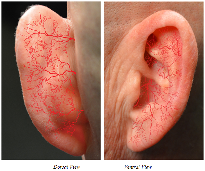 | Figure 1. Arterial branches bifurcate in a self-similar (fractal) manner to the cellular level |
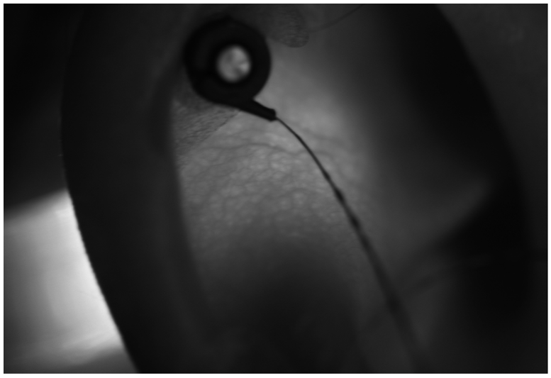 | Figure 2. Transillumination of auricle isolating neurovascular bundles, ventral view |
6. Fractal Design of the NSS
- ArraysThe NSS (Neuro-Stim System) electrode arrays were designed to decrease sympathetic stimulation of targeted neuro-vascular bundles while creating less shear stress on the vascular walls. The device consists of a battery activated generator, a wire harness which connects to the generator, and four percutaneously implantable electrodes. One of the wire leads is connected to a single pin ground electrode. The other three electrodes consist of individual pin arrays that vary from one to four individual pins depending upon device design. All pins are 2.1 mm in length to assure when the trans-illumination guided percutaneous implantation technique is precisely followed the electrical neuro-modulating signal is within the accepted 1-1.5 mm range of the neurovascular bundles assuring proper energy transfer in accordance with Ohm’s and Coulomb’s laws. [19] The functioning surface area of the pins is over 5.15 sq. mm creating a substantial electrode/tissue interface.The draft of the pins is designed to help reduce tissue displacement and as a result minimize discomfort to the patient. The draft also allows the signal to interface with the neurovascular bundles that along with the pin length allows for maximum neurovascular interfacing while reducing vascular impedance and resulting signal scatter. The resulting reduction in tissue “resistance” not only increases efficacy but helps with reduction of discomfort for the patient.Electrical signalThe NSS produces a 1-10 Hz neuro-modulating +/- signal, with a 1ms pulse width, a 3.2v amplitude, and an impulse interval of 2 sec. The generator of the NSS EAD ® has a programmed duty cycle of 2 active on and 2 hrs rest, for a total of 120 hrs. The other devises previously mentioned have similar impulses with differing “off” times to avoid attenuation. These impulses are sufficient to change the action potential of the cranial nerves (V, VII, IX, X) present in the outer ear and stimulate predictable areas of the brain. [20, 21]
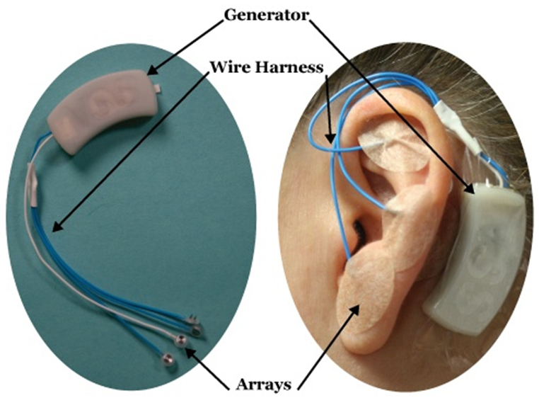 | Figure 3. NSS Device |
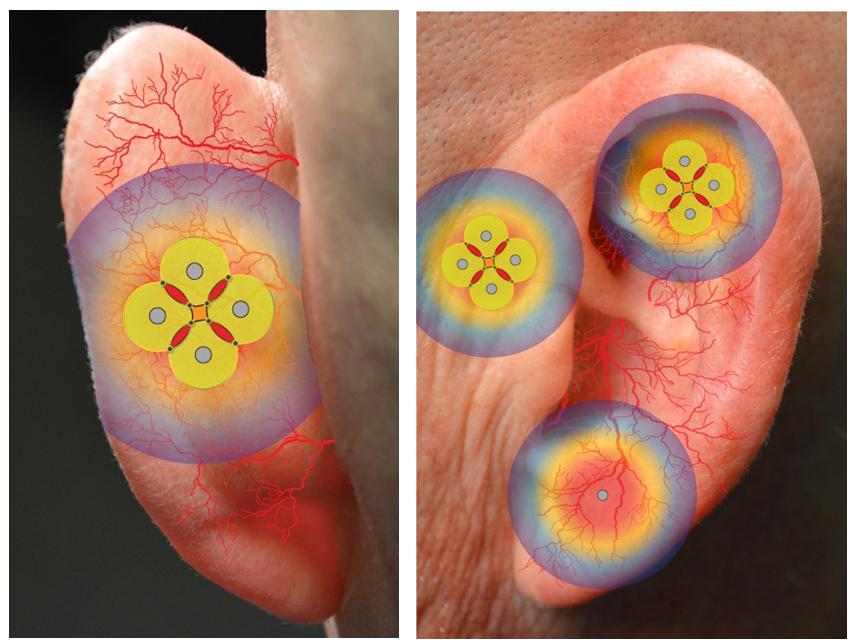 | Figure 4. Array design and proper placement allows the neuro-modulating signals to be distributed in a self-similar (fractal-like) manner (artist’s conception) |
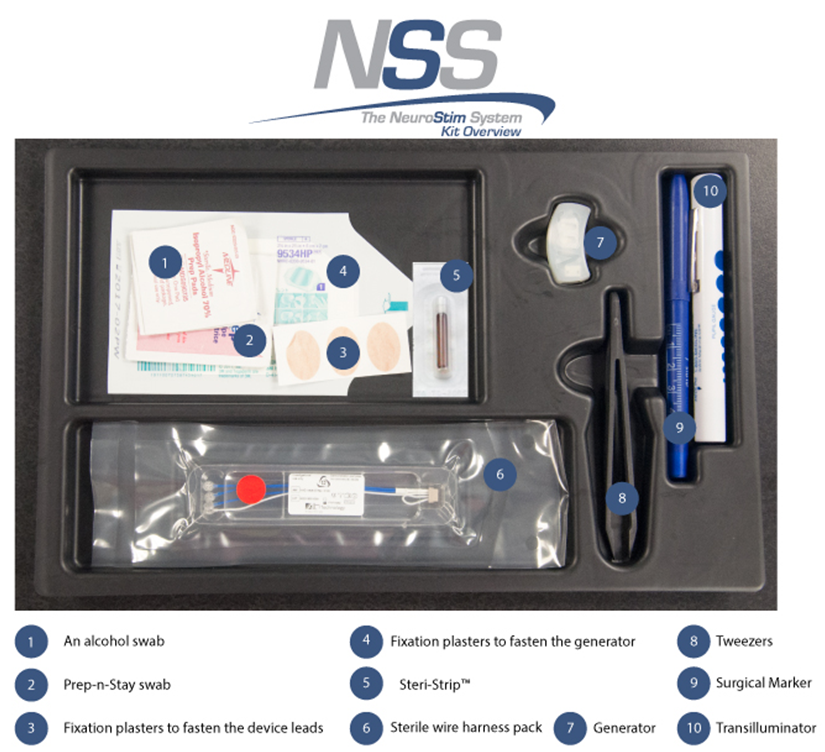 | Figure 5. NSS Surgical Kit |
7. Summary
- Percutaneously implanting the NSS® electrode arrays into the three dimensional mass of continually bifurcating, self-similar arterial branches of the outer ear following the patented IHS trans-illumination technique results in:1. Exponentially increasing the amount of arterial endothelial tissue exposed to the NSS ® neuro-modulating signal.2. Minimizes vascular resistance, deflection and minimizes vessel shear stress.3. Allows for the neuro-modulating signal to create potential for stimulation of neural, vascular and co-joined neurovascular tissue.4. Tissue deflection of the NSS® neuro-modulation signals creates kinetic energy resulting in changes in enthalpy, stimulating changes in permeability of vessel walls affecting osmotic pressure imbalances helping tissues reach equilibrium/homeostasis. Both ischemia and edema would be affected by these changes.5. Thermodynamic changes in enthalpy will further assist in reduction of sympathetic stimulation and reciprocating parasympathetic reactions.6. Restoration of normal tissue perfusion provides hemodynamic “cues” to help modulate sympathetic/parasympathetic feedback systems. [12] The NSS ® family of device therapy is unique in design and function due to the fractal design of the electrical signal as a result of the original signal generated and the pin/array design. When implanted utilizing transillumination the NSS combines the neuromodulation and targeting of neurovascular bundles previously available only with surgically implantable devices with the safety and minimally invasiveness of percutaneous implantation. The overall effect is an efficacious, non-surgical alternative for targeting percutaneous neurovascular field stimulation. [25, 26]