-
Paper Information
- Next Paper
- Paper Submission
-
Journal Information
- About This Journal
- Editorial Board
- Current Issue
- Archive
- Author Guidelines
- Contact Us
Clinical Medicine and Diagnostics
p-ISSN: 2163-1433 e-ISSN: 2163-1441
2016; 6(3): 45-48
doi:10.5923/j.cmd.20160603.01

Prevalence and Incidence of Uterine Fibroid at King Abdulaziz University Hospital Saudi Arabia
 Abstract
 Reference
 Full-Text PDF
 Full-text HTML
Hanan Y. Abbas 1, Ibrahim A. Awad 1, Ebtihal Alharbi 1, Halaiem Alaameri 1, Shaima Althubaiti 1, Layla Ashkar 2
1Department of Diagnostic Radiology, Faculty of Applied Medical Sciences, King Abdulaziz University, Jeddah, Saudi Arabia
2Department of Radiology, King Abdulaziz University Hospital, KSA
Correspondence to: Hanan Y. Abbas , Department of Diagnostic Radiology, Faculty of Applied Medical Sciences, King Abdulaziz University, Jeddah, Saudi Arabia.
| Email: |  |
Copyright © 2016 Scientific & Academic Publishing. All Rights Reserved.
This work is licensed under the Creative Commons Attribution International License (CC BY).
http://creativecommons.org/licenses/by/4.0/

Aim of the work: Extensive study has been made to get specific ratio of the women who had uterine fibroid and the total range of incidence of the leiomyoma. Methods and Material: Retrospective study of (1111) women patients referred from obstetrics and gynecology clinics (Women’s’ age between 15 and 79 years, mean was 52yrs). Data was collected from January 2013 to December 2014 at the Hospital of king Abdulaziz University. Results: A total of 236(21.2%) of 1111 patients were cases of uterine fibroid, while the remaining 875(78.8%) cases had normal ultrasound findings. According to the clinical symptoms that related to fibroid, bleeding which included 65(27.5%) was the commonest symptom for the patient’s then abdominal pain 32(13.6%). The 123(52%) of the women presented with solitary fibroids and 113(48%) women with multiple leiomyomata. Conclusions: Uterine fibroid highly related with reproductive age by (56.3%) and this result is matching to the result published in previous studies.
Keywords: Uterine fibroid, Incidence, Prevalence, Reproductive age
Cite this paper: Hanan Y. Abbas , Ibrahim A. Awad , Ebtihal Alharbi , Halaiem Alaameri , Shaima Althubaiti , Layla Ashkar , Prevalence and Incidence of Uterine Fibroid at King Abdulaziz University Hospital Saudi Arabia, Clinical Medicine and Diagnostics, Vol. 6 No. 3, 2016, pp. 45-48. doi: 10.5923/j.cmd.20160603.01.
Article Outline
1. Introduction
- Uterine leiomyomata are benign soft uterine muscle tumors that known as fibroids. The highest prevalence range of fibroids estimates is 3-20%, with African American and older women [1-4].In the United States women's fibroids comprise the most common cause of gynecological surgery, which means about one third of hysterectomies each year [5]. A range from 25% to 50% was estimated for Lifetime spread [6]. Ultrasound is recommended as the primary imaging modality because it is the least invasive technique and cheaper examination compared to other modalities [7]. Uterine fibroids are the most common female reproductive system tumors. Ultrasound image provides us the information of fibroids number and location completely safe to the patient without complication [8].The goal of the current study is to make extensive static to have specific ratio of the women who had uterine fibroid and the total range of incidence of the leiomyoma in women’s in King Abdul-Aziz University.
2. Material and Methods
- In radiology department, pelvic sonography was obtained for all women for detection of uterine fibroids, with ultrasound machine (IU22 Philips, Healthcare) by using 5-1MHz convex array transducer. The ultrasound reports and medical records were collected and reviewed. This study had a retrospective nature, and the data was obtained from PACS. This study was done after obtaining ethical approval from committee of Biomedical Ethics, Faculty of Medicine, King Abdul-Aziz University.
2.1. Patient Populations
- The population of the study was 1111 women with suspected uterine fibroids attending the obstetric and gynecology clinics at King Abdul Aziz University hospital, Jeddah, KSA. (Women’s’ age between 15 and 79 years, mean was 52yrs). Data was collected from January 2013 to December 2014.
2.2. Statistics
- The data obtained was statistically analyzed by descriptive statistic, using SPSS (version 16) and Microsoft Office Excel 2010.
3. Results
- Among the total number of 1111 women patient participated in this study, 236 of patients (21.2%) were cases of uterine fibroid, while the remaining 875(78.8%) cases had negative ultrasound findings (Fig 1).
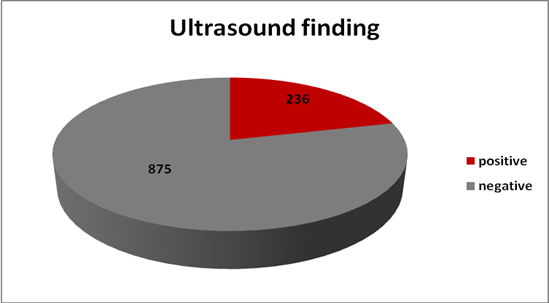 | Figure 1. Incidence of uterine fibroid among 1111 women patients en-rolled in the study. 21.2% were cases of uterine fibroid, while the remaining 78.8% cases had negative ultrasound findings |
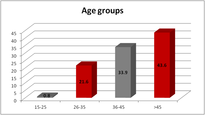 | Figure 2. Frequency and percentage frequency of the masses fibroid appearance in the age groups of 236 patients with uterine fibroid. Age group of 26- 35 years with frequency of 21.6% and 36-45 years with frequency of 33.9%, which are representing the reproductive ages had the greater percentage of uterine fibroid |
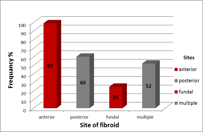 | Figure 3. Distribution of 236 patients with uterine fibroids according to their location in the uterus found by ultrasound. The fibroid nodules were located in different sites in the uterus with the most common location is anterior by 41.9%, while posterior in 25.4%, fundal in 10.6%, and multiple positions in 22% of cases |
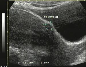 | Figure 4. Longitudinal ultrasound scan of the uterus showing a round hypoechoic fibroid located in anterior aspect of the uterus (Cursors) |
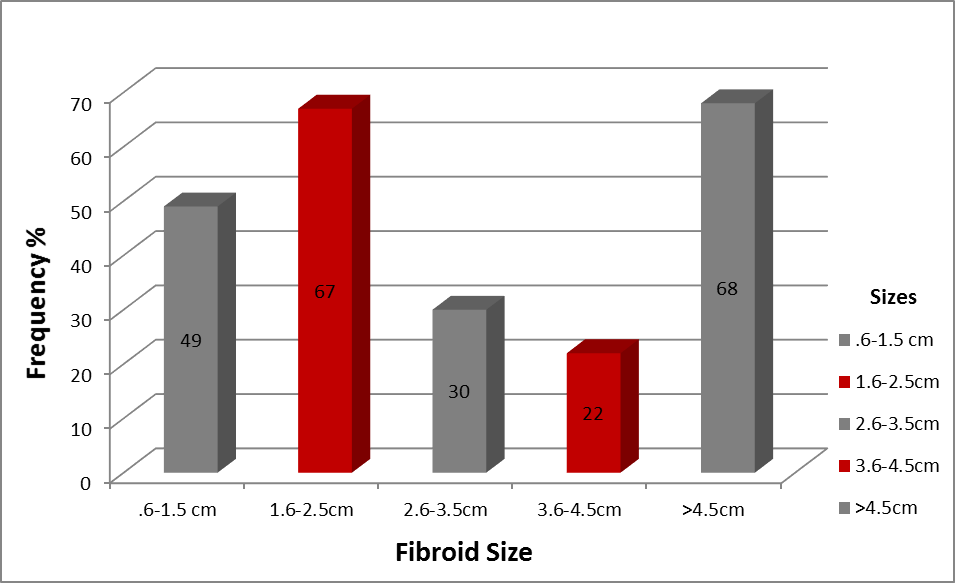 | Figure 5. Distribution of fibroid related to size in 236 women patients. The sizes of the uterine fibroid ranged from less than 0.6 cm to >4.5cm, with the most common sizes of fibroid above 4.5 cm with 6.1%. With significant difference between the single and multiple fibroid sizes |
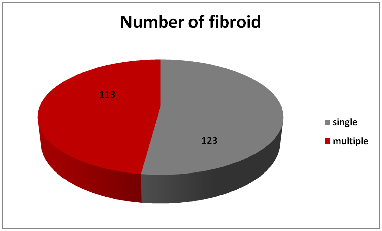 | Figure 6. Distribution of fibroid numbers among 236 women patients enrolled in the study. 52.1% cases had single uterine fibroid nodules, while the remaining 47.9% patients showed multiple uterine fibroid nodules |
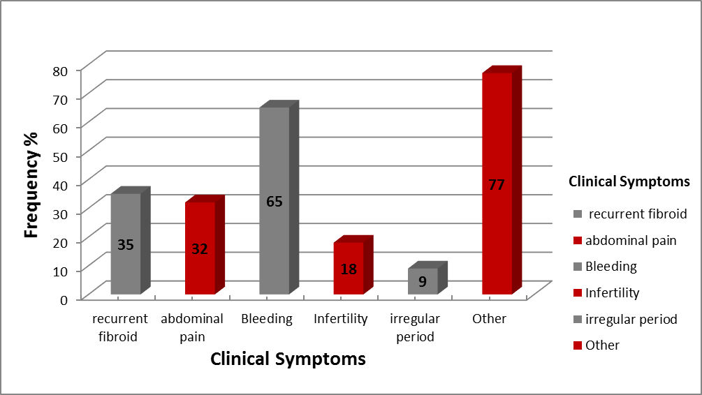 | Figure 7. Distribution of number of 236 patients with uterine fibroid in relation to the Clinical Symptoms. The commonest symptom for the patients was Bleeding included 27.5%, then recurrent fibroid 14.8%, abdominal pain 13.6. %, infertility 7.6%, and irregular menstrual period 3.8%. Other 32.6% had at least two symptoms |
4. Discussion
- In previous study, 4536 women in their reproductive age were taken during five years. There were 896 of the women shown positive uterine fibroid with an incidence of 19.75%. The period for 5 years showed that, there was 23.36% (96 cases) in 2008, 19.78% (120 cases) in 2009, 14.56% ((150 cases) in 2010, 18.91% (230 cases) in 2011 and 23.59% (300 cases) in 2012. It was found that with the progress of time the cases of uterine fibroid increased [9, 10]. Where in our study 1111 women presented vary between reproductive and menopausal age during the last two years, from the beginning of 2013 to the end of 2014 shows that 236 women had positive uterine fibroid with an incidence of 21.24%, which represents higher percentage than that of the previous study for five years (19.75%). Compared to our lesser and recent years of our study, we agreed that the incidence of fibroid boosted with the time progressed.And in a study of 21,746 women across 8 countries (based survey) shows that women at fertile age have a common concern of Uterine fibroid associated with symptoms of pain and multiple bleeding [11] Although prevalence increased with age [12], and our study shows an agreement and prove that the incidence of fibroid is increasing during reproductive age and with women grow older with multiple bleeding and pelvic pain symptoms.Our study results showed higher prevalent rate of leiomyomata in reproductive age group with incidence percentage of (21.2%) which is consistent with previous study (Marino JL et al., 2004) who reported that, the total fibroid spread rate was 21.4% in non pregnant women that is before menopausal [13].
5. Conclusions and Recommendations
- A higher prevalence of uterine fibroid in reproductive age at king Abdul-Aziz University Hospital with an incidence of 21.24% Uterine fibroid usually occur with bleeding, abdominal pain, infertility and irregular period. Future prospective work recommended covering several hospitals in Makkah Governorate.