-
Paper Information
- Paper Submission
-
Journal Information
- About This Journal
- Editorial Board
- Current Issue
- Archive
- Author Guidelines
- Contact Us
Clinical Medicine and Diagnostics
p-ISSN: 2163-1433 e-ISSN: 2163-1441
2014; 4(6): 107-112
doi:10.5923/j.cmd.20140406.01
An Infrequent Case of Intestinal Obstruction: Gallstone Ileus
Giovanni Iannetti1, Vincenzo D’Addetta1, Simona Sestili2, Giorgio Costa1, Cosima Schiavone3
1UOSD Ecografia Clinica, AUSL Pescara, Italy
2Department of Hematology, AUSL Pescara, Italy
3DMSI University G. D’Annunzio Chieti-Pescara, Italy
Correspondence to: Giovanni Iannetti, UOSD Ecografia Clinica, AUSL Pescara, Italy.
| Email: |  |
Copyright © 2014 Scientific & Academic Publishing. All Rights Reserved.
Gallstone ileus (GI) is an infrequent and severe complication of gallbladder disease and an uncommon cause of mechanical intestinal obstruction that occurs in 1%-3% of all cases of bowel disease [1]. It is caused by a single or multiple stones moving through a biliary-enteric fistula, which is formed between the inflamed gallbladder and, in most cases, the duodenum. Clinically, GI is characterized by signs and symptoms of intestinal obstruction, such as constipation, abdominal pain, nausea, vomiting and abdominal distension, which are usually preceded by symptoms of acute cholecystitis. In other cases fistula can directly end in a colonic tract determining symptoms of low intestinal obstruction, otherwise in gastric antrum with a high intestinal obstruction. Mortality is very high in elderly people with an incidence of 20-40%. Delayed diagnosis and presence of other chronic disease contribute to increase mortality [2, 3]. Therefore, early diagnosis is crucial. Plain abdominal X-ray, ultrasound and Computed Tomography (CT) are fundamental for the diagnosis [4]. Surgical management of acute GI is controversial. Most authors prefer two-stage strategy: firstly removing stones in gastrointestinal tract through enterotomy, and then treating biliary-enteric fistula [5]. We report a case of GI where criteria of preoperative diagnosis such as pneumobilia, intestinal obstruction and an aberrantly located gallstone (Rigler’s triad) were initially detected by ultrasound (US). Consequently, the patient underwent urgent surgical intervention with a good recovery.
Keywords: Gallstone ileus, Sonography, Biliary stones
Cite this paper: Giovanni Iannetti, Vincenzo D’Addetta, Simona Sestili, Giorgio Costa, Cosima Schiavone, An Infrequent Case of Intestinal Obstruction: Gallstone Ileus, Clinical Medicine and Diagnostics, Vol. 4 No. 6, 2014, pp. 107-112. doi: 10.5923/j.cmd.20140406.01.
1. Case Report
- An 80-year-old woman was admitted to the Emergency Department (ED) of our hospital with central abdominal pain irradiated to the upper quadrants and vomiting during last 4 days. Past medical history was significant for atrial fibrillation, hypertensive cardiomyopathy, duodenal ulcer associated with Helicobacter Pylori infection treated with antibiotics and proton pump inhibitors (PPI), cholelithiasis untreated for 20 years. Patient reported weight loss during last months, dyspepsia after meal and a progressive worsening of constipation. A physical examination revealed abdominal distension and pain. Laboratory showed an increase of creatinine (3,8 mg/dl), blood urea (2,8 mg/dl), gamma-glutamyltransferase (152 U/l), aspartate aminotrasferase (44 U/l) and alanine aminotransferase (48 U/l). Other blood values were normal. Nephrology consultation was required to correct the acute kidney failure. First plain abdominal radiograph performed in E.D. and the second one performed during hospitalization were both normal. US didn’t show gallbladder in its presumable site. Pneumobilia of intrahepatic biliary system and dilatation of main biliary duct without endoluminal spots were present (Fig.1). Collaterally, US showed gastric and small intestine distension in central abdomen (Fig.2,3) and an hyperechoic mass, approximately 3,5 cm in diameter, in lower right abdominal quadrant characterized by distal shadowing (Fig.4,5). This formation was mobile within a dilated small bowel loop, according to sonographic aspect of the wall. Afterwards the patient was investigated with CT that confirmed small bowel obstruction due to two stones sizing 3 cm in diameter, within proximal ileum (Fig.6,7). The patient underwent to exploratory laparotomy that demonstrated a massively dilated loops upstream proximal ileum and, at the same time, enterotomy showed two large gallstones. The patient had an uneventful postoperative course and was discharged home after two post-operative days.
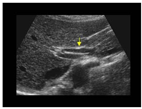 | Figure 1. Dilatation of main biliary duct |
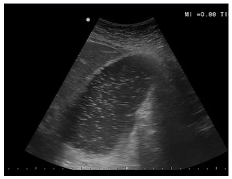 | Figure 2. Gastric distension |
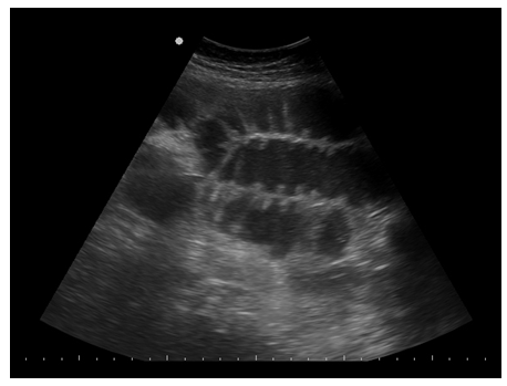 | Figure 3. Small intestine distension |
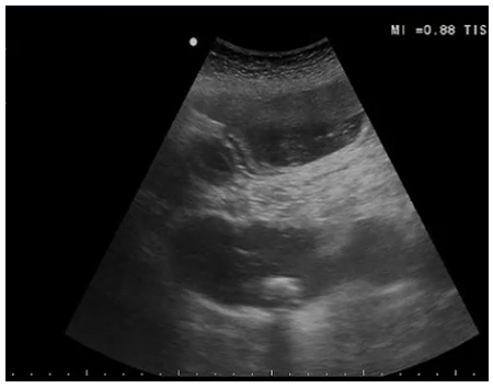 | Figure 4. Hyperecoic formation in intestinal loop with distal shadowing |
 | Figure 5. Hyperecoic formation in intestinal loop with distal shadowing |
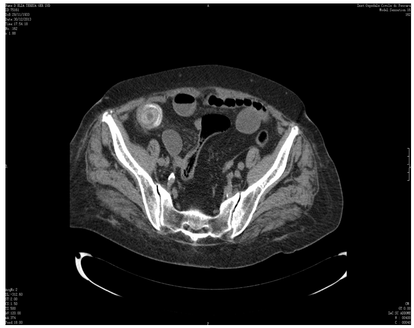 | Figure 6. Calcific stone in ileum on CT transversal scan |
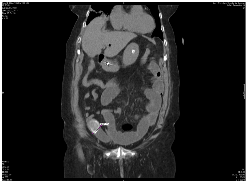 | Figure 7. Calcific stone in ileum on CT longitudinal scan |
2. Discussion
- Necessary but not sufficient condition to realize GI is the presence of communication between any part of extra-hepatic biliary tract and any part of adjacent gastrointestinal tract [6]. Fistula formation can grow up without any symptoms or with mild symptoms, i.e. nonspecific abdominal pain and fever. Other patients present history of biliary tract disease, frequently multiple cholecystitis, that leads to chronic inflammation of gallbladder wall. One or more stones impacted in the gallbladder reduce the arterial supply of the gallbladder wall and venous and lymphatic drainage [7]. Concurrently inflammatory and mechanical factors determine an impairment of gallbladder wall and adhesions with serous surface of the adjacent viscus. The rising intraluminal pressure of the gallbladder causes the penetration of its necrotic wall and the wall of the adherent viscus, resulting in formation of the biliary enteric fistula. However, it must be considered that neoplastic process or penetrating peptic ulcers can be the reason of displacement of the stones [8]. The literature shows that biliary-enteric fistula is more frequently with duodenum (70% of cases), ileum (20%), colon (8,5%) and stomach (1,5%), but impaction of stones depends on their diameter [9]. Small stones (80% of cases) can pass spontaneously and arrive in rectum without any symptoms [10]. Large stones (20% of cases), at least 2-2,5 cm of diameter, induce a “tumbling obstruction” because peristalsis moves them along narrower loops till ileo-cecal valve, where they usually impact [11]. It is also true that very large stones can stop within first tract of small intestine even in duodenum, defining in this last case, the rare Bouveret’s syndrome [12]. GI is a rare disease and accounts for about 1-3 % of mechanic ileus of the small bowel, but for 25% of all small bowel obstructions in patients older than 65 years, with a female to male ratio of 3.5-6.0:1 [13, 14]. Literature data demonstrate that GI has a very high mortality rate especially among elderly people, lying between 20% and 40%. Moreover prevalence of postoperative complications and mortality caused by gallstone ileus have been described in other reports between 7% and 50%, much higher than in any other cause of mechanical ileus [4, 15]. Based on these considerations, it must be important doing early diagnosis and beginning appropriate treatment. Plain abdominal radiograph not always shows the pathognomonic signs of GI: pneumobilia, intestinal obstruction and an aberrantly located gallstone that change position respect of a previously observation (Rigler’s triad) [16]. Two signs are necessary for diagnosis, but they are present only in 40% of patients; three signs in 10% [17]. Therefore plain radiography is not sufficient alone, because of its low sensitivity about 17%. Sonography can give additional informations to diagnose GI, in particular it demonstrates, beyond Rigler’s triad, biliary complications such as remaining stones in gallbladder or in common bile duct, with higher sensibility than plain radiography alone. Two techniques combined increase diagnostic sensitivity, from 74% with US alone to 96% [18]. Some Authors suggest that plain radiograph and US of the abdomen are usually enough to diagnose GI when the bowel is obstructed by a radiopaque calcified stone and no further studies are required [19]. CT with contrast medium remains the gold standard for diagnosis of GI with sensibility, specificity and diagnostic accuracy of 93%, 100% and 99% respectively [9]. CT can determine with precision diameter and position of stone impacted along gastrointestinal tract, degree of abdominal distension and any ischemia of dilated loops [20]. Furthermore CT permits to evaluate the entire abdomen with static images while US gives dynamic images of abdomen, scanning small areas in succession. However CT presents high cost and high doses of ionized radiation and, with this technique, detection of lithiasis depends on its calcified content. US is a repeatable exam, it has low cost and lithiasis always appears hyperechoic with strong acoustic shadowing, regardless of their content, but US depends strictly by experience of the sonographer [18]. In our case, we performed US, after a normal plain radiography, demonstrating clearly the presence of hyperecoic formation of 3,5 cm in diameter with distal shadowing, within proximal ileus, associated with intestinal distension and pneumobilia. These results make us strongly to hypothesize the presence of GI, because of typical ultrasound signs of lithiasis in the lumen of intestinal loop, according to clinical symptoms [15, 21-24]. Sonography gives last informations to surgeon immediately before surgery, marking exactly the position of the stone [25]. Subsequently these signs have been confirmed by abdominal CT and the diagnosis of GI was definitive. Management of gallstone ileus is controversial. Recent literature reviews are concordant for surgical treatment if abdominal obstruction is determined by stones of more than 2 cm in diameter. Small stones can be supervised without surgical treatment because they can be eliminated with natural way. Two-times surgical strategy remains most widely practiced [5-9]. Most of Authors prefer enterotomy with gallstones extraction postponing surgical treatment of bilioenteric fistula, reducing risk of postoperative complications. Patients usually have poor clinical conditions when presenting at surgical table and one-time surgical treatment is contraindicated in these cases. Enterotomy alone is minimally invasive and the risk of recurrent gallstone ileus is very low in presence of remaining stones (10% of cases) [26]. Spontaneous fistula closure occurs in more than 50% of cases [27]. In literature non-surgical treatments are described to treat GI especially when the stone is impacted in duodenal bulb (Bouveret’s syndrome) or in a colonic tract, through endoscopic approach. One of these techniques is the Endoscopic Electrohydraulic Lithotripsy (EEHL) that allows to fragment a large calcified stone introducing one or more specific endoscopic probes and shocking the surface of the stone, with normal saline instillation like a medium for EHL [28-30]. Based on these considerations, the patient of our case underwent to enterotomy with stones extraction, postponing surgical treatment of fistula at a later time.
3. Conclusions
- Even if sonography is operator dependent and so CT is useful to pre-operative diagnosis, we have demonstrated in this study that a correct abdominal sonography with a study of gastrointestinal tract can give more informations to diagnose an infrequent and particular case of intestinal obstruction: gallstone ileus.
 Abstract
 Reference
 Full-Text PDF
 Full-text HTML