-
Paper Information
- Paper Submission
-
Journal Information
- About This Journal
- Editorial Board
- Current Issue
- Archive
- Author Guidelines
- Contact Us
Clinical Medicine and Diagnostics
p-ISSN: 2163-1433 e-ISSN: 2163-1441
2012; 2(5): 60-64
doi: 10.5923/j.cmd.20120205.04
Epithelial-Like Cells Containing Leucocytes (Nurse Cells) of Human Palatine Tonsils, Uterus Cervix and Pleural Fluid
 Abstract
 Reference
 Full-Text PDF
 Full-Text HTML
1Department of Human Anatomy of Novosibirsk State Medical University
2Municipal Hospital № 25, Novosibirsk, Russia
Correspondence to: Alexander Kuznetsov , Department of Human Anatomy of Novosibirsk State Medical University.
| Email: |  |
Copyright © 2012 Scientific & Academic Publishing. All Rights Reserved.
Nurse cells (NCs) are detected in lymph-epithelial organs. The aim was to test the hypothesis that NCs are among tonsil epithelium (group 1), vagina epithelium (2) and pleural fluid (3) and to define the diagnostic value of the palatine tonsil (PT) NCs in leukaemia. NCs were found in patients of group 1 in leukaemia, in group 2 – in dysplasia and cancer, in group 3 – in AIDS; NCs were not obtained from practically healthy patients (group 1- 3), in cancer (groups 1, 3) and different infections and inflammation diseases (group 1-3). Conclusions. NCs are abnormal cells. The PT NCs can be used for early diagnosis of leukaemia.
Keywords: Palatine Tonsil Nurse Cells, Uterus Cervix, Pleural Fluid
Cite this paper: Alexander Kuznetsov , "Epithelial-Like Cells Containing Leucocytes (Nurse Cells) of Human Palatine Tonsils, Uterus Cervix and Pleural Fluid", Clinical Medicine and Diagnostics, Vol. 2 No. 5, 2012, pp. 60-64. doi: 10.5923/j.cmd.20120205.04.
Article Outline
1. Introduction
- Nurse cells (NCs) perform important functions and are generally detected in lymphoid organs in normal and in pathology[1-13]: in thymus[1],[2],[3],[4],[5],[6], tonsils and adenoids[4],[5]. The authors attach important features to NCs. Some subtypes of NCs were found. Reference [5] shows that follicular dendritic cells have been isolated from human tonsils and adenoids and characterized at the ultrastructural level. Follicles were dissected and digested with different hydrolytic enzymes. Follicular dendritic cells enveloping lymphocytes with their cytoplasmic extensions in a way analogous to that described for isolated thymic nurse cells were obtained. The ultrastructural features of isolated follicular dendritic cells are similar to those observed in situ [5].NCs show neither phagocytic ability, nor alpha-naphthyl acetate esterase (ANAE) and peroxidase cytochemical reactions. The majority of NCs from adenoids and tonsils react with the monoclonal antibody (McAb) OKIa [4]. In vitro cultured epithelial reticular cells from foetal thymic explants and thymic nurse cells (TNC) demonstrate the presence of keratin filaments in the cytoplasm. Keratin-positive cells of cultured thymic epithelium are heterogeneous in size and shape as are TNC [11]. Thymic epithelial cells often form lymphoid-epithelial cell (LEC) complexes, thought to contribute both to normal T-cell differentiation and to leukemogenesis. The distribution of the nerve growth factor (NGF) and NGF immunoreactivity modulation of complex-forming thymus epithelial cells were studied in mice with experimental acute L1210 leukemia. Immunoperoxidase and immunogold labelling showed subcapsular and subseptal overexpression of NGF by epithelial cells in leukemic thymus. NGF immunopositive epithelial cells were closely associated with lymphoid cells [12]. Bombesin-like peptides mediate premature thymic maturation and thymic nurse-cell depletion, leading to autoreactive T cells that could contribute to bronchopulmonary dysplasia [10]. Electron microscopic investigations reveal the epithelial character of the large thymic nurse cells. Thymic macrophages identified by phagocytosis marker were serologically identical to thymic nurse cells [1]. Nurse-like cells (NLCs) belong to the wound-healing macrophage subset (so-called 'M2 subset'), but most of all, unsupervised clustering analysis positions NLCs as chronic lymphocytic leukaemia -specific tumor associated macrophages (TAMs). Chemokinome assays confirmed that NLCs secrete prototypical M2 cytokines and chemokines. NLCs are not just nursing, but actively participate in the setting of an environment-mediated drug resistance (EM-DR) and immune escape for chronic lymphocytic leukaemia cells [9]. Tumor-associated macrophages (TAM) have multifaceted roles in tumor development but they have been associated particularly closely with tumor angiogenesis [8]. Tumour-associated macrophages (TAMs) foster tumour progression by several mechanisms, including the promotion of angiogenesis, tissue remodelling, and immunosuppression. Such pro-tumoural activities are thought to be executed by TAM subtypes that harbour features of alternatively activated (or M2-polarized) macrophages. While VEGF-A recruits monocytes from the peripheral circulation, IL-4 induces their differentiation into tumour-promoting, M2-like macrophages. IL-4 signalling blockade was sufficient to reprogram TAMs away from the M2-like phenotype and inhibited tumour angiogenesis and growth. This study attests to the potential of reprogramming TAMs to abate their pro-angiogenic and pro-tumoural functions in tumours [7]. Nurse cells are defined as those that provide for the development of other cells. Human monocyte-derived macrophages can behave as nurse cells with functional capabilities that include de novo generation of CD4+ T-lymphocytes and a previously unknown small cell with monocytoid characteristics. These novel cells have been named "self-renewing monocytoid cells" (SRMC), because they could develop into nurse macrophages that produced another generation of SRMC. SRMC were not detectable in blood. Their transition to nurse behavior was characterized by expression of CD10, a marker of thymic epithelium and bone marrow stroma, typically absent on macrophages. Confocal microscopy revealed individual HIV-1-expressing nurse macrophages simultaneously producing both HIV-1-expressing SRMC and non-expressing CD3+ cells, suggesting that nurse macrophages might be a source of latently infected CD4+ T-cells. Real-time PCR experiments confirmed this by demonstrating 10-fold more HIV-1-genome-harboring T-cells, than virus-expressing ones. These phenomena have far-reaching implications, and elicit new perspectives regarding HIV pathogenesis and T-cell and hematopoietic cell development [13].The large number and size of TNCs containing viable thymocytes in the female transgenic thymus suggest that TNC function is not limited to the removal of apoptotic thymocytes. The selective uptake of viable double-positive thymocytes by TNCs in C57BL/6 and HY-TCR transgenic female mice provides evidence that this interaction occurs during the process of major histocompatibility complex restriction [2].The aims of the research were to test the hypothesis that nurse cells containing leukocytes - NCs can be not only in tonsils but also among epithelium of uterus cervix and pleural fluid and to define the diagnostic value of the NCs in leukaemia.
2. Materials and Methods
- The cytological material was obtained by scraping from the tonsil, uterus cervix and by pleural puncture and studied by light microscopy in smears stained by Giemsa (method of scraping of tonsil – revealing of metastases of malignant tumor in tonsils: Patent № 2293987, Russian Federation). Cytological material of three groups of patients have been examined: group 1- tonsil cells of 1300 patients (male, female, aged 13 - 85), group 2 – uterus cervix/vagina cells of 11520 patients (female, aged 6 months - 80 years) and group 3 - pleural fluid cells of 1288 patients (male and female, aged 25-80). 280 patients of group 1 had malignant solid tumors of different localization, 19 patients had leukaemia (chronic lymphatic leukemia and acute lymphoblastic leukemia), 30 were practically healthy, the rest had different infections and inflammation diseases. 24 patients of group 2 had dysplasia and cancer, 570 patients were practically healthy, the rest had different infections and inflammation diseases. In group 3 there were 18 patients in cancer, 1 – in AIDS, the rest had pneumonia.
3. Results and Discussion
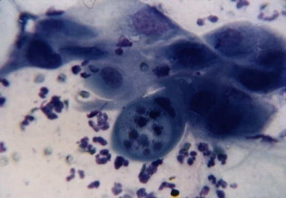
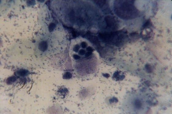
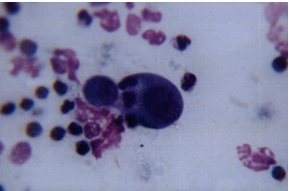
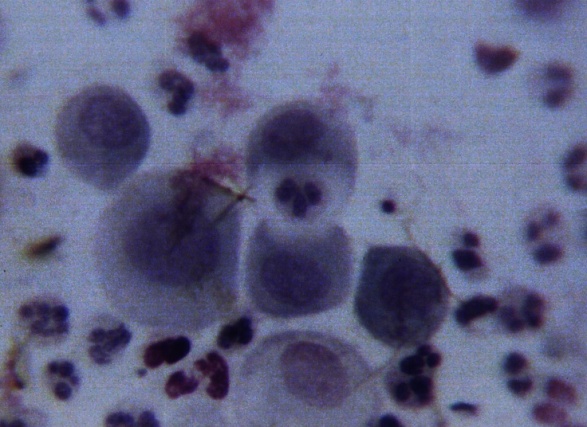
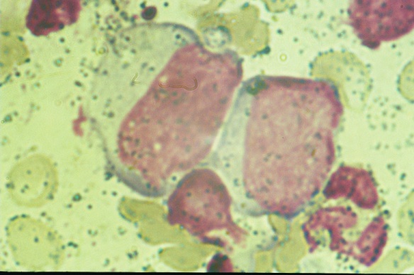
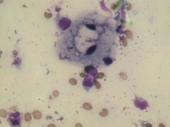
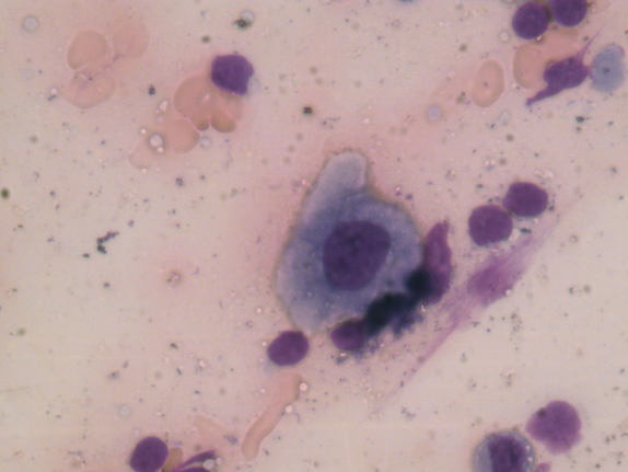
- NCs - cells containing leucocytes are found among tonsil epithelium and uterus cervix epithelium and in pleural fluid.NCs have been obtained from 19 patients of group 1 in leukaemia (Figure 1), in group 2 – from 11 patients with dysplasia (Figure 2) / cancer (Figure 3) and in group 3 – one patient had atypical cells in pleural fluid in AIDS (Figure 4, 5).
 | Figure1. Epithelium - like nurse cell containing leucocytes of palatine tonsil in leukemia. Smear stained with Giemsa, magnification x 1000 |
 | Figure 2. Epithelium - like nurse cell containing leucocytes of uterus cervix in dysplasia. Smear stained with Giemsa, magnification x 1000 |
 | Figure 3. Epithelium - like nurse cell containing leucocytes of uterus cervix in cancer. Smear stained with Giemsa, magnification x 1000 |
 | Figure 4. Epithelium - like nurse cell containing leucocytes of pleural fluid in AIDS. Smear stained with Giemsa, magnification x 1000 |
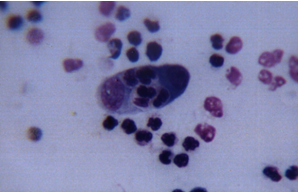 | Figure 5. Epithelium - like nurse cell containing leucocytes of pleural fluid in AIDS. Smear stained with Giemsa, magnification x 1000 |
 | Figure 6. One leucocyte is being surrounded with atypical cell cytoplasm of pleural fluid in AIDS. Smear stained with Giemsa, magnification x 1000 |
 | Figure 7. One leucocyte is in cytoplasm of epithelium of uterus cervix in dysplasia. Smear stained with Giemsa, magnification x 1000 |
 | Figure 8. Atypicall cells of tonsil in stomach cancer. Smear stained with Giemsa, magnification x 1000 |
 | Figure 9. The abnormal ring–like cell of tonsil in trichomoniasis and anemia. Smear stained with Giemsa, magnification x 1000 |
 | Figure 10. Dysplasia of tonsil epithelium in trichomoniasis and anemia. Smear stained with Giemsa, magnification x 1000 |
4. Conclusions
- NCs in vagina and pleural fluid are revealed for the first time. NCs–complexes suggest that NCs can be generated among epithelial organs having lymph tissue. In our research NCs have been revealed only in severe pathology: dysplasia, AIDS (smears of cells from uterus cervix and pleural fluid correspondingly). NCs can reflect an obstinate course of the mentioned diseases. The NCs revealed in our work can be of different subtypes. In this aspect two variants are suggestive. Firstly, NCs can be abnormal and envelope normal leucocytes. Thus, the represented NCs are abnormal cells complexes. Secondly, NCs can appear in epithelium and envelope abnormal leucocytes. NCs have been found only in leukaemia in PT cells smears. In both variants patients have damaged immunity. The appearance of the NCs in PT might reflect protective reaction of human organism. Taking the above mentioned into account it is thought that tonsil NCs can be used for early diagnosis of leukaemia. NCs from uterus cervix and pleural fluid can be used as marks of the severity of the described diseases.
AKNOWLEDGEMENTS
- We would like to thank academician Y.I. Borodin, A.N. Mashak, S.V. Astrakov, O.V. Popkova providing only general support and G.I. Kuznetsova for writing assistance.