American Journal of Chemistry

p-ISSN: 2165-8749    e-ISSN: 2165-8781

2026;  16(3): 55-60

doi:10.5923/j.chemistry.20261603.01

Received: Oct. 19; Accepted: Nov. 3, 2026; Published: Nov. 7, 2026

 

Synthesis, Structure Analysis and Antibacterial Activity of New Pyridoxal-Isothiosemicarbazone- Zn (II) Complex

Sarah Awadh Alreshidi1, Sahar Yahia Rajeh2, Violeta Jevtovic2

1Master Student at Department of Chemistry, University Hail, City, Hail, Kingdom of Saudi Arabia

2Department of Chemistry, University Hail, City, Hail, Kingdom of Saudi Arabia

Correspondence to: Sarah Awadh Alreshidi, Master Student at Department of Chemistry, University Hail, City, Hail, Kingdom of Saudi Arabia.

Email:

Copyright © 2026 The Author(s). Published by Scientific & Academic Publishing.

This work is licensed under the Creative Commons Attribution International License (CC BY).
http://creativecommons.org/licenses/by/4.0/

Abstract

The aim of the research was to synthesize a new zinc complex with the pyridoxal-iso thiosemicarbazone ligand (PLITSC), and to examine the newly synthesized complex structurally and biologically. This paper presents the synthesis, structure, and biological activity of the new complex Zn (II) with PLITSC ligand. A single crystal of the newly synthesized Zn (II) complex was structurally characterized by X-ray analysis, using a Bruker D8 Venture APEX diffractometer. Complex is with molecular formula [Zn (PLITSC) (H2O) (SO4)] and with brutto formula C10 H16 N4 O7 S2 Zn. The synthesized complex was structurally characterized, and biological activity was performed on several microorganisms. The zinc complex has the structure of square pyramid, coordination number 5, with the (O, N, N) pyridoxal-isothiosemicarbazide ligand occupying three sites, while the remaining two are occupied by two oxygens, one from water, the other from the sulfate group. The newly synthesized Zn (II) complex is biologically active, shown antibacterial activity against strains of the bacteria Escherichia coli and Bacillus subtilis.

Keywords: Pyridoxal S-methyl-isothiosemicarbazone (PLITSC), Zn (II) complex, Synthesis, Crystal structure, Antibacterial activity

Cite this paper: Sarah Awadh Alreshidi, Sahar Yahia Rajeh, Violeta Jevtovic, Synthesis, Structure Analysis and Antibacterial Activity of New Pyridoxal-Isothiosemicarbazone- Zn (II) Complex, American Journal of Chemistry, Vol. 16 No. 3, 2026, pp. 55-60. doi: 10.5923/j.chemistry.20261603.01.

1. Introduction

Pyridoxal-derived carbazone ligands have attracted considerable attention in coordination chemistry due to their ability to coordinate with various transition-metal ions and form complexes with diverse structural, biological, and catalytic properties. Among these compounds, pyridoxal S-methyl isothiosemicarbazone (PLITSC) is a Schiff-base ligand formed by the condensation of pyridoxal with S-methyl isothiosemicarbazone (ITSC). [1]
PLITSC acts as a tridentate ONN donor ligand, coordinating to the metal center through the phenolic oxygen atom, hydrazine nitrogen atom, and nitrogen atom of the isothioamide group. This tridentate coordination results in the formation of two chelate rings: a six-membered ring involving the pyridoxal moiety and a five-membered ring involving the isothiosemicarbazone fragment. [1,2]
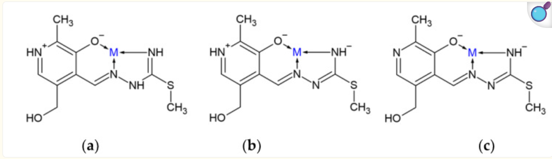
An important feature of PLITSC is its ability to coordinate with metal ions in three different forms: neutral (L), monoanionic (L⁻), and dianionic (L²⁻). In the neutral form (L), the ligand adopts a zwitterionic structure in which the phenolic hydroxyl group is deprotonated, while the pyridine nitrogen is protonated. The monoanionic form (L⁻) is produced by deprotonation of the hydrazine nitrogen atom. Further deprotonation of the protonated pyridine nitrogen results in the formation of the dianionic form (L²⁻). Despite these differences in protonation state, PLITSC maintains its tridentate ONN coordination mode through the phenolic oxygen, hydrazine nitrogen, and isothioamide nitrogen atoms. These different forms provide PLITSC with considerable versatility in the formation of transition-metal complexes. [1,2]
Complexes containing PLITSC with several transition-metal ions have been reported in the literature. Copper complexes represent one of the most extensively reported groups, while PLITSC complexes containing iron, cobalt, nickel, vanadium, and molybdenum have also been described [3-6]. The ability of PLITSC to coordinate different transition-metal ions and adopt different protonation states contributes to the structural diversity of its coordination compounds. In addition, PLITSC-containing complexes have attracted attention because of their reported biological and catalytic properties, including antitumor, antiviral, anti-inflammatory, and electrocatalytic activities [7].
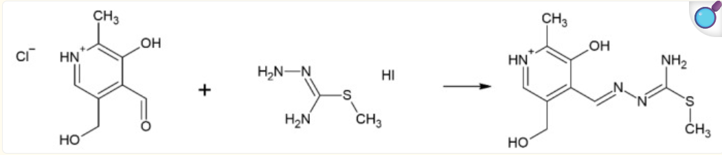
Scheme 1. Synthesis of pyridoxal–isothiosemicarbazide (PLITSC) [1]
Scheme 2. Binding modes of differently protonated PLITSC [1]

2. Experimental Part

Commercial pyridoxal hydrochloride (PL·HCI, "Aldrich") and thiosemicarbazone (TSC, "Merck") were used for ligand synthesis.

2.1. Synthesis of Ligand PLITSC·H2O

PLITSC·H2O Mixture of 2.0 g (10 mmol) PL·HCl and 2.30 g (10 mmol SMeTSC·HI) was perfused with 20 cm3 H2O and heated until complete dissolution of reactants. Warm solution was added to 3.0 g (10 mmol) Na2CO3, previously dissolved in 20 cm3. The deposits of pale-yellow fibrous crystals of the ligands appear extremely fast; 5 hours later, these deposits were filtered off and washed with H2O and EtOH. Yield: 2.40 g (96%).

2.2. Synthesis of [Zn (PLITSC) (H2O) (SO4)] crystal

In 15 cm3 water, 0.01 mol of PLITSC ligand was heated and dissolved. An aqueous solution of 0.01 mol of ligand was added 0.1 mmol of ZnSO4. A clear yellow solution was obtained and left at room temperature to crystallize using the slow evaporation method. After a few hours, yellow crystals appeared. Yield: 0.25 g (85%).

2.3. X-ray Crystallography

A suitable yellow needle-shaped single crystal of the Zn (II) complex was selected for single-crystal X-ray diffraction analysis. A single crystal of size 0.14 × 0.10 × 0.16 mm was obtained and submitted for the X-ray diffraction analysis.
The crystal was mounted on a glass fiber and examined at 296 K using a Bruker D8 Venture APEX diffractometer (Bruker, Billerica, MA, USA) equipped with a Photon 100 CCD detector, employing graphite-monochromatized Mo-Kα radiation [λ = 0.71073 Å].
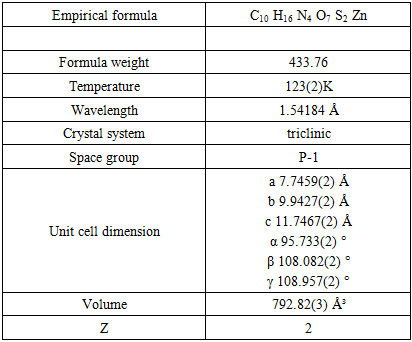
Data collection, cell refinement, and data reduction were performed using CrysAlisPro. The crystal structure was solved using SHELXT-2018/2 and refined using SHELXL-2018/3. The structure was refined as a two-component twin. Crystallographic data have been deposited with the Cambridge Crystallographic Data Centre (CCDC, 12 Union Road, Cambridge CB2 1EZ, UK; e-mail: deposit@ccdc.cam.ac.uk) under deposition number CCDC 2586352. Details regarding crystallographic data collection and structure refinement are presented in Table 1.
Table 1. Crystal Data
     
Crystal data collection and structure refinement are given in Table 1.

3. Results and Discussion

3.1. Crystal Structure of [Zn (PLITSC)(H₂O) (SO₄)]
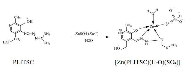
Scheme 3. Synthesis of the Zn (II) complex
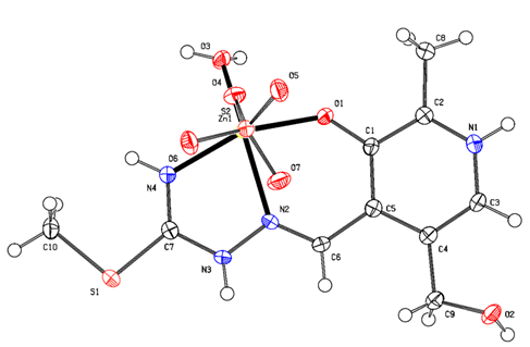
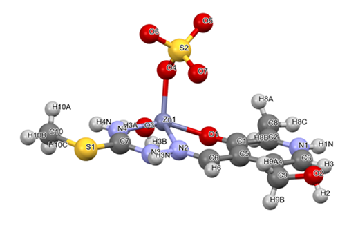
The molecular structure of the new Zn (II) complex, [Zn (PLITSC)(H₂O) (SO₄)], is shown in Figure 1. The complex crystallizes in the triclinic crystal system with the space group P−1. The coordination environment around the central Zn (II) ion can be best described as a distorted square-pyramidal geometry. The zinc center has a coordination number of five and is coordinated by the tridentate PLITSC ligand through the phenolic oxygen atom O1 and two nitrogen atoms, N2 and N4. The remaining coordination sites are occupied by the oxygen atom O3 of one coordinated water molecule and the oxygen atom O4 of the coordinated sulfate group.
Figure 1. Crystal structure of the Zn (II) complex [Zn (PLITSC)(H₂O) (SO₄)]
The PLITSC ligand is coordinated in its neutral zwitterionic form, as indicated by the presence of hydrogen on the pyridine nitrogen N1 and the deprotonated phenolic oxygen O1. The sulfate group coordinates directly to the Zn (II) center through one oxygen atom, O4. Thus, the coordination sphere of Zn (II) consists of an ONN donor set from PLITSC, one oxygen atom from H₂O, and one oxygen atom from SO₄²⁻, resulting in a five-coordinate distorted square-pyramidal structure.
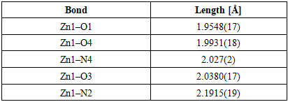
The Zn (II) ion is five-coordinate, and the coordination environment corresponds to a distorted square-pyramidal geometry, as shown in Figure 2. The Zn (II) center is coordinated by the tridentate ONN donor set of the PLITSC ligand (O1, N2, and N4), together with the oxygen atom O3 from the coordinated water molecule and the oxygen atom O4 from the coordinated sulfate group. The Zn–donor bond lengths are Zn1–O1 = 1.9548(17) Å, Zn1–O4 = 1.9931(18) Å, Zn1–N4 = 2.027(2) Å, Zn1–O3 = 2.0380(17) Å, and Zn1–N2 = 2.1915(19) Å. Thus, the shortest Zn–donor bond is Zn1–O1, whereas the longest is Zn1–N2.
Table 2. Bond lengths [Å] of the Zn (II) complex
     
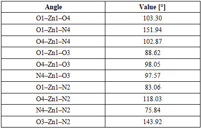
The bond angles around the Zn (II) center further confirm the distorted square-pyramidal geometry. The largest angle around the zinc atom is formed by O1–Zn1–N4, with a value of 151.94°, while the smallest angle is formed by N4–Zn1–N2, with a value of 75.84°. The selected bond angles around the Zn (II) center are summarized in Table 3.
Table 3. Bond angles [°] of the Zn (II) complex
     
Complexes of transition metals with a coordination number of 5 most often adopt the geometry of a trigonal bipyramid or a square pyramid, which are very often distorted.
The main reason why transition metals form a distorted square pyramid structure is the Electronic Effect (Jahn-Teller Distortion). When the transition metal has an electronic configuration that causes the highest energy d-orbitals (d x2-y2 and dz2) to be unevenly filled, the system becomes unstable. To reduce the total energy and remove this degeneracy, the complex spontaneously distorts.
This is extremely common with copper (II) complexes (d⁹ configuration), which, due to the second order Jahn-Teller effect, prefer distorted forms instead of perfect geometric forms. However, this is not the reason the Zn (II) complex has a distorted square pyramid structure (Figure 2).
Figure 2. Appearance of a distorted square environment around the central Zn (II) atom
While in copper (II) the distortion comes "from within" (from the d-electrons themselves), in zinc (II) it comes exclusively "outside" – due to the shape, size and way of binding the ligands that surround it.
Zinc's d10 configuration has no electronic preference (stabilization of the ligand field is zero), so the geometry is completely dictated by the structural properties of the ligand itself and steric relationships [8].
The distortion of the square pyramid in this complex is caused by the following factors such as chelation effect and rigidity of the PLITSC ligand, and asymmetry of the base of the pyramid, Position of zinc above the plane of the base and hydrogen bonds in the crystal.
The PLITSC ligand forms two rings, six- and five-membered, when coordinated. These rings are organic molecules that are quite rigid and have their natural bond angles. To bind to zinc at all, they force it into coordination angles that are significantly less than ideal. For example, the angles within the chelate rings are about 75° to 83°, far from the ideal 90° required for a perfect square pyramid. Three donor atoms from PLITSC (O-phenolic, N-hydrazine, N-amide) form the base of the pyramid with oxygen from coordinated water, which is a chemically completely different mixture of atoms and promotes the asymmetry of the base of the pyramid.
Due to the steric pressure of the three bonded atoms from the PLITSC ligand, the zinc (II) ion cannot fit exactly in the plane of the base of the square pyramid. Zinc is slightly raised above the plane formed by the four base atoms, "looking" towards the ligand at the top of the pyramid. This automatically changes all ideal 90° angles and turns the structure into a warped (distorted) one.
The base of the pyramid consists of O1, N2, N4, and N3 in the corners of the rectangular base with the Zn atom in the middle. The values of those angles are given in table 3. O1-Zn-N2, O1-Zn-O3, N4-Zn-N2, N4-Zn-O3, 83.06, 88.62, 75.84, 97.57 respectively, when added together they amount to 345.09°, which is less than a full circle of 360 degrees. It is evident that there is a large voltage and pressure inside the base of the pyramid, which results in the Zn atom jumping above the level of the base of the pyramid (Figure 2).

3.2. Biological Activity

Isothiosemicarbazides, particularly when coordinated with transition metal ions, constitute a class of compounds exhibiting exceptional biological activities. The potential of these ligands is enhanced upon the formation of transition-metal complexes—primarily through increased lipophilicity, improved cellular uptake, and the ability to modulate electronic properties via metal-ligand interactions. These systems with isothiosemicarbazides and semi-, and thiosemi- chains have garnered significant attention in medicinal chemistry, particularly regarding the development of novel therapeutic and diagnostic agents [9].
The anticancer and antimicrobial activities of isothiosemicarbazide complexes—reported for various metal ions, including copper, nickel, cobalt, and iron—are particularly noteworthy. The biological response of the complex is strongly influenced by the nature of the metal center; coordination frequently results in enhanced reactivity and improved interaction with biological targets. Isothiosemicarbazide-based complexes have demonstrated activity against a range of cancer cell lines and pathogenic microorganisms, highlighting their potential as multifunctional bioactive agents [1].
One of the challenges facing the healthcare sector is the emergence of resistance to many previously effective antibiotics and other drugs that suppress bacterial activity [10–12]. This has created a demand for new classes of active antibacterial agents to combat bacterial resistance.
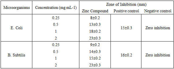
The antimicrobial efficacy of zinc compounds was evaluated against two pathogenic bacterial species: E. coli and B. subtilis. The concentration range tested was 0.25 to 2 mg mL⁻¹, and the zones of inhibition are presented in Table 4. The zone of inhibition increased with concentration, ranging from 8 to 23 mm for E. coli and from 9 to 25 mm for B. subtilis. For the positive control (streptomycin), the zone of inhibition was 15 mm for *E. coli* and 16 mm for B. subtilis at the lowest tested concentration.
Table 4. Zone of inhibition of Zinc compound and standard drug against tested bacteria
     
Surprisingly, the activity of this compound is higher than that of other Co (III), Zn (II), Ni (II), and Cu (II) complexes with similar ligands, as reported in previous studies [10,11,12]. Complexes containing thiosemicarbazone and semicarbazone ligands have been shown to exhibit moderate activity against E. coli and B. subtilis bacteria.
The higher biological activity of isothiosemicarbazones (especially S-alkylated or S-arylated derivatives) compared to classical thiosemicarbazones and semicarbazones primarily results from key structural and electronic modifications that directly affect their pharmacokinetics and interaction with biological targets.
Compared to semicarbazones [13] (which have oxygen in place of sulfur), the presence of sulfur (even modified in isothiosemicarbazones) provides significantly better electronic delocalization. According to Pearson's HSAB theory ("soft and hard acids and bases"), molecules with sulfur show a much stronger affinity for biologically important "soft" transition metal ions and biomolecules with thiol groups (such as glutathione), which semicarbazones cannot achieve to that extent.
The moderate antibacterial activity of complexes with thiosemicarbazone and semicarbazone ligands against E. coli (Gram-negative) and B. subtilis (Gram-positive) bacteria is based on the chelation effect that increases the lipophilicity of the complexes and allows them to pass through bacterial membranes, where they then block key enzymes and respiratory chains. The reasons for this activity and mechanism of action lie in the specific chemical structure and physiology of the bacteria themselves, Overton theory and chelation (Increasing permeability) and Mechanism of action inside the bacterial cell [14].
Another thing is that the effectiveness of the Zn (II) complex is compromised due to the structural differences between these two bacteria (Escherichia coli and Bacillus subtilis) [15].
Escherichia coli (a Gram-negative bacterium) has a complex outer lipopolysaccharide membrane that serves as an additional physical barrier and efflux pumps that actively expel foreign molecules from the cell [16]. Therefore, Gram-negative bacteria often show greater resistance (or lower or moderate sensitivity) to these complexes. On the other hand, Bacillus subtilis (a Gram-positive bacterium) does not have an outer membrane but only a porous peptidoglycan wall, which facilitates the entry of the complex. However, the moderation of action in both strains depends on the stability of the complex itself - if the complex is too stable, it will not release the active components, and if it is too unstable, it will disintegrate before it enters the bacterium.
Also, various levels of antibacterial activity of semicarbazones and isothiosemicarbazones are observed. The difference in their structure is manifested in the presence of a sulfur atom in isothiosemicarbazones, while in semicarbazones sulfur is not present in the chain [17].
The presence of sulfur (even when modified in isothiosemicarbazones, where the atom is alkylated or otherwise transformed) provides significantly better electronic delocalization compared to oxygen or nitrogen due to the specific properties.
Sulfur possesses a larger atomic radius and diffuse orbitals. This greater "spread" of the electron cloud allows for easier overlap with neighboring pi-systems.
The lower electronegativity of sulfur results in a less strong attraction of its valence electrons, which its "shares" much more easily with the rest of the molecule, which directly favors delocalization (resonance) along the entire isothiosemicarbazone skeleton.
Due to the weaker influence of the nucleus on the outer electrons, the electronic shell of sulfur is very polarizable. This means that it is easily deformed under the influence of surrounding charges, which further facilitates the movement of electrons through the conjugated system, i.e., it penetrates the bacterial cell wall more easily.
That is why the presence of sulfur in molecules often drastically increases their antibacterial activity because it enables specific chemical reactions that directly destroy the vital cellular structures of bacteria. Sulfur has unique electronic and structural properties that nature and the pharmaceutical industry use to make effective antibiotics (such as penicillin and sulfonamides).
Here are the main ways sulfur improves the antibacterial activity of molecules [18].
Formation and breaking of disulfide bonds - Redox stress is the main way in which sulfur improves the antibacterial activity of molecules. Many sulfur-containing antibacterial molecules work by disrupting the internal balance of the bacterial cell. Sulfur molecules (like those in garlic - allicin) can react with thiol groups (-SH) within bacterial proteins. When these molecules bind to key bacterial enzymes, they change their three-dimensional structure and permanently deactivate them, leading to the death of the bacteria.
Sulfur has an extremely strong chelating affinity for transition metals, especially iron and zinc. Many important bacterial enzymes depend on these metals to function properly. Molecules with sulfur can "trap" (chelate) these metals, leaving the bacteria without key nutrients and cofactors for survival.
In the case of the newly synthesized complex [Zn (PLITSC) (H2O) (SO4)], both conditions are met, the presence of sulfur and the highly desirable transition metal zinc. Accordingly, it is to be expected that the mentioned complex exhibits antibacterial activity.

4. Conclusions

The paper describes the synthesis of a new Zn (II) complex with the pyridoxal-isothiosemicarbazone ligand. The Zn (II) complex structure was analyzed by X-ray analysis on a single crystal, and the biological activity of the newly synthesized complex was investigated in terms of its antibacterial effect. The synthesis was done by the direct method. The crystal structure showed that it is a penta-coordinated central zinc, with a square pyramid geometry. The Zn (II) complex was investigated as an antibacterial agent, was shown to suppress the growth and kill the bacteria Escherichia coli and Bacillus.

ACKNOWLEDGEMENTS

Author Sarah Awadh Alreshidi, a Master student at University Hail, along with her mentors, would like to thank University Hail for its support and opportunities to use the university's laboratory services.
Also, she expresses great gratitude for the analyzes done at the University of Monash, Victoria, Australia, especially to Dr. Dragoslav Vidovic and his research group of PhD students, who helped a lot in creating this research work.

References

[1]  Abdulaziz, F.; Alabbosh, K.F.; Alshammari, O.A.O.; Tuwalah, W.M.B.; Alanazi, T.Y.A.; Rakić, A.; Barić, M.; Marković, M.; Jevtovic, V.; Dimić, D. Crystallographic Structure and Quantum-Chemical Analysis of Biologically Active Co (III)-Pyridoxal–Isothiosemicarbazone Complex. Inorganics 2023, 11, 466. DOI: 10.3390/inorganics11120466.
[2]  Jevtovic, V. Cu, Fe, Ni and V Complexes with Pyridoxal Semicarbazones: Synthesis, Physical and Chemical Properties, Structural Analyses and Biological Activities; Lambert Academic Publishing 2010.
[3]  Leovac, V.M.; Jevtović, V.S.; Bogdanović, G.A. Transition metal complexes with thio-semicarbazide-based ligands. XLIV. Aqua (3-hydroxy-5-hydroxymethyl-2-methylpyridine-4-carboxaldehyde 3-methylisothiosemicarbazone-κ³O, N¹, N⁴) nitratocopper (II) nitrate. Acta Crystallogr. Sect. C 2002, 58, m514–m516.
[4]  Jevtovic, V.; Cvetkovic, D.; Vidovic, D. Synthesis, X-ray characterization and antimicrobial activity of iron (II) and cobalt (III) complexes with the Schiff base derived from pyridoxal and semicarbazide or S-methylisothiosemicarbazide. J. Iran. Chem. Soc. 2011, 8, 727–733.
[5]  Leovac, V.M.; Divjaković, V.; Joksović, M.D.; Jovanović, L.S.; Vojinović-Ješić, L.S.; Češljević, V.I.; Mlinar, M. Transition metal complexes with thiosemicarbazide-based ligands. Part 57. Synthesis, spectral and structural characterization of dioxovanadium(V) and dioxomolybdenum (VI) complexes with pyridoxal S-methylisothiosemicarbazone. J. Serbian Chem. Soc. 2010, 75, 1063–1074.
[6]  Jevtovic, V.; Vidovic, D. Synthesis, characterization, and X-ray crystal structure of the triaqua Ni (II) nitrate complex with pyridoxal S-methylisothiosemicarbazone. J. Chem. Crystallogr. 2010, 40, 794–798.
[7]  Al-Zahrani, S.; Jevtovic, V.; Alenezi, K.; El, M.; Haque, A.; Vidovic, D. Electrocatalytic hydrogen evolution upon reduction of pyridoxal semicarbazone and thiosemicarbazone-based Cu (II) complexes. J. Serbian Chem. Soc. 2022, 87, 345–354.
[8]  Altamimi, A., Al-Zahrani, A.S., Jevtovic, V., Synthesis, Structure Analysis and Antibacterial Activity of Zn (II) and Co (III) Complexes, American Journal of Chemistry, 2021; 11(3): 43-48, doi: 10.5923/j.chemistry.20211103.01.
[9]  Mir, I.A.; Ain, Q.U.; Qadir, T.; Malik, A.Q.; Jan, S.; Shahverdi, S.; Nabi, S.A. A Review of Semicarbazone-Derived Metal Complexes for Application in Biomedicine and Related Fields. Journal of Molecular Structure 2024, 1295, 136216, doi: 10.1016/j.molstruc.2023.136216.
[10]  Vrdoljak, V.; Pisk, J.; Prugovečki, B.; Matković-Čalogović, D. Novel Dioxomolybdenum (VI) and Oxomolybdenum (V) Complexes with Pyridoxal Thiosemicarbazone Ligands: Synthesis and Structural Characterisation. Inorganica Chimica Acta 2009, 362, 4059–4064, doi: 10.1016/j.ica.2009.05.057.
[11]  Jevtovic, V.; Cvetkovic, D.; Vidovic, D. Synthesis, X-Ray Characterization and Antimicrobial Activity of Iron (II) and Cobalt (III) Complexes with the Schiff Base Derived from Pyridoxal and Semicarbazide or S-Methylisothiosemicarbazide. Journal of the Iranian Chemical Society 2011, 8, 727–733, doi: 10.1007/BF03245904.
[12]  Jevtovic, V.; Alshammari, N.; Latif, S.; Alsukaibi, A.K.D.; Humaidi, J.; Alanazi, T.Y.A.; Abdulaziz, F.; Matalka, S.I.; Pantelić, N.Đ.; Marković, M.; et al. Synthesis, Crystal Structure, Theoretical Calculations, Antibacterial Activity, Electrochemical Behavior, and Molecular Docking of Ni (II) and Cu (II) Complexes with Pyridoxal-Semicarbazone. Molecules 2022, 27, 6322, doi: 10.3390/molecules27196322.
[13]  Raven, F.; Maia, P.; Leite, S.; Deflon, V.; Batista, A.; Sato, D.; Francblau, S.; Leite, C.; Thiosemicarbazones, semicarbazones, dithiocarbazates and hydrazide/hydrazones: Anti – Mycobacterium tuberculosis activity and cytotoxicity. Europian Journal of Medicinal Chemistrty, 2010, 5, 1898-1905, https://doi.org/10.1016/j.ejmech.2010.01.028.
[14]  Sharma, N., Singh, H., Spectroscopic, DFT, anti-bacterial, and in-silico investigations of novel semicarbazone and thiosemicarbazone synthesised from asymmetrical chalcones, Next Materials 7 (2025) 100511 10.1016/j.nxmate.2025.100511.
[15]  Muniz, D., Luiz Silva Pereira, R., Ramos Freitas, P., de Araújo, A., Dos Santos Barbosa, C., Alencar de Menezes, I., de Melo Coutinho, H., Tintino, S., Soares Costa, V., da Silva, L., José Brondani, D., de Morais Oliveira-Tintino, C., da Silva, T., Semicarbazone and thiosemicarbazone derivatives associated with antibiotics inhibit β-lactamases and efflux pumps in Staphylococcus aureus strains, Arch Biochem Biophys 2025, 771:110512. doi: 10.1016/j.abb.2025.110512.
[16]  Krishnamoorthy, G., Woscheck, D., Weeks, J., Croft, C., Rybenkov, V., Zgurskaya, H., Breaking the Permeability Barrier of Escherichia coli by Controlled Hyperporination of the Outer Membrane, Antimicrobial Agents and Chemotherapy, 2016. 60 (12) https://journals.asm.org/doi/pdf/10.1128/aac.01882-16.
[17]  Jevtovic, V., Hamoud, H., Al-Zahrani, S., Alenezi, K., Latif, S., Alanazi, T., Abdulaziz, F., Dimić, D., Synthesis, Crystal Structure, Quantum Chemical Analysis, Electrochemical Behavior, and Antibacterial and Photocatalytic Activity of Co Complex with Pyridoxal-(S-Methyl)-isothiosemicarbazone Ligand, Molecules. 2022 Jul 27; 27(15): 4809. doi: 10.3390/molecules27154809.
[18]  Yuan, T., Wang, R., Ma, Y., Chen, F., Zhou, S., Lu, L., Sulfur-induced oxidative stress enhances horizontal transfer of antibiotic resistance genes, Desalination and Water Treatment, 2026. 327, 101836. https://doi.org/10.1016/j.dwt.2026.101836.