American Journal of Chemistry

p-ISSN: 2165-8749    e-ISSN: 2165-8781

2023;  13(1): 11-25

doi:10.5923/j.chemistry.20231301.02

Received: Dec. 12, 2022; Accepted: Jan. 5, 2023; Published: Jan. 13, 2023


Quantitative Nuclear Magnetic Resonance Spectroscopy in Pharmaceutical Chemistry: A Boon for Real Time Drug Detection!

Sudhaunshu S. Purohit

ETS Laboratories, USA

Correspondence to: Sudhaunshu S. Purohit, ETS Laboratories, USA.

Email:

Copyright © 2023 The Author(s). Published by Scientific & Academic Publishing.

This work is licensed under the Creative Commons Attribution International License (CC BY).
http://creativecommons.org/licenses/by/4.0/

Abstract

Throughout the history of mankind numerous kinds of diseases played a contributing part in shaping the humanity. Since, the beginning of 21st century, the persistent efforts in the medical sciences by the teams of research scientists have witnessed the fruition in the form of vaccinations as well as various lifesaving pharmaceutical drugs with confirmed capability against several ailments. The development of a remedial drug, is equally important to that of its delivery system in human body at the intended location and subsequently nano-particles emerged as an excellent option surpassing the conventional ways. It is equally crucial to study the pharmacokinetics in order to establish the release profile of a drug from its nano-particle encasing along with the encapsulation capacity of the later. A few spectroscopic techniques have been proven to be effective for the quantitative analysis study and the best of them being NMR spectroscopy. This scientific review article successively describes an interdepartmental collaborative translational research including the extensive use of both solid state and solution state Nuclear Magnetic Resonance spectroscopy for the quantitative determination (qNMR) of an anti-HIV, phosphorous containing pharmaceutical drug (Tenofovir) encased in spray dried, mucoadhesive, pH sensitive, nano-particle casing composed of bio-degradable material such as alginate & thiolated chitosan along with the invitro pharmacokinetic study of its subsequent release profile when in contact with simulated human body fluids.

Keywords: Bacterial & Viral Diseases, Quantitative Solid State NMR Spectroscopy, Phosphorous, Drug Delivery & Detection Systems, Real Time Analysis, in-vitro Pharmacokinetics of Release Profile, Encapsulation Efficiency, Mathematical Models, Spray Dried, Mucoadhesive, pH Sensitive, Mannose Responsive, Alginate, Thiolated Chitosan, Hyaluronic acid, Nano-Formulations, HIV-AIDS, Tenofovir, International Conference of Harmonization, Method Development & Validation, Translational Research

Cite this paper: Sudhaunshu S. Purohit, Quantitative Nuclear Magnetic Resonance Spectroscopy in Pharmaceutical Chemistry: A Boon for Real Time Drug Detection!, American Journal of Chemistry, Vol. 13 No. 1, 2023, pp. 11-25. doi: 10.5923/j.chemistry.20231301.02.

1. Introduction

It is a well-accepted fact that for millennia various types of bacterial and viral diseases played a devastating role in the evolution of human society. Several infectious and autoimmune diseases have afflicted human race since, the dawn of civilization in the known history. In 14th century epidemics such as plague commonly known as Black Death factually demonstrated the potential to cause mass extinction of human population in Europe. In the past two decades of 21st century the global human population witnessed a myriad of major viral disease’s pandemics caused by multitude of viruses such as flavi, alpha, filo, myxo, noro and corona to name a few. The recent COVID-19 situation which was imposed impromptu; factually demonstrated its influence to not only wipe out the lives of millions but also severely cripple the world economy.
A significant development in the medical sciences promised greater life expectancy as either a cure or acute preventive therapies were invented to triumph over large number of medical conditions thus marking an introduction of a new era in the beginning of 20th century. The commendable amount of development in medical sciences since the early 20th century introduced a large number of lifesaving drugs which succeeded in putting a leash on the dispersal of majority of life threatening diseases. In spite of these accomplishments, to the date, a therapy is still far from curing the notorious diseases such as Alzheimer, Cancer, Hepatitis, AIDS etc. A successful vaccine is yet to be developed, which renders, an execution of precautionary measures to inhibit from infection to be the best strategy of survival. Substantial levels of physical and emotional alteration and a possible distortion to the ailing person’s personality as well as perspective on life are the direct repercussions ascribed to the contraction of these dire diseases.
In the beginning of current century the scientific community worldwide remarkably got engaged in an intense research to invent measures, if not cure, to prevent the infection from these diseases and their further spread in human body after being diagnosed. Since then, an investigation to discover and develop an effectively functional drug against these diseases along with an efficient drug delivery system to administer the drug in infected human body is incessantly underway. The perseverance demonstrated by the scientific teams around the globe have now resulted in formulating certain drugs with proven ability against these diseases. Interestingly, majority of these drugs in pharmaceutical-medicinal field contain some of the most frequently characterized heteroatoms such as Phosphorous, Fluorine, Sulfur, & Boron along with commonly observed Nitrogen & Oxygen.
As a matter of fact, it is not only the drugs but also their delivery systems in human body that are exclusively being researched and are under development. The extensive research in the field of nanotechnology has provided scientific community with a plausible solution to overcome the problems faced while developing suitable drug delivery system. Years of tenacious research has finally yielded in the development of a means to resolve problems associated with delivery of therapeutic compound to the target site. Nano-formulations are now being synthesized using numerous types of materials which are designed to release the encased drug strictly when triggered by change in the pH of the surrounding media. Hence, it is also crucial to understand the encapsulation capacity of these nano-formulations in their solid form along with the pharmacokinetics of their release profile from their nano-particle placebo-casing.
A. The direst diseases of current era mentioned above and the pharmaceutical drugs contributing in their preventive treatment are discussed herein.
A.1. Alzeimer’s Disease - Alpha GPC
A.1.a. Severity of Alzeimer’s Disease
Alzeimer’s disease was first identified, studied and reported by a German psychiatrist, Alois Alzheimer during years 1901-1906 [1-3]. It is a chronic neurodegenerative disease and is a major cause of dementia. In early stages, it indicates symptoms like short term memory loss which slowly but eventually culminates in permanent dementia. Other symptoms such as problems with language, disorientation, mood swings, loss of motivation, lack of self-care and many behavioral issues are observed in affected people during the advancement of the disease in body. People suffering from Alzeimer’s disease are often seen withdrawing from family and society as their condition exacerbates. Ultimately, by losing their bodily functions, they succumb to death [4-6]. In 2010, about 21-35 million cases of Alzheimer’s disease were identified worldwide, along with, about 486000 deaths that were reported as a result of dementia whereas, in 2020, approximately 50 million people worldwide were identified with Alzheimer's disease [5,7,8]. Even though, the exact cause of Alzheimer’s disease is unknown, 5% of the cases have been identified as genetics, whereas, several competing hypothesis such as cholinergic, amyloid, tau hypothesis etc. exist, trying to explain the causes for the remaining cases [9].
A.1.b. Role of Alpha GPC and its Administration
Figure 1. Alpha-GPC
[(2)-2,3-Dihydroxypropyl] 2-trimethylazaniumylethyl phosphate, also known as alpha-GPC is a parasympathomimetic acetylcholine precursor and a natural choline compound found in brain. It is a biosynthetic precursor of acetylcholine which rapidly delivers choline to the brain and thus has proven potential for the treatment of Alzeimer’s disease [10,11]. It is one of the widely accepted, currently available drug therapies that are based on cholinergic hypothesis which proposes that Alzeimer’s disease is caused by reduced synthesis of the neurotransmitter acetylcholine [12]. Alpha-GPC can be administered intravenously as well as orally with recommended dose of 1200mg/day [13].
A.2. Cancer Ifosfamide
A.2.a. Severity of Cancer
Cancer has existed for all of human history with earliest records found in 1600 BC on papyrus in Egypt [14]. Cancer is a group of diseases involving abnormal cell growth with the potential to invade or spread to other parts of the body. It is also known as malignant tumor [15]. Till the day more than hundred types of cancers have been discovered that affect humans [16,17]. In 2012, about 8.2 million deaths were reported due to cancer all over the globe. In 2015, about 90.5 million people worldwide had cancer. In 2019, annual cancer cases grew by 23.6 million people including 10 million deaths worldwide, representing over the previous decade increases of 26% and 21%, respectively [18-21]. Origin of cancerous growth in human body can be attributed to excessive consumption of tobacco and alcohol products, lack of physical activity, obesity, poor diet, exposure to ionizing radiation and environmental pollutants. Certain infectious diseases such as Hepatitis B & C as well as the genetic defects inherited from the parents are potential causes of the cancer. It is not quite possible to detect the exact cause of the cancer as it can be due to a combined effect of the various reasons. Based on the type of cell that the tumor cell resembles, cancers are categorized as carcinoma, sarcoma, blastoma, lymphoma & leukemia and germ cell tumor. Chemotherapy, radiation, surgery and various treatments under palliative care are employed depending on the type, grade and location of the cancer1 [5-18].
A.2.b. Role of Ifosfamide and its Administration
Figure 2. Ifosfamide
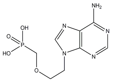
N-3-bis(2-chloroethyl)-1,3,2-oxazaphosphinan-2-amide-2-oxide, classified as alkylating agent is an anti-cancer chemotherapy drug also known as Ifosfamide (trade name Ifex). This cell cycle non-specific drug is administered intravenously in conjunction with Mesna to avoid internal bleeding and gets eliminated from patient’s body by renal elimination [22]. Ifosfamide has proven effectiveness in the treatment of variety of cancers which seized our attention.
A.3. Hepatitis B Adefovir & Lamivudine
A.3.a. Severity of Hepatitis and HBV
Hepatitis was first recorded in 1885 and has yet known to be one of the deadliest epidemic of 20th and 21st century. By 2010, China, India and Indonesia were reported to have 120 million, 40 million and 12 million infected people, respectively. WHO has estimated that 600,000 people die every year as a result of this disease [23,24]. At least 296 million people, or 3.8% of the world's population, had chronic HBV infection as of 2019. This disease can be summarized as a medical condition involving inflammation of the liver which can often lead to jaundice, poor appetite, malaise along with fibrosis and cirrhosis which can develop into liver cancer. Even though, the most common cause of Hepatitis is viral infection involving five types of viruses (A, B, C, D, E), other causes such as toxic & drug induced, alcoholic, autoimmune, ischemic, giant cell, cholestasis and non-alcoholic fatty liver dieses have also contributed considerably. All these causes ultimately result in liver failure [25-30]. The causes of transmission of Hepatitis B Virus (HBV) are very similar to that of the Human Immunodeficiency Virus (HIV) viz. sexual intercourse and blood transfusion, but is observed to be 50 to 100 times more infectious than HIV. HBV primarily interferes with the functions of the liver by replicating in hepatocytes where a functional receptor is NTCP. During HBV infection, the host immune response causes both hepatocellular damage and viral clearance [31-35]. As of now, drugs such as Lamivudine, Adefovir, Telbivudine, Entcavir are being effectively utilized in treating Hepatits B [36]. Even though, a vaccine has been discovered in the decade of 80s, none of the available drugs can completely cure the infection but can only stop the virus from replicating, thus minimizing liver damage.
A.3.b. Role of Adefovir (AFV) and its Administration
Figure 3. Adefovir
{[2-(6-amino-9H-purin-9-yl)ethoxy]methyl}phosphonic acid, also known as Adefovir (AFV) is a drug marketed by Gilead Sciences (trade name Hepsera) that belongs to the category of nucleotide reverse transcriptase inhibitor (NtRTI). It is administered orally as the pivoxil prodrug called as Adefovir dipivoxil and is observed to get eliminated from patient’s body by renal elimination. Adefovir has proven effectiveness against HBV as it blocks the reverse transcriptase enzyme, crucial for HBV to reproduce in human body and has been approved by US FDA in the treatment for Hepatitis B since 2002 [38-40].
A.3.c. Role of Lamivudine and its Administration
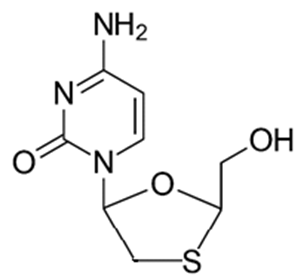
Figure 4. Lamivudine
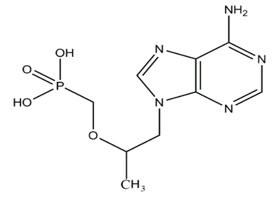
(−)-L-2′,3′-dideoxy-3′-thiacytidine also known as Lamivudine, patented in 1995 and marketed by Galxo-Smith-Kline (trade name Epivir) is an antiretroviral drug typically used in combination with other antiretroviral drugs such as zidovudine and abacavir for the preventive treatment against AIDS and Hepatitis-B. Lamivudine is a nucleoside reverse transcriptase inhibitor (NTRI) administered orally as a liquid or tablet form. Some common side effects observed are nausea, diarrhea, headaches, feeling tired, and cough along with few serious side effects which include liver disease and lactic acidosis [39-42].
A.4. AIDS - Tenofovir
A.4.a. Severity of AIDS and HIV
AIDS undoubtedly remains the deadliest epidemic of our time. WHO/UNAIDS/UNICEF global report of 2021 has estimated an average of 1.5 million new infections and 0.65 million AIDS related deaths along with 38.4 million people living with HIV. Since its discovery, AIDS has yet caused an estimated 40 million deaths worldwide [43]. HIV is a single stranded RNA retrovirus responsible for AIDS. It uses reverse transcriptase enzyme to replicate single stranded DNA in a host cell through the process of reverse transcription. Body fluids such as semen, vaginal fluid and blood are the crucial media for transmitting HIV. There is yet no cure or vaccine invented. The five major types of drugs are usually used in combination, to treat HIV infection. One of these types of drugs known as ‘reverse transcriptase inhibitors’ (RTI) have been specifically designed to disrupt the process of reverse transcription and thereby suppresses the growth of HIV. A study has shown that combinations comprising of three drugs from at least two different types can slow the course of the disease and may lead to a near normal life expectancy. This type of combination treatment is referred as anti-retroviral therapy (ART), combination anti-retroviral therapy (cART) or highly active anti-retroviral therapy (HAART) [44]. Since 2001, expanded access to antiretroviral therapy and declining incidences of HIV infection have led to a steep fall globally by 33% in the number of adults and children dying from HIV-related causes [43].
A.4.b. Role of Tenofovir (TFV) and its Oral Administration
Figure 5. Tenofovir
Reverse transcriptase inhibitors, further categorized in three forms consist of their respective nucleoside and nucleotide analogues (NRTI & NtRTI) and non-nucleoside reverse transcriptase inhibitor (NNRTI). In 1987, US FDA approved the first effective therapy against HIV which was the nucleoside reverse transcriptase inhibitor named Azidothymidine (AZT) [45]. Since, then number of drugs have been synthesized under RTI class. Tenofovir (TFV), a drug marketed by Gilead Sciences (trade name Viread) belongs to the category of NtRTI has proven to be effective against HIV as it blocks the reverse transcriptase enzyme [46,47]. It is administered as the fumarate salt of its disoproxil prodrug called as Tenofovir disoproxil fumarate (TDF) and alafenamide prodrug called as Tenofovir alafenamide fumarate (TAF).
The recommended oral dose (tablet/powder form) of {[(2R)-1-(6-amino-9H-purin-9-yl)propan-2-yl]oxy}methyl phosphonic acid, also known as Tenofovir in adults is 300 mg/day. Tenofovir gets eliminated from patient’s body by renal elimination. Pharmacokinetic studies following oral administration have shown the oral bioavailability of Tenofovir to be 39% along with food and 25% in fasted state based on urinary recovery data and intravenous data in patients [48]. Certain adverse effects such as lactic acidosis and liver problems are commonly incorporated with oral route with typical symptoms including stomach and muscle pain, nausea, headache, diarrhea and flatulence [49]. The treatment of HIV disease was realized to be quite difficult considering the problems associated with delivery of therapeutic compound to the target site. As oral administration of Tenofovir proved to be less efficient as a drug delivery method, the urgent need for alternative pre-exposure prophylaxis (PrEP) methods [50] is a self-indicative of antiretroviral therapy being still far from curing the disease and a successful HIV vaccine is yet to be developed.
B. Drug Delivery Systems-
B.1. Topical Gels as Drug Delivery System- Advantages, Limitations & Improvements
The idea of PrEP methods though started with the oral application of antiretroviral drugs was later focused on the vaginal/rectal application of anti-HIV substances, known as microbicides which are currently the principal focus for HIV prevention strategies [51,52]. To the date a great variety of HIV microbicides candidates have been studied and tested in clinical trials for their safety and microbicidal effects [53-57]. Infections caused by HIV and other enveloped viruses and sexually transmitted pathogens can be prevented by these microbicides which are the agents used topically within the vagina or rectum. In terms of formulation, a lot of the emphasis has been put on to the first generation gel formulation. According to some of the recent studies Tenofoir is approved as a microbicide in its effectiveness and safety for the prevention of HIV infections and its formulations as solid- lipid nanoparticles, vaginal gel, and vaginal ring have already been developed [58-63]. A decade ago, 1% vaginal gel formulation of Tenofovir was developed. It has been proven effective in clinical trials as, HIV incidence was reduced by 54% in the high gel adherence (>80%) group. In the light of this promising results emanated the prospect of a possible total protection against HIV, if a sustainable concentration of the active drug can be maintained. Unfortunately, this topical gel formulation was observed to suffer from several disadvantages such as leakage, poor patient compliance and the most significant one being its low retention time within the vaginal cavity. Thus, the coital dependence not only proved to limit the effectiveness, acceptability and adherence of aqueous gel system but also demanded a high dosing frequency [58]. These limitations compelled a microbicide formulation that could be administered in a coital independent fashion to become an ideal prevention strategy against sexually-acquired HIV.
The requirement to overcome the practical limitations pushed the scientific study in a direction to prepare a formulation that essentially possess high vaginal retention time and can release a high dose of microbicide when sexual intercourse occurs. Nano-formulations such as hydrogels, mucin like polymers and spray dried mucoadhesive microspheres based on polymethacrylate salt were invented and reported. These topical microbicide formulations designed for vaginal delivery are site retentive, pH and temperature sensitive, non-cytotoxic and non-immunogenic to vagina epithelium and vaginal flora and can be triggered to burst release in the presence of human semen [64-69]. Collectively, these data presented potentially effective strategy for intra-vaginal delivery of microbicides for the prevention of HIV transmission.
B.2. Delivery of Pharmaceutical Drugs through Nano-Formulations System
Since the beginning of current century the extensive research in the field of nanotechnology has provided scientific community with a plausible solution to overcome the problems faced while developing suitable drug delivery system. Modern research showed that the problems such as limited effectiveness, poor bio-distribution, and lack of selectivity encountered during conventional ways of application of drug can be overcome by controlling the drug delivery [70]. Recent developments in nanotechnology revealed that the drug molecule encapsulated in nano-formulation are able to be delivered directly to the site of action and accordingly, nanoparticles (structures smaller than 100 nm) are considered to have a great potential as drug carriers. Liposomes, solid lipids, emulsions, suspensions, wafers, micelles, dendrimers, polymers, silicon/carbon materials, and magnetic nanoparticles are the categories of nano-carriers that have been tested as drug delivery systems. Nano-fibers such as albumin, gelatin, alginate, thiolated chitosan (TCS), Hyaluronic acid (HA), Eudragit, Polyethylene glycol (PEG), Polylactic acid (PLA) and Poly(Lactide-co-Glycolide) (PLGA) are some of the most prevalently used examples to synthesize the encasing of the various pharmaceutical drugs. The nanostructures exhibit unique physicochemical and biological properties due to their small size [71,72]. Properties such as an enhanced reactive area as well as an ability to cross cell and tissue barriers made nanoparticles a favorable material for biomedical applications. Nano-carriers have effectively proven a great deal of applications in solubilizing poorly soluble drug candidates, reducing drug toxicity, prolonging circulation times, controlling drug release kinetics, drug targeting, and monitoring drug delivery to enhance therapeutic efficacy [73−78]. Thus, in recent years interest in nanoparticle drug delivery systems has grown dramatically as they have shown improvement in the pharmacological and therapeutic properties of conventional drugs. Nevertheless, in vivo delivery process exhibited the challenges such as minimal drug leakage during the initial period and triggered drug release at the target site which were needed to overcome.
C. Importance of Pharmacokinetics and Pharmacodynamics
Pharmacokinetics and pharmacodynamics are undoubtedly the main pillars that must be thoroughly studied while designing any drug. Pharmacokinetics deals with the quantitative aspect of drug handling by a body which in turn defines the temporal relationship between dose of the drug to be administered and a respective drug concentration in the body. On the other hand, pharmacodynamics establishes relationship between drug concentration at the pharmacological receptor and its respective pharmacological response. The new drug designed to be considered useful as a medicine must be capable of being delivered to its site of action achieving concentrations sufficient to initiate and maintain the appropriate pharmacological response. ADME, stands for absorption, distribution, metabolism and excretion, describing the qualitative process by which the body handles the drug as soon as it is administered in the body. The thorough knowledge of ADME provides a systematic podium to understand relationship between pharmacokinetics and pharmacodynamics. The studies of ADME process have suggested that once the administered drug reaches to equilibrium within the body, the time profile of drug-plasma concentration is observed to be quite parallel to that of drug-tissue concentration if not of absolutely equal magnitude. This makes the issue of time required to achieve equilibrium, rather critical in establishing relationship between pharmacokinetics and pharmacodynamics [79]. Hence, learning pharmacokinetics is vital when launching new drug-body profile as it offers a quantitative outline for the prospective optimization of therapeutic dosage treatments.
D. Analytical Methods Implemented for Detection & in vitro Quantification of Drug
Prior to the actual administration of a drug in human body the quality control of drug delivery system is an extremely important step which makes understanding the factors influencing drug release, from both in vivo and in vitro perspective, quintessential. Hence, a variety of meaningful in vitro drug release testing methods were developed and implemented to predict the in vivo pharmacokinetics and performance specifications. Methods introduced in earlier years such as ultrafiltration and ultracentrifugation proved to be inefficient [80-82] as they incorporated significant errors in measurement due to premature drug release as well as continued drug release during the run time of the separation process. The most prevalently used method to establish the kinetics of drug release from nanoparticles was dynamic dialysis where the amount of drug released from nanoparticle was assumed to be directly proportional to the depletion of drug from donor compartment and its appearance in receiver compartment. Also, the elimination of the additional step of separating nanoparticles from free drug at various time points during the kinetic study surpassed the popularity of dynamic dialysis over ultrafiltration and ultracentrifugation. Soon, potential sources of errors discovered in the experimental data obtained, put the authenticity and reliability of dynamic dialysis method to question. The rate of the drug release in receiving chamber was attributed to the driving force for drug transport across the dialysis membrane and its barrier properties. The driving force on the other hand was influenced by the reversible drug binding to the nano-carrier and the issue of drug partitioning between the phases present in dispersed systems [83-87]. The idea of compensating for the misinterpretation of release rate of dialysis method by implementing the reverse dialysis method and other similar techniques like rotating dialysis cell, Franz diffusion cell, and dialysis bag method were also proven to be not so effective due to its poor sensitivity and specificity [88-92]. Even though, the methods such as (High Performance Liquid Chromatography) HPLC and (Liquid Chromatography Mass Spectroscopy) LCMS utilized for the quantification of drug released in receiving chamber proved to be highly accurate, the experimental demonstration of the practical limitations incorporated within the dialysis method rendered it to be far from being tagged flawless [93]. Thus, the demand for an ideal solution compelled scientific community to search for the direct, accurate, and if possible, real time method for the detection and quantification of drug molecule. Establishing the pharmacokinetics of the drug release, in vitro, imitating the in vivo scenario is very important as it can provide sufficient information about the actual performance of the drug before being tested on animals or humans. In order to achieve the purpose of quality control and chemical standardization in a variety of sectors, qNMR has undoubtedly established its value as an orthogonal method for quantification and a persuasive substitute to chromatographic techniques [94]. Taking into account the proven track record in pharmaceutical-medicinal field, it was certain for Nuclear Magnetic Resonance; NMR technique to become the primary choice as analytical method to be developed which can effectively establish the rate of release of the drug under study, encapsulated in nano-formulation.
E.1. Significance of Quantitative NMR Spectroscopy: A Technique with Upper Hand!
In early 1950s since the discovery of nuclear magnetic resonance (NMR) spectroscopy, it is feasibly the most essential, authoritative, and widespread form of spectroscopy in both academic and industrial research. It probes atomic environments based on the different resonance frequencies exhibited by nuclei in a strong magnetic field. NMR has proven to be one of the most important analytical tools in changing our view of the possibilities of performing analysis on micro-gram level. An increase of sensitivity due to stronger static magnetic fields allowed not only independent and intrinsically reliable determination of chemical purity but also the determination of amount of substance in the sample under study by utilizing NMR spectroscopy. Around six decades ago the potential of NMR spectroscopy for the quantitative analysis of organic chemicals was first demonstrated whereas, the first quantitative measurements have been described in the literature in 1963 [95-99] that included the structural elucidation of organic compounds for determining the number of protons on each site of a molecule by measuring the integrals. NMR is an effective spectroscopic tool for quantitative analysis, as the intensity of a resonance line is directly proportional to the number of resonant nuclei or spins. In contrast to chromatography, quantitative NMR (qNMR) employs a universal reference standard as an internal standard for majority of chemical products assayed, as NMR response can be made the same for all chemical components including an internal standard by optimizing certain instrumental parameters. Thus, qNMR while maintaining greater standards of sensitivity, speed, precision, and accuracy contests commendably with chromatographic methods by avoiding a need for a reference standard for each analyte. These facts enabled qNMR to find applications in various fields such as but not limited to Physics, Chemistry, Bio-Chemistry, Pharmacy, Food Science, Medicine, Veterinary etc. [100-104]. Albeit, qNMR is almost as old as NMR itself, it is still a fresh field as it has yet to find applications in research areas such as geochemistry, environmental and forensic sciences. A destructive technique such as Mass Spectrometry with higher sensitivity has been usually observed to beat qNMR in the fields where the scope and application of qNMR needs a significant boost by overcoming challenges such as resolution and sensitivity.
In the last two decades, in pharmaceutical research quantitative NMR spectroscopy has become increasingly important for the analysis of both active pharmaceutical ingredient (API) and excipients as it has been primarily used in classical organic chemistry framework with its typical investigation focused on (i) structural determination and chirality studies of drug substances, (ii) studies including cellular metabolism and proteins. Quantitative NMR being universal and highly specific proved to be a remarkably successful analytical tool in structural analysis. Systematic validations of qNMR method, performed in a manner consistent with the Good Laboratory Practice guidelines of the United States and international government regulatory agencies, have been reported [105-108]. United States Pharmacopoeia [109], the British Pharmacopoeia [110], Japanese Pharmacopoeia and European Pharmacopoeia and few other pharmacopoeias around the world have also reported the general qNMR as a primary ratio method of measurement in which the analyte can be correlated directly to the calibration standard and hence to be compendial. The prevalent recognition of NMR for quantitative analysis can be credited to specific benefits like, (i) the prospect to define structures at a molecular level, (ii) calibration of intensity is deemed unnecessary in case of determination of ratios as signal area is directly proportional to the number of nuclei, (iii) measuring times are comparatively short, (iv) technique is non-destructive in nature, (v) sample preparation is rather easy as a prior isolation of the analyte in a mixture is not required [111-114].
Even though, most of the early qNMR studies have been performed with solution state NMR technique, for past three decades solid state NMR spectroscopy has come to the forefront of quantitative analytical techniques in pharmaceutical research. The Food and Drug Administration (FDA) has also recognized the need for solid state NMR characterization of drug substances as approximately 80–90% of pharmaceutical products on the market exist in the solid form. The possibility of decrease in stability and increase in interaction with excipients of active pharmaceutical ingredient (API) due to its alteration, caused by the extreme conditions of processing the formulation into dosage form makes it critical to study pharmaceutical compounds in the solid state both at the bulk level and for the final dosage form [115-118]. Thus, SS NMR spectroscopy has also been successfully applied to the study of polymorphism in pharmaceutical drugs at both the qualitative and quantitative levels.
E.2. Theory of Quantitative NMR Spectroscopy
One of the very important fundamental aspect of qNMR is the signal intensity for a compound X (Ix) being directly proportional to the number of nuclei (Nx) producing the NMR signal [101].
Ix = Ks Nx (Ks is an independent spectrometer constant).
Usually the NMR signal of a single substance consists of several resonance lines. Therefore, either the raw integrated signal (Ix) or a ratio of relative integrated peak areas for two compounds X and Y (Ix/Iy) could be used to quantify a molecular concentration. If multiple NMR experiments are performed, Ks being a spectrometer constant across the spectrum, the relative integrated signal area is equivalent to the concentrations.
The determination of relative area ratios Ix/Iy is the easiest way to obtain quantitative results. For single pulse NMR experiments with correct acquisition parameters Ks is the same constant for all resonance lines within the same spectrum, such that it cancels for the ratio-
The molar ratio Nx/Ny of two compounds X and Y can be calculated straightforward using-
Consequently, the amount fraction of a compound X in a mixture of m components is given by-
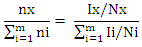
without any need to consider the solvent signal in which the mixture is dissolved as the only sample preparation step. For the purity determination of a substance an internal standard with known purity is needed. The purity of the analyte Px can be calculated as follows-
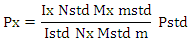
Here, Mx and MStd are the molar masses of the analyte and the standard, respectively, ‘m’ the weighed mass of the investigated sample, mStd and PStd are the weighed mass and the purity of the standard and NStd and IStd correspond to the number of spins and the integrated signal area of a typical NMR line of the standard, as described above.
Taking into account the theory & principle of quantitative NMR an advancement in the research project involving the study of a pharmaceutical drug encased in a nano-formulation can be based on certain definite aims-
Aim 1: Development and validation of a method focused on a specific NMR active nuclei constituting a pharmaceutical drug molecule of interest for the quantitative analysis study utilizing both solid state and solution state NMR spectroscopic techniques.
Aim 2: Application of solution state NMR spectroscopic technique to establish pharmacokinetics of in vitro drug release from the nano-formulation in various simulated human body fluids.
Aim 3: Application of solid state NMR spectroscopic technique to determine the encapsulation efficiency of a drug and nano-formulation pair.
Aim 4: Continue and expand with an investigation of a variety of pharmaceutical drug molecules with proven ability in the treatment of various calamitous diseases of current century such as Alzheimer, Cancer, Hepatitis, AIDS etc. utilizing proposed qNMR spectroscopic methods.
E.3. Advances in Detection Technique
Research has been carried out where quantitative nuclear magnetic resonance (qNMR) spectroscopy was employed to analyze the drug under study by taking advantage of the NMR active nuclei with larger % natural abundance such as Hydrogen, Phosphorous or Fluorine atom in the drug molecule. 1H solution state NMR method to perform a real-time detection and quantification of Tenofovir have been developed and validated [119]. The drug release profile was established in simulated vaginal fluid solution and vaginal-seminal fluid solution by integrating the two peaks (δ = 8.00 & 8.09 ppm) generated from the aromatic protons in Tenofovir to quantify its respective amount. Their results have demonstrated a good sensitivity as well as specificity that allowed the direct quantification of in vitro Tenofovir release from a spray-dried mucoadhesive and pH-sensitive MS formulation based on polymethacrylate salt without the membrane diffusion technique.
Advancement in the research was focused primarily on the development and implementation of a general qNMR method specifically focused on 31P which is NMR active nuclei to achieve direct, real time quantification of in vitro drug release. Researcher stood up in an effort aimed to provide medical field with a better understanding of the drug loading and drug releasing profiles of the large number of phosphorus containing drugs [120-123]. Both solution-state & solid-state NMR spectroscopic techniques were utilized to establish the pharmacokinetics of drug release and for the determination of encapsulation efficiency of nano-formulation for a particular drug. Tenofovir, an antiretroviral topical microbicide with proven mettle against HIV/AIDS was chosen as model phosphorous containing drug. The proven effectiveness of a specific type of spray dried, mucoadhesive, pH sensitive, mannose responsive nano-formulations composed of bio-degradable material, alginate & thiolated chitosan, intended to serve as an encasing for Tenofovir was selected.
E.3.a. Synthesis of Nano-Particle Casings-
The NPs were synthesized by alternatively coating alginate and thiolated chitosan over Tenofovir core by a layer by-layer method. Sprayed alginate NPs to be suspended into the solution of CaCl2 and TCS and stirred. The product was collected by centrifugation and washed thoroughly with water. NPs were then suspended in aqueous solution of alginate and the respective product to be collected by centrifugation and washed thoroughly with water. This process was repeated ‘n’ times in order to obtain, single, double and triple layered NPs (SLNP, DLNP, TLNP) as the final product.
The research is primarily focused on the development and validation of a 31P-qNMR method that can be implemented to determine the amount of drug released from its placebo-encasing in the simulated human body fluids such as simulated plasma, vaginal & seminal fluids. Considering the drawbacks of 1H-NMR & 13C-NMR assays, 31P-qNMR is an excellent technique for studying phosphorus containing compounds. Phosphorous with nuclear spin ½, comparatively higher gyromagnetic ratio and natural isotopic abundance of 100%, produces high signal-to-noise (S/N) ratio, thus making 31P-qNMR method considerably economical and 379 times more sensitive.
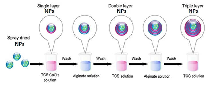
Figure 6. Schematic representation of preparation of multilayer nano-particles (NPs) of thiolated chitosan and alginate alternately layered over a core containing Tenofovir by electro-spinning method
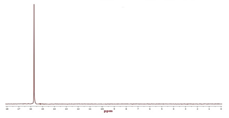
Figure 7. 31P solution state NMR spectra of pure Tenofovir (δ = 15.7 ppm) in simulated vaginal fluid
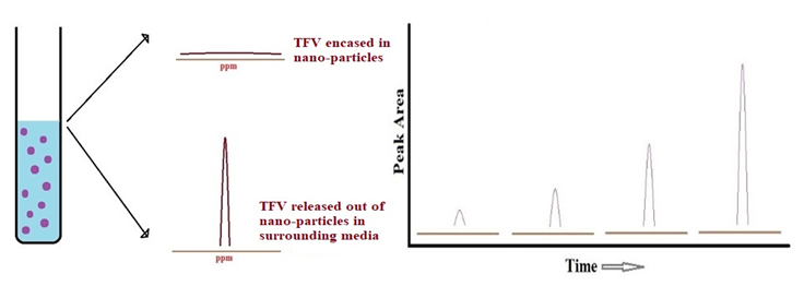
Figure 8. Schematic representation of the release of Tenofovir from its nano-particle casing in simulated human body fluids by 31P solution state qNMR spectroscopy
The experimental procedure for qNMR analyses is substantially different from the conventional use of NMR. O-phosphoric acid was used as an external standard (ES) and its chemical shift (δ) value was set to 0 ppm. A single 31P nuclei in Tenofovir molecule typically gives rise to a single sharp peak (δ = 15.7 ppm); the peak area of which being directly proportional to the amount of Tenofovir (encased or released) can be estimated. The method was validated according to International Conference of Harmonization (ICH Q2:R1) guidelines [124]. Each experiment comprised of three replicate quantitative measurements, from which a mean percent purity and its standard deviation was obtained. The variation within the 31P NMR peak areas were presented as percentage of relative standard error (%RSE) for each validation parameters such as specificity, linearity, LOD & LOQ, accuracy & precision, robustness (through variations in temperature, relaxation delay, pulse width) etc. The experimental parameters were fixed between all spectra (especially the number of experimental scans), the effects of too short a relaxation delay were constant in all experiments making the integral calculation consistent. The primary reason to apply a short relaxation delay is that acquisition time for each experiment during the in vitro drug release study is going to be limited. All of the experimental measurements were carried out at 37°C (human body) temperature. The kinetics models such as Zero & First order, Korsmeyer-Peppas, Higuchi, Weibull were utilized to elucidate mechanism of the in vitro release of Tenofovir from its nano-particle encasing [121].
E.3.b. Experimental Procedure for Solid State 31P qNMR-
The 31P-P45 MAS SS NMR (magic angle spinning solid state nuclear magnetic resonance) spectra was acquired on a Tecmag Apollo console (Houston, TX) with 8.45 T magnet operating at 357.2 MHz and homebuilt, 2-channel, wide-bore 3 mm NMR probes. The Larmor frequency for 31P was 144.596 MHz. The MAS spinning frequency, relaxation delay and 45° pulse length was, 8 KHz, 15 s and 2 μs, respectively. Typically, 5K acquisitions was added using 2K data points with spectral width of 200000 Hz, and an acquisition time of 10.24 ms. Data processing included phase correction, to be performed manually for each replicate, and baseline correction over the entire spectral range. Areas of the peaks will be determined by electronic integration of expanded regions around diagnostic resonances. The amount of the sample and ES were adjusted so that approximately equal intensities are obtained in the spectrum. The compatibility of the chosen ES and an analyte were verified prior to the quantitative analysis. All experiments were performed at ambient temperature without any corrections for sample heating. Around 30 mg of sample of pure TFV, blank NPs and drug loaded NPs were used for each analysis.
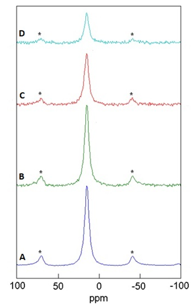
Figure 9. Determination of encapsulation efficiency of nano-particle (NP) casings by 31P solid state qNMR spectra of Tenofovir (δ = 15.7 ppm) encapsulated in NPs. A) Pure Tenofovir (control) = 100 %, B) Single Layer NP = 17.8 %, C) Double Layer NP = 12.4 %, D) Triple Layer NP = 7.4 %.; Peak areas are normalized w.r.t. that of Pure Tenofovir; Side-Bands are denoted by Asterix
This analytical method signifies a huge scope as it can be extensively applied to all of the phosphorous containing drug molecules which can in turn provide a great deal of information about the actual performance of a drug prior being tested on animals or humans. In the similar fashion methods can be developed and validated for several other NMR active nuclei such as but not limited to (11B) Boron, (19F) Fluorine, Nitrogen (15N), Sulfur (33S) etc. [125-127] which prevalently are the constituents of various pharmaceutical drug molecules. This research contribution would potentially aid scientific community in building improved versions of drug delivery systems that will further decrease the probability of infection, act as containment measure or spreading of the disease in human body, saving greater number of human lives.

2. Conclusions

Solution state NMR spectroscopic technique can be implemented to establish real time kinetics of in vitro drug release from its nano-formulation casing in simulated human body fluids such as seminal & vaginal fluids and blood plasma. On the other hand, solid state NMR spectroscopic technique can be employed to determine the encapsulation efficiency of a nano-formulation for the drug under study. In general, these particular research projects are very motivating as they contribute towards that one step which brings scientific community closer towards inventing effective treatment on HBV-Hepatitis B, HIV-AIDS and other similar dire diseases. Hence, working on such projects for any scientist proves to be the principle key in achieving the greatest goal of their career that they incessantly pursued throughout many years which is a progressive role in the future of humanity through scientific inventions. In 21st century, the scientific institutions in many developing nations are positively impacting the health of millions around the globe as they contribute by investing in the research & development of such innovative technologies. Human species have been deeply affected by multiple life threatening diseases for ages; a need of time it is now to rattle their cage.

ACKNOWLEDGEMENTS

The NMR experiments of an interdepartmental collaborative translational research presented herein were conducted at University of Missouri Kansas City (UMKC). The author of this manuscript cordially appreciates the research work contributed by Dr. Vivek Agrahari, Dr. Jianing Meng, Dr. Fohona Coulibaly and Prof. Bi-Botti Youan of School of Pharmacy, UMKC and is also immensely grateful to Prof. Nathan Oyler of Department of Chemistry, UMKC for his invaluable guidance.

References

[1]  A. Alzheimer; “Über eine eigenartige Erkrankung der Hirnrinde”, Allgemeine Zeitschrift fur Psychiatrie und Psychisch-Gerichtlich Medizin, 1907, 64(12), 146-148.
[2]  N.C. Berchtold, C.W. Cotman; "Evolution in the conceptualization of dementia and Alzheimer's disease: Greco-Roman period to the 1960s", Neurobiology of Aging, 1998, 19(3), 173–189. [DOI: 10.1016/s0197-4580(98)00052-9].
[3]  U. Maurer, K. Maurer; “Alzheimer: The Life of a Physician and the Career of a Disease” New York: Columbia University Press. 2003, p. 270. (ISBN: 978-0-231-11896-5).
[4]  “Alzheimer's Disease Fact Sheet"; National Institute on Aging, 2021. https://www.nia.nih.gov/health/alzheimers-disease-fact-sheet.
[5]  “Dementia: A public health priority” Alzheimer's Disease International, World Health Organization, 2012, 1-102. (ISBN: 978-92-4-156445-8).
[6]  Basics of Alzheimer's disease and dementia, National Institute on Aging. https://www.nia.nih.gov/health/alzheimers/basics.
[7]  H.W. Querfurth, F.M. LaFerla; "Alzheimer's Disease", The New England Journal of Medicine. 2010, 362(4), 329-344. [DOI: 10.1056/NEJMra0909142]
[8]  Z. Breijyeh, R. Karaman; “Comprehensive review on Alzheimer's disease: Causes and treatment”, Molecules, 2020, 25(24), 5789(1-28). [DOI: 10.3390/molecules25245789]
[9]  J. Gaugler, B. James, T. Johnson, J. Reimer, M. Solis, J. Weuve, R.F. Buckley, T.J. Hohman, Special Report, “Special Report: More Than Normal Aging: Understanding Mild Cognitive Impairment; 2022 Alzheimer’s Disease Facts and Figures”, Alzheimer's Association Report, Alzheimer's & Dementia: The Journal of The Alzheimer's Association, 2022, 18(4), 1-118 or 700-789 (ISSN:1552-5260, eISSN:1552-5279). [DOI: 10.1002/alz.12638].
[10]  M. De Jesus, M. Moreno; “Cognitive improvement in mild to moderate Alzheimer's dementia after treatment with the acetylcholine precursor choline alfoscerate: A multicenter, double-blind, randomized, placebo-controlled trial”, Clinical Therapeutics, 2003, 25(1), 178-193. [DOI: 10.1016/S0149-2918(03)90023-3]
[11]  L. Parnetti, F. Mignini, D. Tomassoni, E. Traini, F. Amenta; “Cholinergic precursors in the treatment of cognitive impairment of vascular origin: Ineffective approaches or need for re-evaluation?”, Journal of the Neurological Sciences, 2007, 257(12), 264-269. [DOI: 10.1016/j.jns.2007.01.043]
[12]  P.T. Francis, A.M. Palmer, M. Snape, G.K. Wilcock; 1999, 66(2), 137-147. [DOI: 10.1136/jnnp.66.2.137]; T. Babic; 1999, 67(4), 558. [DOI: 10.1136/jnnp.67.4.558]. “The cholinergic hypothesis of Alzheimer's disease: A review of progress”, Journal of Neurology, Neurosurgery, and Psychiatry.
[13]  M.C. Falk; “Generally Recognized as safe (GRAS): Determination for the Use of Alpha Size® Alpha-Glycerylphosphoryl Choline”, Life Sciences Research Organization, Inc., US Food and Drug Administration, 2012, 1-136. (GRAS Notice: GRN 419) https://www.fda.gov/food/food-ingredients-packaging/generally-recognized-safe-gras.
[14]  A. Jemal, F. Bray, M.M. Center, J. Ferlay, E. Ward, D. Forman; “Global Cancer statistics”, CA: a Cancer Journal for Clinicians, 2011, 61(2), 69-90. [DOI: 10.3322/caac.20107].
[15]  “Cancer Fact Sheet”; International Agency for Research on Cancer, World Health Organization, 2020. https://gco.iarc.fr/today/fact-sheets-cancers.
[16]  “National Cancer Institute (NCI)”; National Institutes of Health, U.S. Department of Health and Human Services, 2022. https://www.nih.gov/about-nih/what-we-do/nih-almanac/national-cancer-institute-nci.
[17]  “Suspected cancer: recognition and referral, (NG12)”; National Institutes for Health and Care Excellence, published: 2015, updated: 2021, 1-98.
[18]  C.P. Wild, E. Weiderpass, B.W. Stewart; “World Cancer Report: Cancer Research for Cancer Prevention”, International Agency for Research on Cancer, World Health Organization, 2020. (ISBN: 978-92-832-0447-3, 978-92-832-0448-0).
[19]  C.J.L. Murray, A.D. Lopez, T. Vos (Disease and Injury Incidence and Prevalence Collaborators); “Global, regional, and national incidence, prevalence, and years lived with disability for 310 diseases and injuries, 1990–2015: A systematic analysis for the Global Burden of Disease Study 2015”, The Lancet, 2016, 388(10053), 1545-1602. [DOI: 10.1016/S0140-6736(16)31678-6]
[20]  M. Sciacovelli, C. Schmidt, E.R. Maher, C. Frezza; “Metabolic Drivers in Hereditary Cancer Syndromes”, Annual Review of Cancer Biology, Annual Reviews, 2020, 4, 77-97. [DOI: 10.1146/annurev-cancerbio-030419-033612]
[21]  Global burden of disease study 2019 Cancer collaboration: J.M. Kocarnik; et. al.; “Cancer incidence, mortality, years of life lost, years lived with disability, and disability-adjusted life years for 29 Cancer groups from 2010 to 2019: A systematic analysis for the global burden of disease study 2019”, JAMA Oncology. 2022, 8(3), 420-444. [DOI: 10.1001/jamaoncol.2021.6987]
[22]  R. Pazdur, L.D. Wagman, K.A. Camphausen, W.J. Hoskins; “Cancer Management: A Multidisciplinary Approach” 12th edition, Cmp Healthcare Media, 2009, 1-1050. (ISBN: 978-1-891483-63-9).
[23]  E. Thomas, M. Yoneda, E.R. Schiff; “Viral Hepatitis: Past and Future of HBV and HDV”, Cold Spring Harbor Perspective Medicine, 2015, 5(2), 1-11. [DOI: 10.1101/cshperspect.a021345]
[24]  G.S. Burns, A.J. Thompson; “Viral Hepatitis B: Clinical and Epidemiological Characteristics”, Cold Spring Harbor Perspective Medicine, 2014, 4(12), 1-14. [DOI: 10.1101/cshperspect.a024935]
[25]  W. Bernal, J. Wendon; “Critical care medicine: Acute liver failure”, The New England Journal of Medicine 2013, 369(26), 2525-2534. [DOI: 10.1056/NEJMra1208937]
[26]  Part 10, Section 3, Chapters 329-338; “Liver and Biliary Tract Disease”, in J.L. Jameson, A.S. Fauci, D.L. Kasper, S.L. Hauser, D.L. Longo, J. Loscalzo; Harrison's Principles of Internal Medicine, 20th Edition. New York, NY: McGraw-Hill Education Medical; 2018. (ISBN: 978-1-25-964403-0).
[27]  Part IX, Chapters 38-40, Pages 1003-1052; “Infectious and Granulomatous Disease”. in E.R. Schiff, W.C. Maddrey, K.R. Reddy; Schiff’s Diseases of the Liver, 12th Edition, John Wiley & Sons Ltd. 2017, 1-1213. (ISBN: 978-1-11-925122-4, 978-1-11-925131-6). [DOI: 10.1002/9781119251316]
[28]  Part III, Chapters 10-19, Pages 269-487; “Consequences of Liver Disease” in E.R. Schiff, W.C. Maddrey, K.R. Reddy; Schiff’s Diseases of the Liver, 12th Edition, John Wiley & Sons Ltd. 2017, 1-1213. (ISBN: 978-1-11-925122-4, 978-1-11-925131-6). [DOI: 10.1002/9781119251316]
[29]  R.B. Pinto, A.C.R. Schneider, T.R. da Silveira; “Cirrhosis in children and adolescents: An overview”, 2015, 7(3), 392-405. [DOI: 10.4254/wjh.v7.i3.392]
[30]  E.Y. Cheng, A. Zarrinpar, D.A. Geller, J.A. Goss, R.W. Busuttil; Chapter 31, Pages 1263-1308; “Liver"; in F.C. Brunicardi, D.K. Anderson, T.R. Billiar, D.L. Dunn, J.G. Hunter, J.B. Matthews, R.E. Pollock; Schwartz's principles of surgery, 10th Edition, New York: McGraw-Hill Education Medical, 2015, 1-2098. (ISBN: 978-0-07-180092-1, 978-0-07-179675-0).
[31]  “Viral Hepatitis”, Centers for Disease Control and Prevention, 2021. https://www.cdc.gov/hepatitis/hbv/index.htm.
[32]  C.K. Fairley, T.R.H. Read; “Vaccination against sexually transmitted infections”, Current Opinion in Infectious Diseases, 2012, 25(1), 66-72. [DOI: 10.1097/QCO.0b013e32834e9aeb]
[33]  F. Buddeberg, B.B. Schimmer, D.R. Spahn; “Transfusion-transmissible infections and transfusion-related immunomodulation”, Best Practice & Research in Clinical Anaesthesiology, 2008, 22(3), 503-517. [DOI: 10.1016/j.bpa.2008.05.003]
[34]  R.A. Hughes; “Drug Injectors and the Cleaning of Needles and Syringes” Special Topic Section: Development of Harm Reduction and Methadone Maintenance in Europe, European Addiction Research, 2000, 6(1), 20-30. [DOI: 10.1159/000019005]
[35]  H. Yan, G. Zhong, G. Xu, W. He, Z. Jing, Z. Gao, Y. Huang, Y. Qi, B. Peng, H. Wang, L. Fu, M. Song, P. Chen, W. Gao, B. Ren, Y. Sun, T. Cai, X. Feng, J. Sui, W. Li; “Sodium taurocholate cotransporting polypeptide is a functional receptor for human hepatitis B and D virus”, Biochemistry | Microbiology and Infectious Diseases, eLIFE, 2012, 1:e00049, 1-28. [DOI: 10.7554/eLife.00049.001]
[36]  A. Vittal, M.G. Ghany; “WHO Guidelines for Prevention, Care and Treatment of Individuals Infected with HBV: A US Perspective”. Clinics in Liver Disease, 2019, 23(3), 417-432. [DOI: 10.1016/j.cld.2019.04.008]
[37]  P. Marcellin, T.T. Chang, S.G. Lim, M.J. Tong, W. Sievert, M.L. Shiffman, L. Jeffers, Z. Goodman, M.S. Wulfsohn, S. Xiong, J. Fry, C.L Brosgart, Adefovir Dipivoxil 437 Study Group; “Adefovir dipivoxil for the treatment of hepatitis B e antigen-positive chronic hepatitis B”, The New England Journal of Medicine, 2003, 348(9), 808-816. [DOI: 10.1056/NEJMoa020681]
[38]  S. Manolakopoulos, S. Bethanis, S. Koutsounas, J. Goulis, J. Vlachogiannakos, E. Christias, A. Saveriadis, C. Pavlidis, C. Triantos, A. christidou, G. Papatheodoridis, D. Karamanolis, D. Tzourmakliotis; “Long-term therapy with Adefovir dipivoxil in hepatitis B e antigen-negative patients developing resistance to Lamivudine”, Alimentary Pharmacology & Therapeutics, 2007, 27(3), 266-273. [DOI: 10.1111/j.1365-2036.2007.03567.x]
[39]  P. Reiss, “A randomized placebo-controlled trial of Adefovir dipivoxil in advanced HIV infection: the ADHOC trial”, ADHOC International Steering Committee; HIV Medicine, 2002, 3(4), 229-238.
[40]  D. Thaczuk, S.R. Hosein, B. Ziegler; "3TC (lamivudine, Epivir)", CATIE. 2014.
[41]  G.W. Koszalka, S.M. Daluge, F.L. Boyd; “Chapter 16-Advances in Nucieoside and Nucleotide Antiviral Therapies”, Annual Reports in Medicinal Chemistry, 1998, 30, 163-171. [DOI: 10.1016/S0065-7743(08)61081-7]
[42]  J. Fischer, C.R. Ganellin, C.R.; “Lamivudine”, Analogue-based Drug Discovery, John Wiley-VCH Verlag GmbH & Co. KGaA, 2006, 506. (ISBN: 978-3-52-731257-3, 978-3-52-760800-3). [DOI: 10.1002/3527608001]
[43]  “Global HIV & AIDS Statistics-Fact Sheet”. UNAIDS, 2022. https://www.unaids.org/en/resources/fact-sheet
[44]  “Guidelines for the Use of Antiretroviral Agents in Adults and Adolescents with HIV”. US Department of Health and Human Services, 2021, 1-454.
[45]  I. Molotsky; “US Approves Drug to Prolong Lives of AIDS Patience”. New York Times, 1987-03-21.
[46]  P. Emau, Y. Jiang, M.B. Agy, B. Tian, G. Bekele, C-C. Tsai; “Post-exposure prophylaxis for SIV revisited: Animal model for HIV prevention”, AIDS Research and Therapy, 2006, 3(29), 1-14. [DOI: 10.1186/1742-6405-3-29]
[47]  Gilead Sciences, Inc. “Prescribing Information: DESCOVY® (Emtricitabine and Tenofovir, Alafenamide) tablets, for oral use”, Initial U.S. Approval: 2015, Revised: 2016, 1-28. (Reference ID: 3911541) [©2016 Gilead Sciences, Inc. All rights reserved. GILP0636 04/16 and 208215-GS-000]
[48]  B.P. Kearney, J.F. Flaherty, J. Shah; “Tenofovir disoproxil fumarate: Clinical pharmacology and pharmacokinetics”, Clinical Pharmacokinetics, 2004, 43(9), 595-612. [DOI: 10.2165/00003088-200443090-00003]
[49]  Gilead Sciences Inc. “VIREAD, (Tenofovir disoproxil fumarate) tablets/powder, for oral use”, Initial U.S. Approval: 2001, Revised: 2019, 1-49. [©2019 Gilead Sciences, Inc. All rights reserved. 21356-GS-041 and 21356-GS-022 240908]
[50]  V. Agrahari, S.M. Anderson, M.M. Peet, A.P. Wong, O.N. Singh, G.F. Doncel, M.R. Clark; “Long-acting HIV pre-exposure prophylaxis (PrEP) approaches: recent advances, emerging technologies, and development challenges”, Expert Opinion on Drug Delivery, 2022, 19(10), 1365-1380. [DOI: 10.1080/17425247.2022.2135699]
[51]  L.C. Rohan, A.B. Sassi; “Vaginal drug delivery systems for HIV prevention”, The AAPS Journal, 2009, 11(1), 78-87. [DOI: 10.1208/s12248-009-9082-7]
[52]  V.M.K. Ndesendo, V. Pillay, Y.E. Choonara, E. Buchmann, D.N. Bayever, L.C.R. Meyer; “A review of current intra-vaginal drug delivery approaches employed for the prophylaxis of HIV/AIDS and prevention of sexually transmitted infections”, AAPS Pharm. Sci. Tech. 2008, 9(2), 505-520. [DOI: 10.1208/s12249-008-9073-5]
[53]  J. das Neves, J. Michiels, K.K. Arien, G. Vanham, M. Amiji, M.F. Bahia, B. Sarmento; “Polymeric nanoparticles affect the intracellular delivery, antiretroviral activity and cytotoxicity of the microbicide drug candidate Dapivirine”, Pharmaceutical Research, 2011, 2012, 1468-1484. [DOI: 10.1007/s11095-011-0622-3]
[54]  F.X. Mbopi-Keou, S. Trottier, R.F. Omar, N.N. Nkele, S. Fokoua, E.R. Mbu, M.C. Domingo, J.F. Giguere, J. Piret, A. Mwatha, B. Masse, M.G. Bergeron; “A randomized, double-blind, placebo-controlled Phase-II extended safety study of two invisible condom formulations in Cameroonian women”, Contraception, 2010, 81(1), 79-85. [DOI: 10.1016/j.contraception.2009.07.002]
[55]  S. McCormack, G. Ramjee, A. Kamali, H. Rees, A.M. Crook, M. Gafos, U. Jentsch, R. Pool, M. Chisembele, S. Kapiga, R. Mutemwa, A. Vallely, T. Palanee, Y. Sookrajh, C.J. Lacey, J. Darbyshire, H. Grosskurth, A. Profy, A. Nunn, R. Hayes, J. Weber; “PRO2000 vaginal gel for prevention of HIV-1 infection (Microbicides Development Programme 301): A phase 3, randomised, double-blind, parallel-group trial”, Lancet, 2010, 376(9749), 1329-1337. [DOI: 10.1016/S0140-6736(10)61086-0]
[56]  L. Wan, S. Pooyan, P. Hu, M.J. Leibowitz, S. Stein, P.J. Sinko; “Peritoneal macrophage uptake, pharmacokinetics and biodistribution of macrophage-targeted PEG-fMLF (N-formyl-methionyl-leucyl-phenylalanine) nanocarriers for improving HIV drug delivery”, Pharmaceutical Research, 2007, 24(11), 2110-2119. [DOI: 10.1007/s11095-007-9402-5]
[57]  M.M. Peet, V. Agrahari, S.M. Anderson, H. Hanif, O.N. Singh, A.R. Thurman, G.F. Doncel, M.R. Clark; “Topical inserts: A versatile delivery form for HIV prevention”, Pharmaceutics, 2019, 11(8), 374(1-17). [DOI: 10.3390/pharmaceutics11080374]
[58]  Q. Abdool Karim, S.S. Abdool Karim, J.A. Frohlich, A.C. Grobler, C. Baxter, L.E. Mansoor, A.B. Kharsany, S. Sibeko, K.P. Mlisana, Z. Omar, T.N. Gengiah, S. Maarschalk, N. Arulappan, M. Mlotshwa, L. Morris, D. Taylor; “Effectiveness and safety of Tenofovir gel, an antiretroviral microbicide, for the prevention of HIV infection in women”, Science. 2010, 329(5996), 1168-1174. [DOI: 10.1126/science.1193748]
[59]  L.C. Rohan, B.J. Moncla, N. Kunjara, R.P. Ayudhya, M. Cost, Y. Huang, F. Gai, N. Billitto, J.D. Lynam, K. Pryke, P. Graebing, N. Hopkins, J.F. Rooney, D. Friend, C.S. Dezzutti; “In vitro and ex vivo testing of Tenofovir shows it is effective as an HIV-1 microbicide”, PLoS One. 2010, 5(2), e9310. [DOI: 10.1371/journal.pone.0009310]
[60]  D. Alukda, T. Sturgis, B.-B.C. Youan; “Formulation of Tenofovir-loaded functionalized solid lipid nanoparticles intended for HIV prevention”, Journal of Pharmaceutical Sciences, 2011, 100(8), 3345-3356. [DOI: 10.1002/jps.22529]
[61]  K.H. Mayer, L.A. Maslankowski, F. Gai, W.M. El-Sadr, J. Justman, A. Kwiecien; B. Masse, S.H. Eshleman, C. Hendrix, K. Morrow, J.F. Rooney, L. Soto-Torres, HPTN Protocol Team; “Safety and tolerability of Tenofovir vaginal gel in abstinent and sexually active HIV-infected and uninfected women”, AIDS. 2006, 20(4), 543-551. [DOI: 10.1097/01.aids.0000210608.70762.c3]
[62]  R.K. Rosen, K.M. Morrow, A. Carballo-Dieguez, J.E. Mantell, S. Hoffman, F. Gai, L. Maslankowski, W.M. El-Sadr, K.H. Mayer; “Acceptability of Tenofovir gel as a vaginal microbicide among women in a phase-I trial: A mixed-methods study”, Journal of Womens Health (Larchmt). 2008, 17(3), 383-392. [DOI: 10.1089/jwh.2006.0325]
[63]  B.B. Saxena, Y.A. Han, D. Fu, P. Rathnam, M. Singh, J. Laurence, S. Lerner; “Sustained release of microbicides by newly engineered vaginal rings”, AIDS. 2009, 23(8), 917-922. [DOI: 10.1097/QAD.0b013e32832af57c]
[64]  K.M. Gupta, S.R. Barnes, R.A. Tangaro, M.C. Roberts, D.H. Owen, D.F. Katz, P.F. Kiser; “Temperature and pH sensitive hydrogels: An approach towards smart semen-triggered vaginal microbicidal vehicles”, Journal of Pharmaceutical Sciences, 2007, 96(3), 670-681. [DOI: 10.1002/jps.20752]
[65]  A. Mahalingam, J.I. Jay, K. Langheinrich, S. Shukair, M.D. McRaven, L.C. Rohan; B.C. Herold, T.J. Hope, P.F. Kiser; “Inhibition of the transport of HIV in vitro using a pH- 169 responsive synthetic mucin-like polymer system”, Biomaterials. 2011, 32(33), 8343-8355. [DOI: 10.1016/j.biomaterials.2011.05.001]
[66]  M.R. Clark, H.A. Aliyar, C.W. Lee, J.I. Jay, K.M. Gupta, K.M. Watson, R.J. Stewart, R.W. Buckheit, P.F. Kiser; “Enzymatic triggered release of an HIV-1 entry inhibitor from prostate specific antigen degradable microparticles”, International Journal of Pharmaceutics, 2011, 413(1-2), 10-18. [DOI: 10.1016/j.ijpharm.2011.04.004]
[67]  T. Zhang, T.F. Sturgis, B.-B.C. Youan; “pH-responsive nanoparticles releasing Tenofovir intended for the prevention of HIV transmission”, European Journal of Pharmaceutics and Biopharmaceutics, 2011, 79(3), 526-536. [DOI: 10.1016/j.ejpb.2011.06.007]
[68]  F. Cilurzo, F. Selmin, P. Minghetti, I. Rimoldi, F. Demartin, L. Montanari; “Fast-dissolving mucoadhesive microparticulate delivery system containing Piroxicam”, European Journal of Pharmaceutical Sciences, 2005, 24(4), 355-361. [DOI: 10.1016/j.ejps.2004.11.010]
[69]  T. Zhang, C. Zhang, V. Agrahari, J.B. Murowchick, N.A. Oyler, B.-B.C. Youan; “Spray drying tenofovir loaded mucoadhesive and pH-sensitive microspheres intended for HIV prevention”, Antiviral Research, 2013, 97(3), 334-346. [DOI: 10.1016/j.antiviral.2012.12.019]
[70]  D. Nevozhay, U. Kañska, R. Budzyñska, J. Boratyñski; “Współczesny stan badań nad koniugatami i innymi systemami dostarczania leków w leczeniu schorzeń nowotworowych i innych jednostek chorobowych”, Translated: “Current status of research on conjugates and related drug delivery systems in the treatment of cancer and other diseases”, Postepy Higieny i Medycyny Doswiadczalnej, 2007, 61, 350-360. [PMID: 17554238]
[71]  S.S. Suri, H. Fenniri, B. Singh; “Nanotechnology-based drug delivery systems”, Journal of Occupational Medicine and Toxicology, 2007, 2, 16(1-6). [DOI: 10.1186/1745-6673-2-16]
[72]  I. Brigger, C. Dubernet, P. Couvreur; “Nanoparticles in cancer therapy and diagnosis”, Advanced Drug Delivery Reviews, 2002, 54(5), 631-651. [DOI: 10.1016/s0169-409x(02)00044-3]
[73]  S. Marchesan, M. Prato; “Nanomaterials for (Nano) medicine”, ACS Medicinal Chemistry Letters, 2013, 4(2), 147-149. [DOI: 10.1021/ml3003742]
[74]  P.A. McCarron, M. Hall; “Pharmaceutical Nanotechnology”, Encyclopedia of Nanoscience and Nanotechnology, 2004, 8, 469-487. (ISBN: 1-58883-064-0)
[75]  L. Mei, Z. Zhang, L. Zhao, L. Huang, X.-L. Yang, J. Tang, S.-S. Feng; “Pharmaceutical nanotechnology for oral delivery of anticancer drugs”, Advanced Drug Delivery Reviews, 2013, 65(6), 880-890. [DOI: 10.1016/j.addr.2012.11.005]
[76]  T. Musacchio, V.P. Torchilin; “Recent developments in lipid-based pharmaceutical nanocarriers”, Frontiers in Bioscience Landmark, 2011, 16, 1388-1412. [DOI: 10.2741/3795]
[77]  L. Zhang, F.X. Gu, J.M. Chan, A.Z. Wang, R.S. Langer, O.C. Farokhzad; “Nanoparticles in Medicine: Therapeutic Applications and Developments”, Clinical Pharmacology and Therapeutics, 2008, 83(5), 761-769. [DOI: 10.1038/sj.clpt.6100400]
[78]  N. Nagai, H. Otake; “Novel drug delivery systems for the management of dry eye”, Advanced Drug Delivery Reviews, 2022, 191, 114582(1-16). [DOI: 10.1016/j.addr.2022.114582]
[79]  H.J. Smith, H. Williams “Smith and Williams’ Introduction to the Principles of Drug Design and Action”, 4th edition. (ISBN: 978-1-03-230661-2).
[80]  I. Henriksen, S.A. Sande, G. Smistad, T. Agren, J. Karlsen; “In vitro evaluation of drug release kinetics from liposomes by fractional dialysis”, International Journal of Pharmaceutics, 1995, 119(2), 231-238. [DOI: 10.1016/0378-5173(94)00403-R]
[81]  S.J. Wallace, J. Li, R.L. Nation, B.J. Boyd; “Drug release from nanomedicines: selection of appropriate encapsulation and release methodology”, Drug Delivery and Translational Research, 2012, 2, 284-292. [DOI: 10.1007/s13346-012-0064-4]
[82]  M.Y. Levy, S. Benita; “Drug release from submicronized o/w emulsion: a new in vitro kinetic evaluation model”, International Journal of Pharmaceutics, 1990, 66(1-3), 29-37. [DOI: 10.1016/0378-5173(90)90381-D]
[83]  G. Moreno-Bautista, K.C. Tam; “Evaluation of dialysis membrane process for quantifying the in vitro drug-release from colloidal drug carriers”, Colloids and Surfaces A: Physiochemical and Engineering Aspects; 2011, 389(1-3), 299-303. [DOI: 10.1016/j.colsurfa.2011.07.032]
[84]  D. Friedman, S. Benita; “A mathematical model for drug release from o/w emulsions: Application to controlled release Morphine emulsions”, Drug Development and Industrial Pharmacy, 1987, 13(9-11), 2067-2085. [DOI: 10.3109/03639048709068707]
[85]  C. Washington; “Evaluation of non-sink dialysis methods for the measurement of drug release from colloids: effects of drug partition”, International Journal of Pharmaceutics, 1989, 56, 71-74. [DOI: 10.1016/0378-5173(89)90062-8]
[86]  C. Washington; “Drug release from microdisperse systems: a critical review”, International Journal of Pharmaceutics, 1990, 58, 1-12. [DOI: 10.1016/0378-5173(90)90280-H]
[87]  P. Calvo, J.L. Vila-Jato, M.J. Alonso; “Comparative in vitro evaluation of several colloidal systems, nanoparticles, nanocapsules, and nanoemulsions, as ocular drug carriers”, Journal of Pharmaceutical Sciences, 1996. 85(5), 530-536. [DOI: 10.1021/js950474+]
[88]  X. Xu, M.A. Khan, D.J. Burgess; “A two-stage reverse dialysis in vitro dissolution testing method for passive targeted liposomes”, International Journal of Pharmaceutics, 2012, 426(1–2), 211-218. [DOI: 10.1016/j.ijpharm.2012.01.030]
[89]  V. Agrahari, B-B.C. Youan; “Sensitive and Rapid HPLC Quantification of Tenofovir from Hyaluronic Acid-Based Nanomedicine”, AAPS. Pharm. Sci. Tech. 2012, 13(1), 202-210. [DOI: 10.1208/s12249-011-9735-6]
[90]  S. Sentenac, C. Fernandez, A. Thuillier, P. Lechat, G. Aymard; “Sensitive determination of Tenofovir in human plasma samples using reversed-phase liquid chromatography”, Journal of Chromatography B, 2003, 793(2), 317-324. [DOI: 10.1016/s1570-0232(03)00333-7]
[91]  M. Takahashi, Y. Kudaka, N. Okumura, A. Hirano, K. Banno, T. Kaneda; “Determination of plasma Tenofovir concentrations using a conventional LC-MS method” Biological and Pharmaceutical Bulletin, 2007, 30(9), 1784-1786. [DOI: 10.1248/bpb.30.1784]
[92]  M. Yadav, T. Mishra, P. Singhal, S. Goswami, P.S. Shrivastav; “Rapid and specific liquid chromatographic tandem mass spectrometric determination of Tenofovir in human plasma and its fragmentation study”, Journal of Chromatographic Science, 2009, 47(2), 140-148. [DOI: 10.1093/chromsci/47.2.140]
[93]  S. Modi, B.D. Anderson; “Determination of Drug Release Kinetics from nanoparticles: overcoming pitfalls of the dynamic dialysis method”, Molecular Pharmaceutics, 2013, 10(8), 3076-3089. [DOI: 10.1021/mp400154a]
[94]  B.U. Jaki, A. Bzhelyansky, G.F. Pauli; “Quantitative NMR (qNMR) forpharmaceutical analysis: The pioneering work of George Hanna at the US FDA”, Magnetic Resonance in Chemistry, 2021, (59), 7–15. [DOI: 10.1002/mrc.5099]
[95]  J.N. Shoolery; “Some quantitative applications of 13C NMR spectroscopy”, Progress in NMR Spectroscopy. 1977, 11, 79-93. [DOI: 10.1016/0079-6565(77)80003-4]
[96]  R. Greenhalgh, J.N. Shoolery; “Analysis of organo-phosphorus insecticide and formulations for contaminants by phosphorus-31 Fourier transform nuclear magnetic resonance spectrometry”, Analytical Chemistry, 1978, 50(14), 2039-2042. [DOI: 10.1021/ac50036a025]
[97]  J.W. Turczan, T. Medwick; “Qualitative and Quantitative Analysis of Amygdalin Using NMR Spectroscopy”, Analytical Letters, 1977, 10(7-8), 581-590. [DOI: 10.1080/00032717708059224]
[98]  J.L. Jungnickel, J.W. Forbes; “Quantitative Measurement of Hydrogen Types by Intergrated Nuclear Magnetic Resonance Intensities”, Analytical Chemistry, 1963, 35(8), 938-942. [DOI: 10.1021/ac60201a005]
[99]  D.P. Hollis; “Quantitative analysis of Aspirin, Phenacetin, and Caffeine mixtures by nuclear magnetic resonance spectrometry”, Analytical Chemistry, 1963, 35(11), 1682-1684. [DOI: 10.1021/ac60204a043]
[100]  M. Yang, J. Wang, L. Kong; “Quantitative analysis of four major diterpenoids in Andrographis paniculata by 1H NMR and its application for quality control of commercial preparation” Journal of Pharmaceutical and Biomedical Analysis, 2012, 70, 87-93. [DOI: 10.1016/j.jpba.2012.05.037]
[101]  V. Pieri, A. Belancic, S. Morales, H. Stuppner; “Identification and quantification of major Steviol Glycosides in Stevia rebaudiana purified extracts by 1H NMR spectroscopy”, Journal of Agricultural and Food Chemistry, 2011, 59(9), 4378-4384. [DOI: 10.1021/jf104922q]
[102]  J. Staneva, P. Denkova, M. Todorova, L. Evstatieva; “Quantitative analysis of sesquiterpene lactones in extract of Arnica montana L. by 1H NMR spectroscopy”, Journal of Pharmaceutical and Biomedical Analysis, 2011, 54(1), 94-99. [DOI: 10.1016/j.jpba.2010.08.018]
[103]  G.M. Pinheiro, E.A. Basso, B.C. Fiorin, F. Cendes, R. Rittner, A.N. Oliveira, N.F. Hoehr; “A fast 1H NMR spectroscopy procedure for quantitative determination of N-acetylaspartate in urine samples”, Clinica Chimica Acta, 2009, 404(2), 166-168. [DOI: 10.1016/j.cca.2009.03.008]
[104]  S.K. Bharti, R. Roy; “Quantitative 1H NMR spectroscopy”, Trends in Analytical Chemistry, 2012, (35), 5-26. [DOI: 10.1016/j.trac.2012.02.007]
[105]  Good Laboratory Practice regulations 40 CFR Part 160 established by the Federal Insecticide, Fungicide and Rodenticide Act, and 21 CFR Part 58 established by the Federal Food, Drug, and Cosmetic Act, and the Organization for Economic Cooperation and Development Principles of Good Laboratory Practice as outlined in OECD/GD(92)32, Code of Federal Regulations, revised: 1997, Title 40, Volume 14, Parts 150 to 189, Page 125-137, CITE: 40CFR160.
[106]  G.F. Pauli, T. Godecke, B.U. Jaki, D.C. Lankin; “Quantitative 1H NMR. Development and potential of an analytical method: an update”, Journal of Natural Products, 2012, 75(4), 834-851. [DOI: 10.1021/np200993k]
[107]  F. Malz, H. Jancke; “Validation of quantitative NMR”, Journal of Pharmaceutical and Biomedical Analysis, 2005, 38(5), 813-823. [DOI: 10.1016/j.jpba.2005.01.043]
[108]  G. Maniara, K. Rajamoorthi, S. Rajan, G.W. Stockton; “Method performance and validation for quantitative analysis by 1H and 31P NMR spectroscopy: Applications to analytical standards and agricultural chemicals”, Analytical Chemistry, 1998, 70(23), 4921-4928. [DOI: 10.1021/ac980573i]
[109]  The United States Pharmacopoeia, USP XXII; United States Pharmacopoeia Convention, Inc.: Rockville, MD, 1992, Suppl. 7, 3140.
[110]  British Pharmacopoeia, Medicines and Healthcare Products Regulatory Agency, Her Majesty’s Stationery Office; London, 1988; Vol. II.
[111]  T. Beyer, B. Diehl, U. Holzgrabe; “Quantitative NMR spectroscopy of biologically active substances and excipients”, Bioanalytical Review, 2010, (2), 1-22. [DOI: 10.1007/s12566-010-0016-8]
[112]  M. Malet-Martinoa, U. Holzgrabe; “NMR techniques in biomedical and pharmaceutical analysis”, Journal of Pharmaceutical and Biomedical Analysis, 2011, (55), 1-15. [DOI: 10.1016/j.jpba.2010.12.023]
[113]  U. Holzgrabe ∗, R. Deubner, C. Schollmayer, B. Waibel; “Quantitative NMR spectroscopy-Applications in drug analysis”, Journal of Pharmaceutical and Biomedical Analysis, 2005, (38), 806-812. [DOI: 10.1016/j.jpba.2005.01.050]
[114]  U. Holzgrabe; “Quantitative NMR spectroscopy in pharmaceutical applications”, Progress in Nuclear Magnetic Resonance Spectroscopy, 2010, 57, 229-240. [DOI: 10.1016/j.pnmrs.2010.05.001]
[115]  M. Otsuka, T. Matsumoto, N. Kaneniwa; “Effects of the mechanical energy of multi-tableting compression on the polymorphic transformations of chlorpropamide”, Journal of Pharmacy and Pharmacology, 1989, 41(10), 665-669. [DOI: 10.1111/j.2042-7158.1989.tb06337.x]
[116]  K.A. Connors, G.L. Amidon, V.J. Stella; “Chemical Stability of Pharmaceuticals: A Handbook for Pharmacist” 2nd edition, John Wiley & Sons, New York, 1986. (ISBN: 978-0-471-87955-8)
[117]  R.T. Berendt, D.M. Sperger, P.K. Isbester, E.J. Munson; “Solid-state NMR spectroscopy in pharmaceutical research and analysis”, Trends in Analytical Chemistry, 2006, 25(10), 977-984. [DOI: 10.1016/j.trac.2006.07.006]
[118]  P.A. Tishmack, D.E. Bugay, S.R. Byrn; “Solid-State Nuclear Magnetic Resonance Spectroscopy-Pharmaceutical Applications”, Journal of Pharmaceutical Sciences, 2003, 92(3), 441-474. [DOI: 10.1002/jps.10307]
[119]  C. Zhang, T. Zhang, N.A. Oyler, B.-B.C. Youan; “Direct and real-time quantification of Tenofovir release from pH-sensitive microparticles into simulated biological fluids using 1H-NMR”, Journal of Pharmaceutical Sciences, 2014, 13(4), 1170-1177. [DOI: 10.1002/jps.23886]
[120]  F.S. Coulibaly, M.J. Ezoulin, S.S. Purohit, N.J. Ayon, N.A. Oyler, B-B.C. Youan; “Layer by layer engineered microbicide drug delivery system targeting HIV-1 gp120: physicochemical and biological properties”, Molecular Pharmaceutics, 2017, 14(10), 3512-3527. [DOI: 10.1021/acs.molpharmaceut.7b00555]
[121]  V. Agrahari, J. Meng, S.S. Purohit, N.A. Oyler, B-B.C. Youan; “Real-Time analysis of Tenofovir release kinetics using quantitative phosphorus (31P) nuclear magnetic resonance spectroscopy”, Journal of Pharmaceutical Sciences, 2017, 106(10), 3005-3015. [DOI: 10.1016/j.xphs.2017.03.043]
[122]  J. Meng, V. Agrahari, S.S. Purohit, T. Zhang, M.J. Ezoulin, A. Molteni, D. Dim, N.A. Oyler, B-B.C. Youan; “Spray dried thiolated chitosan coated sodium alginate multilayer microparticles for vaginal HIV microbicide delivery”, The AAPS Journal, 2017, 19(3), 692-702. [DOI: 10.1208/s12248-016-0007-y]
[123]  J. Meng, V. Agrahari, M.J. Ezoulin, C. Zhang, S.S. Purohit, A. Molteni, D. Dim, N.A. Oyler, B.-B.C. Youan; “Tenofovir containing thiolated chitosan core/shell nanofibers: in vitro and in vivo evaluations”, Molecular Pharmaceutics, 2016, 13(12), 4129-4140. [DOI: 10.1021/acs.molpharmaceut.6b00739]
[124]  Harmonization ICo. Geneva, Switzerland: 2005.
[125]  F.S. Coulibaly, A.S. Alnafisah, N.A. Oyler, B.-B.C. Youan; “Direct and real-time quantification of Bortezomib release from alginate microparticles using boron (11B) nuclear magnetic resonance spectroscopy”, Molecular Pharmaceutics, 2019, 16(3), 967-977. [DOI: 10.1021/acs.molpharmaceut.8b00873]
[126]  A.O. Mattes, D. Russell, E. Tishchenko, Y. Liu, R.H. Cichewicz, S.J. Robinson; “Application of 19F quantitative NMR to pharmaceutical analysis”, Concepts in Magnetic Resonance, Part A, 2018, 45A(5), 1-8. [DOI: 10.1002/cmr.a.21422]
[127]  C. Simmler, J.G. Napolitano, J.B. McAlpine, S.-N. Chen, G.F. Pauli; “Universal Quantitative NMR Analysis of Complex Natural Samples”, Current Opinion in Biotechnology, 2014, 51-59. [DOI: 10.1016/j.copbio.2013.08.004]