American Journal of Chemistry

p-ISSN: 2165-8749    e-ISSN: 2165-8781

2019;  9(5): 150-158

doi:10.5923/j.chemistry.20190905.03


Some Antibacterial and Antifungal Compounds from Root Bark of Rhus natalensis

Peter W. Njoroge, Sylvia A. Opiyo

Department of Physical and Biological Sciences, Murang’a University, Murang’a, Kenya

Correspondence to: Sylvia A. Opiyo, Department of Physical and Biological Sciences, Murang’a University, Murang’a, Kenya.

Email:

Copyright © 2019 The Author(s). Published by Scientific & Academic Publishing.

This work is licensed under the Creative Commons Attribution International License (CC BY).
http://creativecommons.org/licenses/by/4.0/

Abstract

Phytochemical studies on Rhus natalensis root bark collected from Iten, Kenya led to isolation and characterization of five compounds namely epicatechin (1), 3β-sitosterol (2), 3β-sitosterol glucoside (3), stigmasterol (4) and lupeol (5). The structures of the isolated compounds were established using spectroscopic techniques as well as by physical methods. The compounds were tested for bacterial and antifungal activities against Staphylococcus aureus (ATCC no. 25923), Bacillus subtilis (local isolate), Escherichia coli (ATCC no. 25922), Pseudomonas aeruginosa (ATCC no. 10622), Candida albicans, Penicillium notatum and Aspergillus Niger. All the compounds showed antibacterial and antifungal activities against the test pathogens. Compounds 1, 3 and 5, exhibited moderate activity while compounds 2 and 3 showed low activity. These results further confirm the efficacy of extracts of Rhus species in treating bacterial and fungal infections.

Keywords: Rhus species, Isolation and characterization, NMR, Antibacterial, Antifungal, Inhibition zone

Cite this paper: Peter W. Njoroge, Sylvia A. Opiyo, Some Antibacterial and Antifungal Compounds from Root Bark of Rhus natalensis, American Journal of Chemistry, Vol. 9 No. 5, 2019, pp. 150-158. doi: 10.5923/j.chemistry.20190905.03.

1. Introduction

Bacteria and fungi are frequent causes of serious opportunistic infections [1]. This is more so with a wide range of hosts with weakened immunity such as cancer patients, premature infants and human immunodeficiency virus (HIV) victims [2]. Many opportunistic bacteria and fungi are resistant to the antibiotics currently available in the market [3]. The increase in drug-resistance cannot be matched by parallel expansion in the anti-infective agents used to treat infections [4]. In addition, side effects and cost of antibiotics limit their usefulness in disease management.
About 80% of the world’s inhabitants still rely on traditional medicines based on herbal plants for their primary healthcare [5,6]. Previous studies have shown that in plants there are bioactive compounds that have gained increasing interest as potential therapeutic agents [7-9]. Anti-infective agents from plant origin are preferred since they have no side effects, have better patient tolerance and are relatively inexpensive [10].
The genus Rhus (sumac) which belongs to the family Anacardiaceae, consists of approximately 250 species which occur mainly in the tropics, subtropics and temperate areas, especially in North America and Africa [11]. In traditional medicine, extracts of Rhus species are used to manage several ailments including influenza, wounds, diarrhea, abdominal pain, indigestion [12], diabetes, malaria, rheumatism [13], toothaches, swollen legs [14], dog bite, peptic ulcer, kidney stones, skin eruptions, bruises and boils [15]. Root extracts of R. natalensis are used to manage venereal diseases, heartburn, cold, cough, diarrhea [16], hernia and stomach ache [17], influenza, abdominal pain and gonorrhea [12]. The leaves are used for treatment of syphilis [18], cough and colds [12].
Previous studied showed that the species posses a wide range of bioactivities including antimicrobial [19-21], antiviral [21], antiinflammation [23], anticancer [24,25], antiplasmodial [26,27] and antioxidant [20,23]. Phytochemical studies revealed that Rhus species are rich in flavonoids [20,23,25,26], biflavonoids [27], anthocyanins and quinones [26], triterpenes [28] sterols and urushiols [20]. We report the antibacterial and antifungal activities of compounds from R. natalensis.

2. Materials and Methods

2.1. General Experimental Procedure

Melting points were determined on a Gallenkamp (Sanyo, West Sussex, UK) melting point apparatus and are uncorrected. The UV spectra were run on PyeUnicam SP8-150 UV–vis spectrophotometer (Cambridge, UK) using acetonitrile as a solvent. IR data were recorded on a Perkin-Elmer FTIR 593 series spectrophotometer (Tokyo, Japan) on KBr pellets. The 1H and 13C NMR data were recorded in CD3OD on a Varian Gemini (Illinois, USA) operating at 400 MHz, and 100 MHz, respectively. The mass spectroscopy data were obtained on a Varian MAT 8200A instrument (Bremen, Germany).

2.2. Plant Material

The root bark of R. natalensis was collected from Kapkonga Iten, 40 km north east of Eldoret Town in Kenya. The plant materials were identified at the National Museums of Kenya where the voucher specimen, reference numbers PKT/R1, was deposited. The plant materials were dried under the shade at room temperature for one month and then ground into coarse using a mill (Christy and Norris Ltd., Chelmsford, England).

2.3. Solvent Extraction

Powdered root bark of Rhus natalensis (1.6 kg) were extracted sequentially with hexane, CH2Cl2, EtOAc and MeOH at room temperature for five days with occasional shaking. The macerates were filtered and concentrated at reduced pressure at 45°C using rotary evaporator to give 12.0, 11.5, 21.5 and 400.0 g of hexane, dichloromethane, ethyl acetate and methanol extracts, respectively. The extracts were stored at 4°C in brown glass bottles.

2.4. Fractionation of Hexane Extract

Hexane extract (11 g) was dissolved in minimum amount of hexane and adsorbed into silica gel and then evaporation to dryness. The resulting material was loaded on silica gel column (3 cm 60 cm, SiO2 250 g, pressure = 1 bar) and eluted with hexane, hexane-DCM (4:1 and 1:1) and finally with DCM. A total of 153 fractions (each 20 ml) were sampled and their homogeneity was monitored by TLC. The fractions that showed similar TLC profiles were combined to give three pools (I–III). Pool I (fractions 20-62, 211 mg), a yellowish-brown oil, did not give any major spot and was discarded. Pool II (fractions 65-89, 156 mg) which resulted from elution with hexane-DCM 4:1, was subjected to further column chromatography over silica gel, eluting with hexane: DCM, 2:1 to yield compound 2 (24 mg). Pool III (fractions 90-128, 125 mg) which resulted from elution with hexane - DCM 1:1 solvent system was subjected to further column chromatography eluting with hexane-DCM 4:1, 3:2 and 1:1 solvent systems to give 46 fractions (15 ml each). The fractions were combined into two pools (IIIa and IIIb) depending on their TLC profiles. Pool IIIb (hexane-DCM 1:1 eluates, 85.4 mg) was subjected to PTLC fractionation using hexane-DCM 3:1 solvent system to yield 15 mg of compound 4.
DCM extract (10.8 g) was fractionated over silica gel column using hexane, gradient hexane-DCM mixture and DCM to give 200 fractions (20 ml each). The fractions were combined into five pools (I-V) depending on their TLC profiles. Pool II (fractions 42-78, 120.0 mg) which resulted from elution with hexane was subjected to further column chromatography eluting with hexane-DCM 2:1 solvent mixture to yield 41 mg of compound 5. Pool III (fractions 80-146, 214 mg) which resulted from elution with hexane- DCM 2:3 solvent system was subjected to further column chromatography and eluted with hexane-DCM 2:3, 1:1, 3:2 and 1:9 solvent systems to give 67 fractions (15 ml each). The fractions were combined into four pools (IIIa-IIId) depending on their TLC profiles. Pool IIIc (fractions 30-42 91.7 mg) which resulted from elution with hexane-DCM 2:3 solvent system crystallized in MeOH to yield 16.9 mg of compound 3.
Ethyl acetate extract (20.5 g) was fractionated over silica gel using DCM, DCM containg increaing amounts of EtOAc and finally with EtOAc to give a total of 100 fractions (50 ml each). The fractions were combined into four pools (I-IV) based on their TLC profiles. Pool II (fractions 15-38, 3.0 g) was further fractionated over Sephadex using DC and DCM-MeOH (9:1, 4:1, 3:2 & 1:1) solvent systems to give 32 fractions which were combined into three pools (IIa-IIc) after monitoring their compositions by TLC. Pool IIc (fractions 25-32, 254 mg) was subjected to PTLC fractionation using DCM-acetone 1:1 solvent system to yield 44 mg of compound 1.

2.5. Physical and Spectroscopic Data

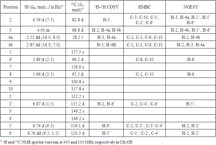
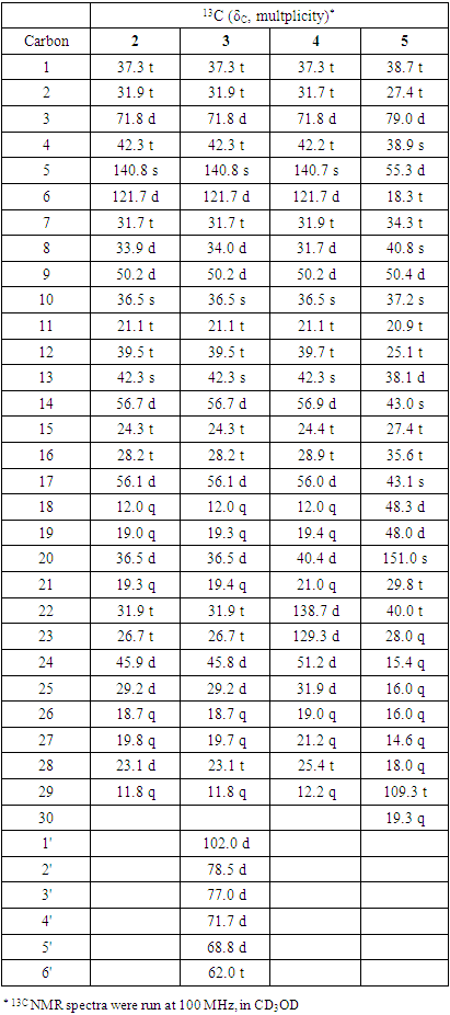
Compound 1: Yellow crystals, mp 115-117°C, Rf 0.68 (acetone: DCM 2:3); 1H-NMR (CD3OD, 400 MHz) δ ppm: 4.59 (1H, d, J = 7.5 Hz), 4.01(1H, m), 2.7 (1H, dd, J = 16.0, 7.0 Hz), 2.53 (1H, dd, J = 16.0, 8.0 Hz), 5.89 (1H, d, J = 2.5 Hz), 5.96 (1H, d, J = 2.5 Hz), 6.87 (1H, d, J = 1.5 Hz), 6.76 (1H, dd, J = 9.5, 1.5 Hz), 6.74 (1H, d, J = 0.5 Hz); 13C NMR (CD3OD, 100 MHZ) δ ppm (Table 1). EIMS (rel. int) m/z: 290, 272, 152 and 139.
Compound 2: White crystalline solid, mp 128-130°C, Rf 0.56 (100% DCM). IR νmax (KBr, cm-1) 3422, 2920, 1640, 1463, 1380, 1048, 1021, 723. 1H NMR (CD3OD, 400 MHz) δ ppm: 5.37 (1H, d, J = 7.2 Hz), 3.53 (m), 1.03 (3H, s), 0.94 (3H, d, J = 8.4 Hz), 0.86 (9H, m), 0.70 (3H, s); 13C NMR (CD3OD, 100 MHZ) δ ppm (Table 2). EIMS (rel. int) m/z: 414, 396, 303, 329 and 213.
Compound 3: White needle-like crystals, mp 156-160°C, Rf 0.65 (DCM: MeOH 3:1). 1H-NMR (CD3OD, 400 MHz) δ ppm: 5.37 (1H, d, J = 7.2 Hz), 3.53 (m), 1.03 (3H, s), 0.94 (3H, d, J = 8.4 Hz), 0.86 (9H, m), 0.70 (3H, s), 5.2, 3.8, 3.2; 13C NMR (CD3OD, 100 MHZ) δ ppm (Table 2).
Compound 4: White feather-like crystals, mp 168-170°C, Rf 0.66 (100% DCM). 1H NMR (CD3OD, 400 MHz) δ ppm: 5.15 (1H), 5.09 (1H, d, J = 1.5, 8.5 Hz), 5.35 (1H, d, J = 3.15 HZ), 3.51(1H, m) 1.01, 0.91, 0.85, 0.81, 0.77, 0.68; 13C NMR (CD3OD, 100 MHZ) δ ppm (Table 2). EIMS (rel. int): m/z 412, 394, 369, 351, 300, 271 and 255.
Compound 5: White needle-like crystals, mp 126-128°C, Rf 0.57 (n-hexane: DCM 1:1). 1H NMR (CD3OD, 400 MHz) δ ppm: 4.6, 4.5, 3.15, 2.3, 1.66, 1.01, 0.95, 0.92, 0.81, 0.77 and 0.74 ppm; 13C-NMR (CD3OD, 100 MHZ) δ ppm (Table 2). EIMS (rel. int): m/z 426, 411, 385, 355, 220, 218, 207, 189.

2.6. Antibacterial Activity Test

Antibacterial test was done using paper disc diffusion method [29]. Staphylococcus aureus (ATCC no. 25923), Bacillus subtilis (local isolate), Escherichia coli (ATCC no. 25922) and Pseudomonas aeruginosa (ATCC no. 10622) used were obtained from the Department of Botany, Kenyatta University in Kenya. Sterile nutrient agar (28 g/l in distilled water, 15 ml) was dispensed in 90 mm diameter sterile petri dishes to yield a uniform depth of 4 mm. Bacteria from stock cultures were spread on the nutrient agar surface using sterile wire loop and incubated at 37°C for 24 hrs. One loop-full of the bacterial strain from the culture was added to sterile nutrient broth medium and incubated at 37°C in a rotary shaker. After 24 hrs, the broth bacteria culture (0.1 ml) was pipetted into nutrient agar media in petri dishes and spread evenly. 100 µl of the isolated compounds at a concentration of 1 mg/ml in DMSO were dispensed on 6 mm sterile paper disc [29]. The discs were oven died at 50°C for 1 hr and then firmly placed on the inoculated petri dishes using sterile forceps. The petri-dishes and their contents were then incubated at 37°C in inverted position. The zones of inhibition were measured after 24 hrs [20,30].

2.7. Anti-fungal Activity Test

Agar-well diffusion method was used as described by Opiyo et al [31]. Three fungal species, Candida albicans, Penicillium notatum and Aspergillus Niger were obtained from the Department of Botany, Kenyatta University in Kenya. 15 ml of sterile potato dextrose agar prepared by dissolving 39 g in 1L of water was dispensed in petri dishes under sterile conditions and left to solidify. Pure cultures of test fungi were introduced in the petri dishes from stock cultures and incubated at 30°C for 7 days to produce a good crop of spores. The fungal inoculums were prepared by harvesting the spores from the crop of spores using a spores-harvesting needle and transferred into a sterile tube containing sterile distilled water. The spore suspension (0.5 ml) was transferred to PDA surface in petri-dishes using a pipette and the petri-dishes were tilted several times to spread the inoculum. After 10 minutes four wells were cut out from the inoculated PDA medium using a sterile cork-borer (6 mm). 100 μl of suspension of the isolated compounds at concentration of 1 mg/ml in DMSO were pipetted into the wells and then incubated at 30°C for 24 hours. Diameters of zones of inhibition were measured as described by [31].

3. Results and Discussion

3.1. Characterization of Compounds

Chromatographic fractionation of root bark extracts of R. natalensis lead to isolation of five compounds (Figure 1). The structures of the compounds were determined using spectroscopic as well as physical methods. Compound 1 was isolated as yellow crystals having Rf value of 0.68 (DCM-acetone 3:2). On spraying with anisaldehyde the spot turned red and then yellow suggesting the compound to be a flavonoid. 1H-NMR data (Table 1 and Figure 2) showed aromatic peaks at δ 6.87 (1H, d, J = 1.5 Hz), 6.76 (1H, dd, J = 9.5, 1.5 Hz) and 6.74 (1H, d, J = 0.5 Hz) representing protons of a trisubstituted benzene ring, and δ 5.96 (1H, d, J = 2.5 Hz) and δ 5.89 (1H, d, J = 2.5 Hz) representing protons of a tetra-substituted benzene ring [32,33].
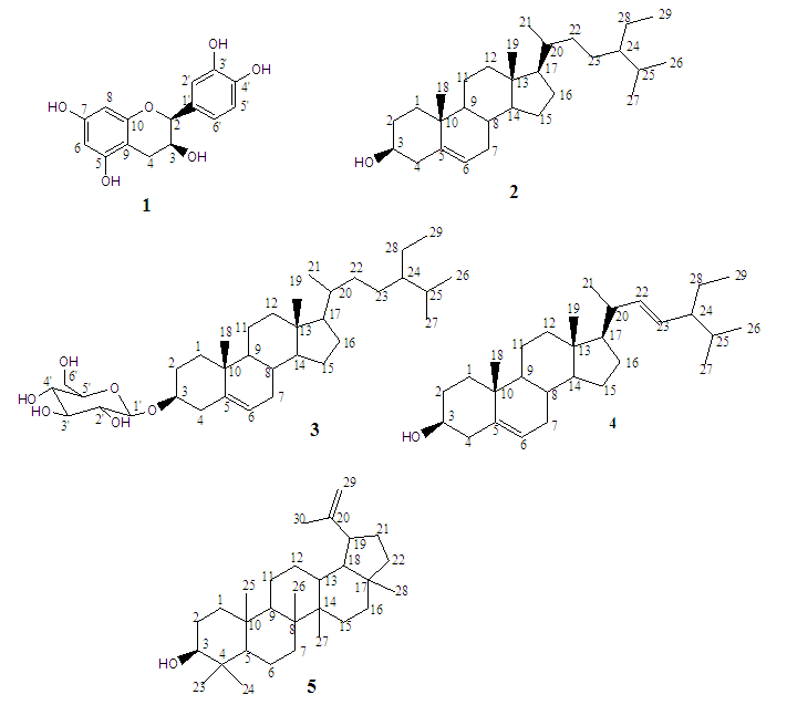
Figure 1. Structures of the isolated compounds
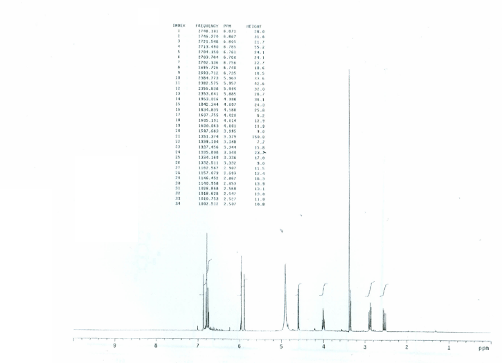
Figure 2. 1H-NMR spectrum of compound 1 (CD3OD, 400 MHz)
Table 1. 1D and 2D NMR data of compound 1
     
The relatively small coupling constant (J = 2.5 Hz) between the two protons indicated meta substitution [32,33]. The pattern of the proton peaks strongly suggested a flavonoid skeleton with hydroxyl groups at positions C5, C7, C3′ and C4′ [32-34]. A doublet at δ 4.59 (J = 7.5 Hz) was assigned to H-2 while a multiplet centered at δ 4.01 was assigned to C-3 [32,33,35]. Two additional signals centered at δ 2.87 (1H, dd, J = 16.0, 7.0 Hz) and 2.53 (1H, dd, J = 16.0, 8.0 Hz) were assigned to the germinal protons at C4. 13C NMR peaks at δ 28.5, 68.8 and 82.8 (Table 1 and Figure 3) were assigned the methylene carbon at C4, hydroxylated carbon at C-3 and oxymethine carbon at C-2 [33]. Signals at δ 157.5, 156.9 146.2 and 146.2 ppm were assigned to the four hydroxylated quaternary carbon atoms at C5, C7, C3’ and C4’ respectively. The connectivity and orientation of the protons were supported by the HMBC, COSY and NOESY experiments (Table 1). EIMS data of the compound gave the molecular ion peak at m/z 290 which corresponded to C15H14O6 formula. Other diagnostic peaks occurred at m/z 272 [M-H2O]+, 152 [C8H8O3]+ and 139 [C7H7O3]+. The spectral data were in agreement with those published for epicatechin [32-35].
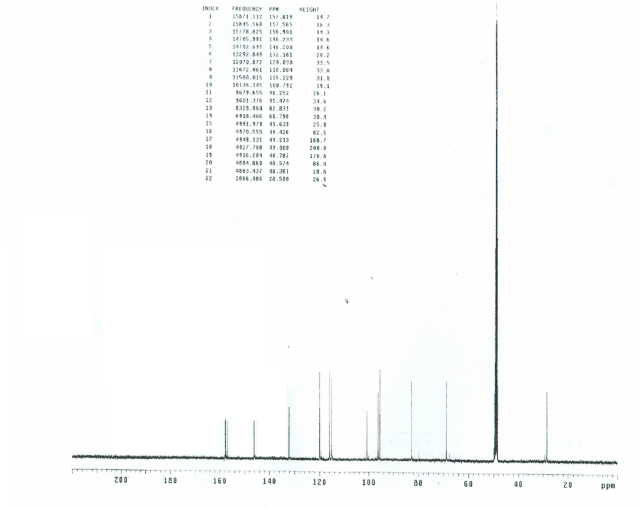
Figure 3. 13C-NMR spectrum of compound 1 (CD3OD, 100 MHz)
Compound 2 was obtained as white crystals having a melting point of 128-130°C and Rf value of 0.56 in 100% DCM.1H-NMR signal at δ 5.37 (1H, d J=7.2 Hz) suggested presence of a trisubstituted double bond while a multiplet centered at δ 3.53 was characteristic of a proton geminal to a hydroxyl group at C-3 in terpenoids (Table 2) [33,36,37]. Six signals representing the methyl groups were observed at δ 1.03 (3H, s), 0.94 (3H, d, J = 8.4 Hz), 0.86 (9H, m) and 0.70 (3H, s) which are characteristic of a modified triterpenoid [38]. 13C-NMR and DEPT data (Table 2) confirmed presence of the six methyl (at δ 11.8, 12.0, 18.7, 19.4, 19.4 & 19.7 ppm), 11 methylene ( at δ 21.1, 23.1, 24.3, 26.7, 28.2, 31.7, 31.9, 31.9, 37.3, 39.5 & 42.3 ppm), nine methine (at δ 29.2, 31.9, 33.9, 36.5, 45.9, 50.2, 56.1, 56.7 & 121.7 ppm) and three quaternary (at δ 36.5, 42.3, 140.8 ppm) carbons. 13C-NMR data further confirmed the presence of olefinic carbons at δ 140.8 and 121.7 ppm with the former peak representing a quaternary carbon atom. The signal at δ 71.8 was assigned to oxymethine carbon at C-3. ESI-MS spectrum of compound 2 showed a molecular ion peak at m/z 437 [M+Na]+ corresponding to the formula C29H50O. EIMS spectrum showed other diagnostic peaks at m/z 396 [M-H2O]+, 329 [M-C6H13]+, 273 [M-C10H21]+ and 255 [M-C10H21-H2O]+. Comparison of spectral data of compound 2 literature data confirmed 2 to be β-sitosterol [36-38].
Compound 3 was isolated as white needle-like crystals, mp 156-160°C and Rf value of 0.65 (DCM-MeOH 3:1). The spot turned blue then green on spraying with anisaldehyde suggesting 3 to be a terpenoid [38]. 1H-NMR data (Table 2) strongly suggested a triterpenoid structure [37,39]. A doublet at δ 5.37 ppm (1H, J =7.2 Hz) suggested the presence of olefinic proton on trisubstituted alkene. A multiplet centered at δ 3.53 ppm was characteristic of oxymethine proton at C-3 [37]. Methyl proton peaks at δ 1.03 (3H, s), 0.94 (3H, d, J = 8.4 Hz), 0.86 (9H, m) and 0.70 (3H, s) were characteristic of a modified triterpenoid [38]. 1H-NMR spectral data closely compared to the spectral data of compound 2. However, there appeared additional peaks between δ 3.0 and 5.0 suggesting presence of a sugar moiety [40]. The olefinic carbon peak appeared at δ 140.8 and 121.7 ppm while the signal due to the C-3 oxymethine carbon appeared at δ 71.8 ppm. 13C NMR data was similar to those of β-sitosterol except for the six peaks at δ 102.0, 78.5, 77.0, 71.7, 68.8 and 62.0 ppm associated with a sugar moiety [40]. The identity of β-sitosterol glucoside was confirmed by Co-TLC with authentic specimen.
Compound 4 was isolated as white feather-like crystals having melting point of 168-170°C and Rf value of 0.66 in DCM. On spraying with anisaldehyde, the spot turned blue/purple, suggesting compound 4 to be a terpenoid [38]. 1H-NMR data (Table 2) displayed several peaks between δ 1.01 and 0.68 ppm which are characteristic of triterpenoid parent. A broad doublet at δ 5.35 ppm suggested presence of a double bond at a quaternary carbon atom. Two signals at δ 5.17 and 5.01 ppm suggested presence of a double bond on the side chain [8,9,33,41]. The multiplet at δ 3.51 ppm represented a proton attached to oxymethine carbon [33]. The diagnostic chemical shift values of the angular methyl protons at C-18 and C-19 appeared as singlets at δ 0.68 and 1.01 ppm, respectively. The protons peaks of the remaining four methyl groups appeared at δ 0.77, 0.81, 0.85 and 0.91 ppm. 13C-NMR and DEPT data showed twenty-nine carbon atoms consisting of six methyl (at δ 12.0, 12.2, 19.0, 19.4, 21.0 & 21.2), nine methylene (at δ 21.1, 24.4, 25.4, 28.9, 31.7, 31.9, 37.3, 39.7 & 42.2), eleven methane (at δ 31.7, 31.9, 40.4, 50.2, 51.2, 56.0, 56.9, 71.8, 121.7, 129.3 & 138.7) and three quaternary ( at δ 36.5, 42.3 & 140.7) carbons atoms, which further confirmed the presence of a sterol. Signals at δ 140.7 and 121.7 ppm were assigned to the olefinic carbons with the deshielded signal assignable to the quaternary carbon at the bridge. Signal at δ 138.7 and 129.3 were assigned to the olefinic carbon on the side chain while δ 71.8 ppm was assigned to the oxymethine carbon at C3 [33,38]. EIMS spectrum gave a molecular ion peak at m/z 412 corresponding to C29H48O. Other diagnostic peaks were observed at m/z 394 [C29H46]+, 369 [M-C3H7]+, 300 [C21H32O]+, 271 [C19H27O]+ and 255 [C19H27]+. Based on the spectral data as well as comparison with literature data [8,9,33,38], compound 4 was identified to be stigmasterol.
Compound 5 was isolated as white needle-like crystals having m.p. of 126-128°C and Rf value of 0.57 in hexane-DCM (1:1). On spraying with anisaldehyde, the spot turned blue/purple then green suggesting compound 5 to be a terpenoid. 1H-NMR data displayed two singlets at δ 4.67 and 4.58 ppm which suggested presence of a terminal olefinic group [9,41,42,44] while a multiplet at δ 3.16 suggested presence of oxymethine carbon at C3 (Table 2). Seven singlets associated with methyl groups of a pentacyclic terpene appeared at δ 1.66, 1.01, 0.95, 0.92, 0.81, 0.77 and 0.74 ppm [9,44]. The 13C-NMR data and DEPT experiment showed the presence of 30 carbon atoms which further suggested compound 5 to be a triterpenoid. Signals at δ 150.8 and 109.3 ppm were assigned to C20-C29 double bond while the signal at δ 79.0 ppm was assigned to the oxymethine carbon atom at C-3 [42]. The seven methyl peaks at δ 14.6, 15.4, 16.0, 16.1, 18.0, 19.3 & 28.0 ppm were assigned to C23-C28 and C30 carbon atoms. EIMS data showed a molecular ion peak at m/z 426 [M+] for the formula C30H50O. Other diagnostic peaks were observed at m/z 411 [M-Me]+, 355 [M-Me- 3H₂O]+, 220 [M-C15H26]+, 207 [M-C16H27]+, 189 and [M-C16H29O]+. Based on comparison with literature data [9,42-44], compound 5 was confirmed to be lupeol.
Table 2. 13C NMR data of compounds 2-5
     

3.2. Antibacterial Activity of Isolated Compounds

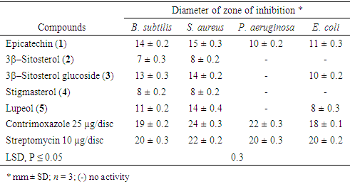
Isolated compounds were tested for antibacterial activity against P. aeruginosa, B. subtilis, S. aureus and E. coli using paper disc diffusion method. All isolated compounds were active against all the gram-positive bacteria tested (Table 3). Compounds 1, 3 and 5, exhibited moderate activity while 2 and 3 showed low activity. Bacillus subtilis and S. aureus were most susceptible (P ≤ 0.05) to compound 1 with zones of inhibitions of 14 ± 0.2 and 15 ± 0.3 mm respectively. Stigmasterol (4) showed equal antibacterial activity against the two gram-positive bacteria. S. aureus was more susceptible to compound 2 than B. subtilis. Only compounds 1, 3 and 5 were active against the gram-negative bacteria. Escherichia coli was most susceptible (P ≤ 0.05) to epicatechin (1) while P. aeruginosa was only susceptible to epicatechin (1). All isolated compound showed lower antibacterial activity compared to standard drugs. These results were consistent with previous studies which reported antibacterial activities of 3β–sitosterol (2), stigmasterol (4) [36], 3β-sitosterol glucoside (3) [45] lupeol (5) and epicatechin (1) [46].
Table 3. Antibacterial activities of isolated compounds
     

3.3. Antifungal Activity of Compounds

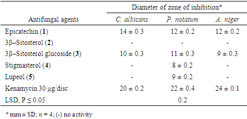
Isolated compounds were tested for antifungal activity against C. albicans, P. notatum and A. niger (Table 4). Epicatechin (1) recorded the highest activity against all the testes fungi having inhibition zones of 14 ± 0.3, 12 ± 0.2 and 12 ± 0.2 mm against C. albicans, P. notatum and A. niger respectively. Lupeol (5) and stigmasterol (4) were only active against P. notatum with inhibition zones of 9 ± 0.2 and 8 ± 0.2 mm, respectively. β-Sitosterol (2) showed no activity against the test fungi. Antifungal activity of β-sitosterol (2) [36,47], stigmasterol (4) [36], lupeol (5) [48] and β-sitosterol glucoside (3) [45] were earlier reported.
Table 4. Antifungal activity of isolated compounds
     

References

[1]  Edwards, J.E., Jr., Lehrer, R.I., Stiehm, E.R., Fischer, T.J., Young, L.S., 1978. Severe candidal infections: clinical perspective, immune defense mechanisms, and current concepts of therapy. Ann. Intern. Med. 89, 91-106.
[2]  Barley, I., Fanaroff, A.A., 2000. Disseminated fungal infections in very low-birth-weight infants: Clinical manifestation and epidemiology. Pediatrics 73, 144 - 152.
[3]  Gordon, M.A., Lapa, E.W., Passero, P.G., 1988. Improved method for azole anti-fungal susceptibility testing. J. Clin. Microbiol. 26, 1874 - 1877.
[4]  World Health Organization/IUATLD Global project on anti-tuberculous drug resistance surveillance. Anti-tuberculous drug resistance in the world. Third global report. WHO/HTM/TB/2004.343. Geneva: World Health Organization, 2004.
[5]  Arama, P., Jeruto P., Anyango, B., Nyunja, R., Taracha, C., Opiyo, S., 2017. Morphometric study of Senna didymobotrya (Fresen.) H. S. Irwin and Barneby in Kenya. J. Nat. Sci. Res. 7 (6), 54-69.
[6]  Opiyo, S.A., Manguro, L.O.A., Okinda-Owuor, P., Ateka, E.M., Lemmen, P. 2011a.7 alpha-Acetylugandensolide and antimicrobial properties of Warburgia ugandensis extracts and isolates against sweet potato pathogens. Phytochem Lett. 4:161-165.
[7]  Ochieng, C.O., Opiyo, S.A., Mureka, E.W., Ishola, I.O., 2017. Cyclooxygenase inhibitory compounds from Gymnosporia heterophylla aerial parts. Fitoterapia, 119, 168-174.
[8]  Ochung, A.A, Manguro, L.A.O., Owuor, P.O., Jondiko, I.O., Nyunja, R.A., Akala, H., Mwinzi, P., Opiyo, S.A., 2015. Bioactive carbazole alkaloids from Alysicarpus ovalifolius (Schumach). J. Korean Soc Appl Biol Chem. 58(6), 839-846.
[9]  Ochung, A.A, Owuor, P.O., Manguro, L.A.O., Ishola, O.I, Nyunja, R.A., Ochieng, C.O., Opiyo, S.A., 2018. Analgesics from Lonchocarpus eriocalyx Harms. Trends Phytochem. Res. 2(4), 253-260.
[10]  Vermani, K., Garg, S., 2002. Herbal medicines for sexually transmitted diseases and AIDS. J. Ethnopharmacol. 80, 49-66.
[11]  Rayne, S., Mazza, G., 2007. Biological activities of extracts from sumac (Rhus Spp): a review. Plant Foods for Human Nutr, 62(4), 165-175.
[12]  Kokwaro, J.O., 1993. Medicinal plants of East Africa. East Africa Literature Bureau, Nairobi.
[13]  Duke, J.A., Ayensu, E.S., 1985. Medicinal Plants of China. 2 Vols. 705 S., 1300 Strichzeichnungen. Reference Publ., Inc. Algonac. Michigan.
[14]  Dafini, A., Yaniv, Z., Paletivitch, D., 1984. Ethinopharmacol- botanical survey of medicinal plants in Northern Israel. J Ethnopharmacol. 103, 295-310.
[15]  Aysen, Y., Jimmy, O., Otto, S., Topul, R., 1998. Triterpenes from Rhus taitensis. Phytochemistry, 48, 863-863.
[16]  Jeruto, P., Lukhoba, C., Ouma, G., Otieno, D., Mutai, C., 2008. An ethnobotanical study of medicinal plants used by the Nandi people in Kenya. Ethnopharmacol. 116: 370-376.
[17]  Geissler, P.W., Harris, S.A., Prince, R.J., Olsen, A., Odhiambo, R.A., Oketch-Rabah, H., Madiega, P.A., Andersen, A., Mølgaard, P., 2002. Medicinal plants used by Luo mothers and children in Bondo District, Kenya. Ethnopharmacol. 83, 39-54.
[18]  Ssegawa, P., Kasenene, J.M., 2007. Medicinal plant diversity and uses in the Sango bay area, Southern Uganda. Ethnopharmacol. 113, 521-540.
[19]  Mahdavi, S., Hesami, B., Sharafi, Y. 2018. Antimicrobial and antioxidant activities of Iranian Sumac (Rhus coriaria L.) fruit ethanolic extract. J. Appl Microbiol Biochem. 2(2), 5-10.
[20]  Abdel-Mawgoud, M., Khedr, F.G., Mohammed, E.I., 2019. Phenolic compounds, antioxidant and antibacterial activities of Rhus flexicaulis Baker. Jordan J. Biol. Sci. 12 (1), 17-21.
[21]  Njoroge, P.W., Opiyo, S.A., 2019. Antimicrobial aactivity of root bark extracts of Rhus natalensisa and Rhus ruspolii (Unpublished).
[22]  Nasar-Abbas, S.M., Halkman, A.K., Al-Haq, M.I., 2004. Inhibition of some food borne bacteria by alcohol extract of sumac (Rhus coriaria L.). J. Food Safety, 24, 257-267.
[23]  Shahat, A.A, Ibrahim, AY, Al-Ghamdi, A.A.M., Alsaid, S.M., 2016. Phytochemical investigation of Rhus tripartita and its activity against cyclooxygenases and acetylcholinesterase. Trop J. Pharm Res., 15(8), 1697-1706.
[24]  Choi, H.S., Kim, M.K., Choi, Y.K., Shin, Y.C., Cho, S.G., Ko, S.G., 2016. Rhus verniciflua Stokes (RVS) and butein induce apoptosis of paclitaxel-resistant SKOV-3/PAX ovarian cancer cells through inhibition of AKT phosphorylation. BMC Complement. Altern. Med, 16, 122-128.
[25]  Varela-Rodríguez, L., Sánchez-Ramírez, B, Rodríguez-Reyna, I S., Ordaz-Ortiz, J.J., Chávez-Flores, D., Salas-Muñoz, E., Osorio-Trujillo, J.C., Ramos-Martínez, E., Talamás-Rohana, P., 2019. Biological and toxicological evaluation of Rhus trilobata Nutt. (Anacardiaceae) used traditionally in Mexico against cancer BMC Complement Altern Med, 19, 153.
[26]  Chhabra, S.C., Uiso, F.C., 1991. Antibacterial activity of some Tanzanian plants used in traditional medicine. Fitoterapia, 62, 499-503.
[27]  Mwangi, H.M., Mabusela, W.T., Abegaz, B.A., Onani, M.O., 2013. Antimicrobial activities of a novel biflavonoid and other constituents from Rhus natalensis. J. Med. Plants Res, 7(10), 619-623.
[28]  Lee, T.H, Chiou, J.L., Lee, C.K., Kuo, Y.H., 2005. Separation and determination of chemical constituents in the roots of Rhus javanica L. var. roxburghiana. J. Chin. Chem. Soc. 52, 833-841.
[29]  Elgayyar, M, Draughon, F.A., Golden, D.A., Mount, J.N., 2000. Antimicrobial activity of essential oils from plants against selected pathogenic and saprophytic microorganisms. J. Food Prod. 64, 1019-1024.
[30]  Opiyo, S.A., Manguro, L.O.A., Owuor, P.O., Ochieng, C.O., Ateka, E.M., Lemmen, P., 2011b. Antimicrobial compounds from Terminalia brownii against sweet potato pathogens. Nat Prod J. 1, 116-120.
[31]  Opiyo, S.A., Manguro, L.O.A., Ogur, J.A., Wagai, S.O., 2010. Bioactive constituents of Conyza floribunda. Research Journal of Pharmacology, 4 (3), 55-59.
[32]  El-Razek, M.H.A., 2007. NMR assignments of four catechin epimers. Asian Journal of Chemistry, 19 (6), 4867-4872.
[33]  Osuntokun, O.T., Idowu, T.O., Cristina, G.M., 2018. Bio-guided isolation, purification and chemical characterization of spigallocatechin; epicatechin, stigmasterol, phytosterol from of ethyl acetate stem bark fraction of Spondias mombin (Linn.). Biochem Pharmacol (LosAngel) 7: 240.
[34]  Harborne, F.B., 1998. Phytochemical Methods. 3rd ed. Chapman J. R., Blackburn H.S. (1973). Introductory Mocrobiolgy. John Willey and Sons Inc. New York pp. 535 – 537.
[35]  Dartini, Nurdin, H., Afrizal, Ekaprasada, M.T., Suciati, D., 2016. Isolation and characterization of flavonoids from the bark of Toona Sureni (Blume) Merr. J. Chem. Pharm. Res., 8(6), 156-159.
[36]  Opiyo, S.A., Manguro, L.A.O., Owuor, P.O., Ateka E.M., 2017.Triterpenes from Elaeodendron schweinfurthianum and their antimicrobial activities against crop pathogens. American Journal of Chemistry, (3), 97-104.
[37]  Phatangare, N.D., Deshmukh, K.K., Murade, V.D., Naikwadi, P.H., Hase, D.P., Chavhan, M.J., Velis, H.E., 2017. Isolation and characterization of β-sitosterol from Justicia gendarussa burm. F.-An anti-inflammatory compound. International Journal of Pharmacognosy and Phytochemical Research, 9(9), 1280-1287.
[38]  Dey, P.M., Harborne, J.B., 1991. Methods in plants biochemistry. Terpenoids. Academic press, New York, London 7, 370-425.
[39]  Mizanur Rahman S.M.M., Mukta, Z.A., M. Hossain, A., 2016. Isolation and characterization of β-sitosterol-D-glycoside from petroleum extract of the leaves of Ocimum sanctum L. As. J. Food Ag-Ind. 2009, 2(1), 39-43.
[40]  Peshin, T., Kar, H.K., 2017. Isolation and characterization of β-sitosterol-3-O-β-D-glucoside from the extract of the flowers of Viola odorata. British Journal of Pharmaceutical Research, 16(4), 1-8.
[41]  Opiyo, S.A., Manguro, L.A.O., Akinyi, D., Ochung, A.A., Ochieng, C.O., 2015. Biopesticidal extractives and compounds from Warburgia Ugandensis against maize weevil (Sitophilus zeamais). Natural Products Journal, 5(4), 236-243.
[42]  Beserra, F.P., Xue, M., Azevedo Maia, G.L.A., Rozza, A.L., Pellizzon, C.H., Jackson, C.J., 2018. Lupeol, a pentacyclic triterpene, promotes migration, wound closure, and contractile effect in vitro: Possible involvement of PI3K/Akt and p38/ERK/MAPK Pathways. Molecules 2018, 23, 2819-2835.
[43]  Muktar, B., Bello, I.A., Sallau, M.S., 2018. Isolation, characterization and antimicrobial study of lupeol acetate from the root bark of Fig-Mulberry Sycamore (Ficus sycomorus LINN). J. Appl. Sci. Environ. Manage. Vol. 22 (7), 1129-1133.
[44]  Silva, A.T.M., Magalhães, C.G., Duarte, L.P., Mussel, W.N., Ruiz, A.L.T.G., Shiozawa, L., Carvalho, J.E., Trindade, I.C., Filho, S.A.V., 2017. Lupeol and its esters: NMR, powder XRD data and in vitro evaluation of cancer cell growth. Braz. J. Pharm. Sci. 53(3): e00251.
[45]  Njinga, N.S., Sule, M.I., Pateh, U.U., Hassan, H.S., Abdullahi, S.T., Ache, R.N., 2016. Isolation and antimicrobial activity of β-sitosterol-3-O-glucoside from Lannea Kerstingii Engl. & K. Krause (Anacardiacea). NUJHS Vol. 6 (1), 4-8.
[46]  Awolola, G.V., Koorbanally, N.A., Chenia, H., Francis, O. Shode, F.O., Baijnath, H., 2014. Antibacterial and anti-biofilm activity of flavonoids and triterpenes isolated from the extracts of Ficussansi barica Warb. Subsp. Sansibarica (Moraceae) extracts. Afr. J. Tradit. Complement Altern Med. 11(3), 124-131.
[47]  Choi, N.H., Jang, J.Y., Choi, G.J., Choi, Y.H., Jang, K.S., Nguyen, V.T., Min, B.S., Le Dang, Q., Kim, J.C., 2017. Antifungal activity of sterol and dipsacussaponins isolated from Dipsacusasper root against phytopathogenic fungi. Pest. Biochem. Physiol, 141, 103-108.
[48]  Aristimuño Ficoseco, M.E., Vattuone, M.A., Audenaert, K., Catalán, C.A., Sampietro, D.A., 2014. Antifungal and antimycotoxigenic metabolites in Anacardiaceae species from northwest Argentina: isolation, identification and potential for control of Fusarium species. J. Appl. Microbiol, 116, 1262 -1273.