American Journal of Chemistry

p-ISSN: 2165-8749    e-ISSN: 2165-8781

2017;  7(3): 97-104

doi:10.5923/j.chemistry.20170703.03


Triterpenes from Elaeodendron schweinfurthianum and Their Antimicrobial Activities against Crop Pathogens

Sylvia Awino Opiyo 1, Lawrence Onyango Arot Manguro 2, Philip Okinda Owuor 2, Elijah M. Ateka 3

1Department of Physical Sciences, Murang’a University, Murang’a, Kenya

2Department of Chemistry, Maseno University, Maseno, Kenya

3Jomo Kenyatta University of Agriculture and Technology, Nairobi, Kenya

Correspondence to: Sylvia Awino Opiyo , Department of Physical Sciences, Murang’a University, Murang’a, Kenya.

Email:

Copyright © 2017 Scientific & Academic Publishing. All Rights Reserved.

This work is licensed under the Creative Commons Attribution International License (CC BY).
http://creativecommons.org/licenses/by/4.0/

Abstract

Phytochemical evaluation of Elaeodendron schweinfurthianum (Loes) extracts led to the isolation of nine compounds which were identified as 3-oxofriedelane (1), 3α-hydroxyfriedelane (2), 3-oxo-29-hydroxyfriedelane (3), 3-oxofriedelan-28-al (4), α-amyrin (5), α-amyrin acetate (6), β-sitosterol (7), stigmasterol (8) and lanosterol (9). The structures of the compounds were determined using spectroscopic and physical methods as well as by comparison with literature data. The in vitro antimicrobial activities of the extracts and isolates were investigated against fungi and bacteria which infect food crops. All the crude extract inhibited the growth of the tested pathogens with EtOAc and n-hexane extracts being the most active with 11.3 and 7.3 mm diameter zone of inhibition respectively. All the compound showed antimicrobial activity except compounds 3 which did not exhibit any visible activity at concentrations ≤ 200 µg/ml. Finding from this study confirm that plant extracts can provide alternative readily available and environmentally safe antimicrobials for managing crop infections.

Keywords: Elaeodendron schweinfurthianum, Celastraceae, Triterpenes, Antibacterial, Antifungal

Cite this paper: Sylvia Awino Opiyo , Lawrence Onyango Arot Manguro , Philip Okinda Owuor , Elijah M. Ateka , Triterpenes from Elaeodendron schweinfurthianum and Their Antimicrobial Activities against Crop Pathogens, American Journal of Chemistry, Vol. 7 No. 3, 2017, pp. 97-104. doi: 10.5923/j.chemistry.20170703.03.

1. Introduction

Globally, food scarcity is the third most pressing problem after poverty [1]. Approximately one billion people are faced by severe hunger worldwide of which 10% actually die from hunger-related complications [1]. This problem arises due to inadequate management methods of pest and microbes-induced spoilages of agricultural produce [2]. Bacterial and fungal infection of agricultural crop can cause up to 100% loses [3-5]. Microbes belonging to several genus including Alternaria, Aspergillus, Fusarium, Rhizopus, Ralstonia and Streptomyces cause infection on crops both in storage and in the field [2]. These pathogens cause disease in a wide range of crops including cereals, tubers, fruits, and vegetables as well as ornamental crops [3-6].
Synthetic chemicals such as dichloronitroaniline have been used to protect crops against microbial infections [7]. However, the use of such chemicals apart from their potential danger to both for humans and environment [8, 9] are unaffordable by most farmers. Moreover, because of pathogens resistance, most chemicals have become ineffective [9]. Most crop infecting microbes have a wide host range which further complicates their control. In order to eliminate agricultural produce spoilage, there is a need to search for affordable, readily available, sustainable, and environmentally friendly means of managing the problems posed by these pathogens.
Plants belonging to the genus Elaeodendron (Celastraceae) are characterized by the presence of terpenoids, steroids and flavonoids [10-15]. Biological activities of Elaeodendron species include antifungal, antibacterial, feeding deterrent, cytotoxic and antiviral [15-18]. E. schweinfurthianum (Loes) which is widely distributed in tropical Africa is used traditionally to manage bacterial and fungal infections including wounds, primary symptoms of syphilis and diarrhea [19, 20]. In this communication, we report the isolation of 3-oxofriedelane (1), 3α-hydroxyfriedelane (2), 3-oxo-29-hydroxyfriedelane (3), 3-oxofriedelan-28-al (4), α-amyrin (5), α-amyrin acetate (6), β-sitosterol (7), stigmasterol (8) and lanosterol (9) from E. schweinfurthianum. These compounds together with their activities are being reported from this plant for the first time.

2. Materials and Methods

2.1. General

Melting points were determined on a Gallenkamp (Loughborough, UK) melting point apparatus and are uncorrected. The UV spectra were run on Pye Unicam SP8-150 UV–vis spectrophotometer (Cambridge, UK) using acetonitrile. IR data were recorded on a PerkinElmer FTIR 600 series spectrophotometer (Waltham, MA, USA) as KBr pellet. The 1H and 13C NMR data were measured in CDCl3 and CDCl3–DMSO-d6 on a Bruker NMR Ultrashield TM (Darmstadt, Germany) operating at 500 and 125 MHz, respectively. The MS data were obtained on a Varian MAT 8200A instrument (Bremen, Germany).

2.2. Plant Materials

Stem bark of E. schweinfurthianum was collected from Shimba Hills (latitude 4° 15' 53.84'' S and longitude 39° 22' 19.61'' E) in September 2008 and voucher specimen (2008/09/04/SAO/CHEMMK) was identified at the Kenya National Museum herbarium after comparison with authentic samples. The plant materials were chopped into small pieces, air dried and ground into fine powder using a mill.

2.3. Extraction and Isolation of Compounds

Powdered plant material (2 kg) was extracted sequentially with n-hexane, EtOAc and MeOH by soaking the material in the solvent for seven days. The mixture was filtered and solvent evaporated at reduced pressure to yield 15 g, 100 g and 210 g of n-hexane, EtOAc and MeOH extracts, respectively. n-Hexane extract (10 g) was chromatographed over silica gel-packed column (2.5 x 60 cm, 150 g) and eluted with n-hexane - ethyl acetate mixture to yield 100 fractions each of 20 ml. Fractions showing similar TLC profiles were combined resulting into three pools (I-III). Pool I (1 g) did not show any major spot on TLC and was discarded. Pool II (3 g) crystallized out to give a white compound which on further purification using n-hexane-ethyl acetate (9:1) gave α-amyrin acetate (6) 56 mg. The mother liquor of this pool was subjected to further column chromatography with n-hexane- EtOAc (9:1) to afford stigmasterol (8) 78 mg. Pool III (3 g) also crystallized out and after re-crystallization (n-hexane-EtOAc, 9:1) afforded further stigmasterol (8) 45 mg.
Ethyl acetate extracts (75 g) was chromatographed over silica gel-packed column (5 x 60 cm, 200 g) eluting with n-hexane-ethyl acetate (10% increment of ethyl acetate), ethyl acetate neat and finally with CH2Cl2-MeOH (with 10% and 20% increment of MeOH) to yield 251 fractions (20 ml each). Fractions showing similar TLC profiles were combined resulting in five pools (I-V). Pool I (8 g) on subjection to further column chromatography eluting with n-hexane-ethyl acetate (95:5, 9:1) gave α-amyrin acetate (6) 30 mg. Pool II (15 g) on further fractionation with n-hexane: ethyl acetate mixture (95:5, 9:1, 4:1) afforded α-amyrin acetate (6) 72 mg, 3-oxofriedelane (1) 65 mg and β-sitosterol (7) 54 mg. Pool III (17g) yielded stigmasterol (8) 78 mg, 3-oxofriedelan-28-al (4) 80 mg and 3α-hydroxyfriedelane (2) 83 mg. Pool IV (13 g) afforded α-amyrin (5) 77 mg and 3-oxo-29-hydroxyfriedelane (3) 93 mg on further fractionation with n-hexane: ethyl acetate mixture (4:1, 7:3). Pool V (9 g) gave lanosterol (9) 74 mg on further column chromatography eluting with n-hexane: ethyl acetate (7:3, 3:2).

2.4. Antimicrobial Assay

Test organisms were isolated from infected farm produce obtained from local market were tested for antimicrobial activity as described by Barry et al., [21]. Inoculation was done by spreading the test pathogen on the surface of the solidified agar. Sterile paper discs (Whatmann No. 1, 5 mm diameter) containing 100 µL of the plant extracts (5 mg/ml) were placed on PDA and NA plates previously inoculated with test fungi and bacteria, respectively and incubated at 28°C for 48 h for fungi and 37°C for 24 h for bacteria. Blitox and streptocycline (10 mg/ml) were used as positive controls while DMSO without plant extract was used as a negative control. The minimum inhibitory concentrations (MICs) of pure isolates were determined according to Kariba et al., [22]. Isolated compounds were tested at concentrations ranging between 1-200 µg/ml. MIC was regarded as the lowest concentration that produced a visible zone of inhibition.

3. Results and Discussion

3.1. Isolated Compounds

Phytochemical evaluation of the plant yielded nine compounds (Fig. 1) whose structures were determined using spectroscopic methods. EIMS spectrum of compound 1 gave a molecular ion peak at m/z 426 corresponding to C30H50O and was supported by the 13C NMR and DEPT spectra which showed the presence of 30 carbons attributed to eight methyl, eleven methylene, four methine and seven quaternary carbon atoms. The 13C NMR spectra showed the presence of a carbonyl carbon at δ 213.24 and eight methyl carbon atoms peaks at δ 6.81, 14.59, 17.91, 18.63, 20.21, 32.05, 31.73 and 34.94 [23-25]. 1H NMR spectrum showed the presence seven singlets (δ 0.70, 0.84, 0.90, 0.90, 0.98, 1.04 and 1.16) and one doublet (δ 0.86, J =7.0 Hz), integrating for three protons each confirmed the presence of the eight methyl groups [24, 26, 27]. EIMS spectrum of 1 further revealed fragmentation pattern typical of a saturated triterpene as evidenced by characteristic daughter ions at m/z 411 [M-15]+, 344, 273, 205 and 123 [24]. Based on the spectral data as well as comparison with literature data, compound 1 was identified as 3-oxofriedelane.
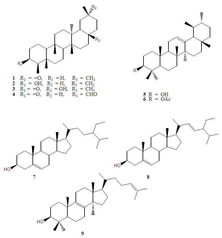
Figure 1. Structures of compounds isolated from E. schweinfurthianum
EIMS spectrum of compound 2 showed a molecular ion peak at m/z 428, corresponding to C30H52O formula. Other diagnostic peaks were at m/z 413 [M-Me]+, 395 [M-Me-H2O]+, 206 [C14H22O]+ and 220 [C16H28]+ which are characteristic peaks commonly found in the spectra of oleanane type of triterpenes with OH at C-3 [23, 28]. 13C NMR spectra showed the presence of 30 carbon peaks attributed to eight methyl, eleven methylene, five methine and six quaternary carbon atoms. The 13C NMR and DEPT spectra further showed the presence of an oxymethine carbon peak (δ 72.72) which was assigned to C-3 and eight methyl peaks (δ 11.61, 16.38, 18.23, 18.64, 20.11, 31.78, 32.08 and 35.02) which confirmed the presence of the eight methyl groups in friedelane type of triterpenes [23, 29]. The 13C NMR data were further supported by the 1H NMR spectrum which showed the presence of an oxymethine proton signal (δ 3.73 d, J = 2.3 Hz) thus confirming the presence of OH group at C-3. The small coupling constant (2.3 Hz) allowed the assignment of α- orientation of the OH group [23]. Other characteristic peaks in the 1H NMR spectrum were seven singlets integrating for three protons each (δ 0.88, 0.96, 0.98, 0.99, 1.00, 1.01 and 1.17) assigned to the tertiary methyl protons and a doublet (δ 0.94, J = 7.0 Hz) assigned to the secondary methyl protons at C-23 (Islam et al., 2014). Based on the spectral data as well as comparison with literature data, compound 2 was identified as 3α-hydroxyfriedelane.
ESI-MS spectrum of compound 3 exhibited a quasi-molecular ion peak at m/z 465 [M+Na]+ corresponding to a molecular weight of 442 and formula C30H50O2 and was supported by 13C and DEPT data which showed the presence of 30 carbon atoms consisting of seven methyl, twelve methylene, four methine and seven non-protonated carbon atoms. The presence of a carbonyl peak (δ 213.19) and an oxymethylene peak (δ 74.74) in the 13C NMR and DEPT spectra suggested the presence of a ketone carbon at C-3 and hydroxyl group at C-28, respectively [12, 23, 27]. Other diagnostic peaks in the 13C NMR were the seven methyl carbon atom peaks at δ 6.81, 14.64, 17.68, 18.43, 20.74, 25.79 and 32.06 [26]. The 1H NMR spectrum peak at δ 3.26 s, confirmed the presence of OH at C-29 while the singlets (δ 0.72, 0.86, 1.02, 1.04, 1.21 and 1.25) and a doublet (δ 0.87 J = 6.5 Hz) confirmed the presence of the six tertiary methyl and one secondary methyl groups [23]. Comparison of these data with the literature data confirmed the structure of compound 3 as 3-oxo-29-hydroxyfriedelane.
ESI-MS spectrum of compound 4 (Fig. 2) showed a quasi-molecular ion peak at m/z 463 [M+Na]+ suggesting a molecular formula of C30H48O2 and was supported by 13C NMR and DEPT data which showed the presence of 30 carbon peaks consisting of seven methyl, eleven methylene, five methine and seven non-protonated carbon atoms. The 13C NMR spectrum (Fig.3) exhibited diagnostic peaks at δ 213.01 for the carbonyl carbon at C-3 and δ 205.47 which suggested the compound to be 3-oxofriedelane with an aldehyde group at C-28 [30]. The spectrum also gave peaks at δ 6.77, 14.64, 16.12, 18.58, 18.92, 26.80, and 32.74 which were assigned to the seven methyl carbon atoms [23]. 1H NMR spectrum of 4 exhibited a singlet at δ 9.39 (-CHO), seven methyl peaks consisting of six singlets (δ 0.70, 0.85, 0.87, 0.89, 0.97 and 1.01) and a doublet (δ 0.88 J = 6.6), thus further supporting the 13C data [23, 27, 30]. Based on the spectral data as well as comparison with literature data, compound 4 was identified as 3-oxofriedelan-28-al.
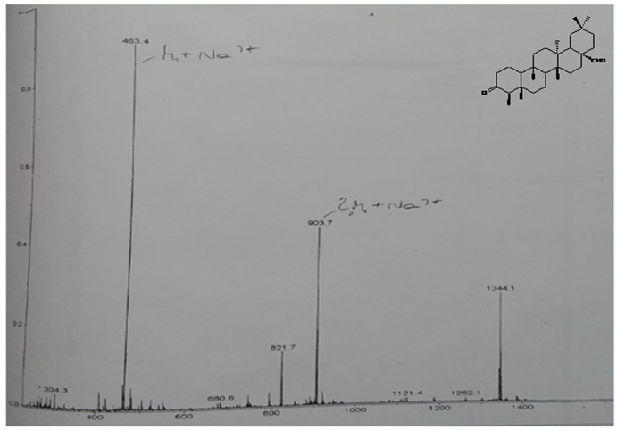
Figure 2. Mass spectrum of compound 4
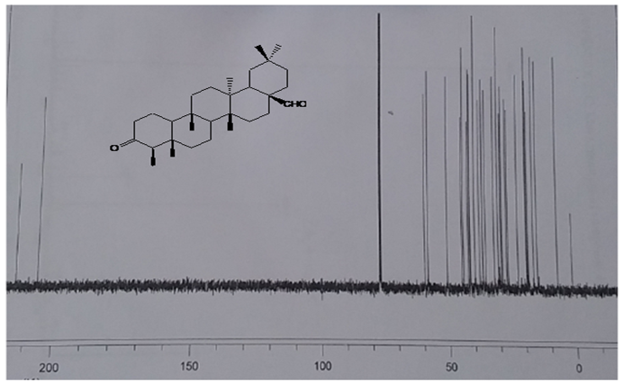
Figure 3. 13C NMR spectrum of compound 4 (CDCl3, 90MHz)
ESI-MS spectrum of compound 5 afforded a quasi-molecular ion peak at m/z 449 [M+Na]+ corresponding to a molecular formula of C30H50O and was supported by 13C NMR and DEPT spectra which showed a total of 30 carbon peaks attributed to eight methyl, nine methylene, seven methine and six quaternary carbon atoms. The 13C NMR spectrum showed the presence of two olefinic carbons (δ 139.84 and 124.42), an oxymethine carbon (δ 79.06) and eight methyl carbon atoms consisting of six tertiary (δ 15.61, 15.67, 16.86, 23.26, 28.10, 28.73) and two secondary (δ 17.46, 21.35) methyl carbon atoms [23, 31, 32]. The 1H NMR spectrum afforded an olefinic peak at δ 5.12 t (J = 3.5 Hz), confirming the double bond to be trisubstituted and an oxymethine peak at δ 3.22 dd (J = 11.5, 4.5 Hz) at C-3. The large coupling constant (J = 11.5 Hz) observed for H-3 showed the OH to be in equatorial position (Morris and Mansor, 1991; Ebajo et al., 2015). Other characteristic peaks at δ 0.79, 0.80, 0.87, 0.93, 0.95, 0.99, 1.01 and 1.07 confirmed the presence of eight methyl groups [23, 33-35]. Based on the spectral the data as well as comparison with literature data, compound 5 was identified as α-amyrin.
ESI-MS spectrum of compound 6 showed a quasi-molecular ion peak at m/z 491 [M+Na]+ corresponding to a molecular formula C32H52O2. The 13C NMR spectrum gave 32 carbon signals attributed to nine methyl, nine methylene, seven methine and seven non-protonated carbon atoms. Peaks at δ 139.62 and 124.31 confirmed the presence of the olefinic carbon atoms at C-12 and C-13, a fact which was further supported by the 1H NMR peak δ 5.12 t (J = 3.6 Hz) assigned to H-12 [23, 35]. Further examination of the 13C NMR spectrum revealed the presence of eight methyl carbon peaks corresponding to six tertiary (δ 28.73, 18.13, 15.44, 16.84, 23.40 and 27.83) and two secondary methyl (δ 21.97 and 21.42) carbon atoms. The additional peaks δ 170.04 and δ 21.36 in the 13C NMR spectrum confirmed compound 6 to be acetylated α-amyrin [35-37]. The 1H NMR spectrum of compound 6 further confirmed the presence of an oxymethine proton at C-3 (δ 4.51 m) and nine methyl groups consisting of seven tertiary (δ 2.05, 0.80, 1.57, 0.98, 1.07, 0.91, 1.01) and two secondary at δ 0.87 d (J = 5.5 Hz) methyl groups. Comparison of these data with those available in the literature confirmed compound 6 to α-amyrin acetate.
ESI-MS spectrum of compound 7 gave a quasi-molecular ion peak at m/z 437 [M+Na]+ for molecular formula C29H50O. 13C NMR and DEPT spectra afforded 29 carbon peaks corresponding to six methyl, eleven methylene, nine methine and three quaternary carbon atoms. The 13C NMR spectrum showed the presence of two olefinic carbon atoms (δ 140.65, 121.81), an oxymethine carbon atom (δ 71.78) and six methyl carbon atoms δ 19.81, 19.40, 19.01, 17.68, 11.97 and 11.79 [35, 37, 38]. The 13C NMR data were supported by the 1H NMR spectrum which showed the presence of one olefinic proton (δ 5.35), oxymethine the proton (δ 3.54), two tertiary, three secondary and one primary methyl groups at δ 1.02(s), 0.94 (d, J = 6.2 Hz), 0.86 (t, J = 7.0 Hz), 0.83 (d, J = 6.5 Hz), 0.81 (d, J = 6.5 Hz) and 0.69 (s) [35, 36]. Based on the spectral data as well as comparison with literature data, compound 7 was identified as β-sitosterol.
EIMS spectrum of compound 8 afforded a molecular ion peak at m/z 412 corresponding to a molecular formula of C29H50O. The EIMS spectrum further showed diagnostic peaks at m/z 369 [M-C3H7]+, 300 [M-C8H17]+ and 271 [M-C10H21]+ which are characteristic of sterols [35, 37]. 13C NMR and DEPT spectra shows the presence of 29 carbon atoms consisting of six methyl, nine methylene, eleven methine and three quaternary carbon atoms. 13C NMR the presence of four olefinic carbon atoms (δ 139.61, 138.10, 127.69 and 117.42), oxymethine carbon atom (δ 71.12) and six methyl carbon atoms (δ 12.09, 21.97, 21.34, 12.14, 19.12 and 12.86) which further suggested the compound to be a sterol [35]. The 1H NMR spectrum showed the presence of three olefinic protons at δ 5.19 m, 5.14 d (J = 15.3 Hz) and 5.04 dd (J = 15.3, 8.1 Hz); an oxymethine proton (δ 3.64 m) and six methyl groups at δ 0.56 s, 0.87 s, 0.79 d (J = 7.1 Hz), 0.84 d (J = 6.5 Hz), 1.00 d (J = 6.5 Hz) and 0.81 t (J = 8.0 Hz) corresponding to two tertiary, three secondary and one primary methyl groups [29, 39]. Based on the spectral data as well at comparison with literature information, compound 8 was identified as stigmasterol.
ESI-MS spectrum of compound 9 showed a quasi-molecular ion peak at m/z 449 [M+Na]+ suggesting the molecular formula of C30H50O. 13C NMR and DEPT spectra showed the presence of four olefinic peaks corresponding to three quaternary (δ 134.05, 134.05, 130.56) and one methine (δ 124.91) carbon atom, a fact which was supported by the presence of one olefinic peak at δ 5.08 t (J = 6.6 Hz) in the 1H NMR spectrum [40-42]. The HMBC peak at δ 78.68 correlating with a proton at δ 3.24 dd (J = 10.7, 4.4 Hz) confirmed the presence of OH at C-3 to be in equatorial conformation [39]. Other characteristic peaks in the 13C NMR spectrum were the methyl signals (δ 15.52, 15.52, 17.80, 19.27, 21.25, 21.90 25.93 and 28.00) which confirmed the presence of the eight methyl carbon atoms [41, 42]. 1H NMR spectrum peaks at δ 0.69 s, 0.81 s, 0.87 s, 0.98 s, 1.01 s, 1.68 s, 1.60 s and 0.95 d (J = 6.3 Hz) further confirmed the eight methyl groups. Based on the spectral data as well as comparison with literature data, compound 9 was identified to be lanosterol.

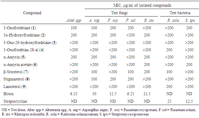
3.2. Antimicrobial Activities of Extracts and Isolates

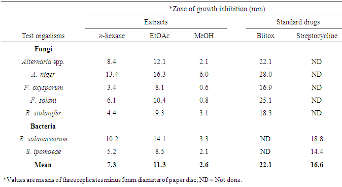
Crude extracts and isolates from stem bark of E. schweinfurthianum were subjected to antimicrobial assays against sweet potato pathogens: Alternaria spp, Aspergillus niger, Fusarium oxysporum, F. solani, Rhizopus stolonifer, Ralstonia solanacearum and Streptomyces ipomoeae. All the extracts were active against the fungi and bacteria species tested (Table 1). Ethyl acetate extract was the most active (p ≤ 0.05) against the pathogens followed by n-hexane extract. The most susceptible fungi to EtOAc extract was A. niger (inhibition zone, 16.3 mm) while F. oxysporum was least susceptible to the extract (inhibition zone, 8.1 mm). In the antibacterial test, R. solanacearum was more susceptible EtOAc extract (inhibition zone, 14.1 mm) than S. ipomoeae.
All the compound were active against one or more of the seven pathogens tested except compounds 3 which did not exhibit any visible inhibition at concentrations ≤ 200 µg/ml (Table 2). 3-Oxofriedelane (1) inhibited the growth of all the pathogens except R. stolonifer and R. solanacearum while α-amyrin (5) inhibited the growth all except F. solani, and R. solanacearum. 3-oxofriedelan-28-al (4) and lanosterol (9) were only active against R. solanacearum. 3-oxofriedelane (1) was the most active against Altenaria spp (MIC = 100 µg/ml) 3-oxofriedelane (1), β-sitosterol (7) and stigmasterol (8) were the most active against A. niger (MIC = 100 µg/ml). F. solani, and R. solanacearum were most susceptible to β-sitosterol (7) MIC = 100 µg/ml) compared to the other isolates. The results from this study confirm that plant infections can be managed using herbal extracts as had also been observed in other studies [43-45]. The herbal extracts are more environmentally safe compared to the synthetic antimicrobial drugs currently used. Further studies to determine the synergism effect of the isolated compound against the test microorganisms is recommended.
Table 1. Antimicrobial activity of crude extracts
     
Table 2. Minimum inhibitory concentration (MIC, µg/ml) of isolated compounds
     

ACKNOWLEDGEMENTS

The authors are thankful to Kenya Medical Research Institute (KEMRI), Kisumu, Kenya for the use of their laboratory to perform the biological activity tests and Biosciences Eastern and Central Africa Network (BecANet) for financial support. Mr. Mutiso of Botany Department, Nairobi University is highly thanked for identification of the plant.

References

[1]  Food and Agriculture Organisation of the United Nations, 1997. Consultative Group on International Agricultural Research technical advisory committee. Report on the inter-centre review of root and tuber crops research in the CGIAR. FAO, Rome.
[2]  Kana, H.A., Aliyu, I.A., Chammang, H.B., 2012. Review on neglected and underutilized root and tuber crops as food security in achieving the millennium development goals in Nigeria. Journal of Agricultural Veterinary Science 4, 27-33.
[3]  Loria, R.; Bukhalid, R.A., Fry, B.A. King, R.R., 1997. Plant pathogenicity in the genus Streptomyces. Plant Disease, 81(8), 836.
[4]  Anukwuorji, C.A., Obianuju, C.M., Ezebo, R.O., Anuagasi, C.L., 2016. Antimicrobial Effects of Four Plant Extracts against Post Harvest Spoilage Fungi of Yam (Dioscorea rotundata Poir). International Journal of Plant and Soil Science 12(3), 1-10.
[5]  Khatoon, A., Mohapatra, A., Satapathy, K.B., 2017. Studies on fungi associated with storage rot of Sweet potato [Ipomoea batatas (L.) Lam.] root tubers in Odisha, India. International Journal of Microbiologyand Mycology, 5, 1-7.
[6]  Sharma, R., 2012. Pathogenecity of Aspergillus niger in plants. Cibtech Journal of Microbiology ISSN: 2319–3867 http://www.cibtech.org/cjm.htm.
[7]  Clark, C.A., Moyer, J.W., 1988. Compendium of Sweet Potato Diseases. The American Phytopathological Society, St. Paul, MN.
[8]  Obagwu, J., Emechebe, A.M., Adeoti, A.A., 1997. Effects of extract of garlic, Allium sativum Bulb and neem, Azadiracha indica Juss seed on mycelial growth and sporulation of Collectotrichum capsicisyde Butler and Bixby. J. Agric. Technol., 5, 51-55.
[9]  Cameron, H.J. Julian, G.R., 1984. The effects of four commonly used fungicides on the growth of Cyanobacteria. Plant Soil, 78, 409-415.
[10]  Anjaneyulu, A.S.R., Rao, N.M., 1980. Elaeodendrol and elaeodendradiol, new nor-triterpenes from Elaeodendron glaucum. Phytochemistry, 19, 1163-1169.
[11]  Weeratunga, G., Kumar, V., Sultanbawa, M.U.S., 1982. Two new angular methyl deoxygenated D:A-friedooleananes. Tetrahedron Letters 23, 2031-2032.
[12]  Weeratunga, G., Kumar, V., 1985. D:B-Friedoolean-5-ene-3β, 29-diol, an angular methyl oxygenated D:B-friedooleanene from Elaeodendron balae. Phytochemistry, 24, 2369-2372.
[13]  Yasuko, T., Jondiko, I.J.O., Hiroyuki, T., Takane, F., Mori, K., 1995. Buchananoside, a steroidal glycoside from Elaeodendron buchananii. Phytochemistry, 40 (3), 753-756.
[14]  Yelani, T., Hussein, A.A., Meyer, J.J.M., 2010. Isolation and identification of poisonous triterpenoids from Elaeodendron croceum . Natural Products Research, 24(15), 1418-1425.
[15]  Tshikalange, T.E., Hussein, A., 2010. Cytotoxicity activity of isolated compounds from Elaeodendron transvaalense ethanol extract. Journal of Medicinal Plants Research, 4(16), 1695-1697.
[16]  Shimada, K., Kyuno, T., Nambara, T., Uchada, I., 1982. Isolation and characterization of cardiac steroids from seeds of Elaeodendron glaucum Pers. Structures of elaeodendrosides A, D, E, H, I, J and elaeodendrogenin. Chemical Pharmacology Bulletin, 30, 4075-4081.
[17]  Shimada, K., Kyuno, T., Nambara, T., Uchada, I., 1985. Structures of elaeodendrosides B, C, F, G, K and L, a series of cardiac glycosides isolated from Elaeodendron glaucum. Phytochemistry, 24, 1345-1350.
[18]  Maregesi, S.M., Hermans, N., Dhooghe, L., Cimanga, K., Ferreira, D., Pannecouque, C., Vanden Berghe, D.A., Cose, P., Maes, L., Vlietinck, A.J., Apers, S., Pieters, L., 2010. Phytochemical and biological investigations of Elaeodendron schlechteranum. Journal of ethnopharmacology. 2010, 129: 319-326.
[19]  Chhabra, S.C., Mahunnah, R.L.A., Mshiu, E.N., 1989. Plants used in tradidional medicine in Eastern Tanzania II. Angiosperm (Capparidaceae to ebenaceae). Ethnopharmacology, 25(3), 339-359.
[20]  Kokwaro, J.O., 2009. Medicinal Plants of East Africa. University of Nairobi Press, Nairobi, Kenya.
[21]  Barry, A.L., Coyle, M.B., Gerlach, E.H., Haw-Kinson, R.W., Thornberry, C., 1979. Methods of measuring zones of inhibition with the Baver-Kirby disc susceptibility test. Clinical Microbialogy, 10, 885-889.
[22]  Kariba, R.M., Siboe, G.M., Dossaji, S.F., 2001. In-vitro antifungal activity of Schizozygia coffaeoides Bail. (Apocynaceae) extracts. Ethnopharmacology, 74, 41-44.
[23]  Mahato, S.B., Kundu, A.P., 1994. 13C NMR spectra of pentacyclic triterpenoids. A compilation and some silent features. Phytochemistry. 37, 1517-1575.
[24]  Costa, P.M.D., Carvalho, M.G.D., 2003. New triterpene isolated from Eschweilera longipes. Annals of Brazillian Academy of Science, 75, 41-48.
[25]  Sousa, G.F., Duarte, L.P., Alcântara, A.F.C., Silva, G.D.F., Vieira-Filho, S.A., Silva, R.R., Oliveira, D.M., Takahashi, J.A., 2012. New Triterpenes from Maytenus robusta: Structural elucidation based on NMR experimental data and theoretical calculations. Molecules, 17, 13439-13456.
[26]  Mann, A.A., Ibrahim, K., Oyewale, A.O., Amupitan, J.O., Majekodumi O Fatope, M.O.; Okogun, J.I., 2011. Antimycobacterial friedelane-terpenoid from the root bark of Terminalia avicennioides American Journal of Chemistry 1(2), 52-55.
[27]  Islam, R., Ahmed, I., Sikder, A.A., Haque, M.R., Mansur, A., Mansoor Ahmed, M., Rasheed, M., Rashid, M.A., 2014. Chemical investigation of Mesua nagassarium (Burm. f.) Kosterm. Journal of Basic & Applied Sciences, 10, 124-128.
[28]  Srinivasan, R., Chandrasekar, M.J.N., Nanjan, M.J., 2011. Phytochemical investigations of Caesalpinia digyna root. E-Journal of Chemistry, 8(4), 1843-1847.
[29]  Mawa, S.; Said, I.M., 2012. Chemical constituents of Garcinia prainiana (Komposisi Kimia, Garcinia prainiana). Sains Malaysiana 41, 585-590.
[30]  Abbas, F.A., Al-Massarany, S.M., Khan, S., Al-Howiriny, T.A., Mossa, J.S., Abouashed, E.A., 2007. Phytochemical and biological studies on Saudi Commiphora opobalsamum L. Natural Products Research, 21, 383-391.
[31]  Seo, S., Tomiata, Y., Tori, K., 1975. Carbon-13-NMR spectra of urs-12-enes and application to structural assignments of compounds of Isodon Japonicas tissue cultures. Tettrahedron Letters, 1, 7-11.
[32]  Morris, M.D., Mansor, H., 1991. Analysis of the non-rubber constituents of latex from alstonia angustiloba. Tropical Forest Science, 4, 225-232.
[33]  Vázquez, L.H., Palazon, J., Arturo Navarro-Ocaña, A., 2012. The Pentacyclic Triterpenes - amyrins: A Review of Sources and Biological Activities, Phytochemicals - A Global Perspective of Their Role in Nutrition and Health, Dr Venketeshwer Rao (Ed.), ISBN: 978-953-51-0296-0. https://0x9.me/WMGVc.
[34]  Fingolo, C.E.; Santos, T.S.; M.D.M.V.; Kaplan, M.A.C., 2013. Triterpene esters: Natural products from Dorstenia arifolia (Moraceae). Molecules 18, 4247-4256.
[35]  Rasoanaivo, L.H., Wadouachi, A., Andriamampianina, T.T., Andriamalala, S.G., Razafindrakoto, E.J.B., Raharisololalao, A., Randimbivololona, F., 2014. Triterpenes and steroids from the stem bark of Gambeya boiviniana Pierre. Journal of Pharmacognosy and Phytochemistry 3 (1), 68-72.
[36]  Koay, L.Y.C., Wong, K.C., Osman, H., Eldeen, I., Asmawi, M.Z., 2013. Chemical constituents and biological activities of Strobilanthes crispus. Rec. Nat. Prod. 7(1), 59-64.
[37]  Ebajo, D., Shen, C., Ragasa, C.Y., 2015. Terpenoids and Sterols from Hoya multiflora Blume Virgilio. Journal of Applied Pharmaceutical Science, 5 (4), 33-39.
[38]  Prachayasittikul, S., Saraban, S., Cherdtrakulkiat, R., Ruchirawat, S., Prachayasittikul, V., 2010. New bioactive triterpenoids and antimalarial activity of rubra lec. EXCLI Journal 9, 1-10.
[39]  Reginatto, H.F., Gosmann, G., Guillaume, D., Kauffmann, C., Schenkel, P.E., Schripsema, J., 2001. Steroidal and triterpenoidal glucosides from Passiflora alata. Brazillian Chemical Society, 12, 32-36.
[40]  Emmons, G.T., Wilson, W.K., Schroepfer Jr, G.J., 1989. 1H and 13C NMR assignments for lanostan-3β-ol derivatives: Revised assignments for lanosterol. MRC, 27, 1012-1024.
[41]  Müller J., 2002. Analytik von freien Sterolen Und Entwicklung einer Screening-Methode zur Charakterisierung des Inhibitionsverhaltens neuer Sterol biosynthese inhibitoren. PhD thesis Universität München.
[42]  Dias, J.R., Gao, H., 2009. 13C Nuclear magnetic resonance data of lanosterol derivatives -Profiling the steric topology of the steroid skeleton via substituent effects on its 13C NMR Spectrochimica Acta Part A: Molecular and Biomolecular Spectroscopy 74, 1064-1071.
[43]  Okigbo, R.N.; Nmeka, I.A., 2005. Control of Yam tuber rot with leaf extracts of Xylopia aethiopica and Zingiber officinale. Afr. J. Biotechnol., 4, 804-807.
[44]  Opiyo, S.A., Manguro, L.O.A., Okinda-Owuor, P., Ateka, E.M., Lemmen, P., 2011a. 7a-Acetylugandensolide and antimicrobial properties of Warburgia ugandensis extracts and isolates against sweet potato pathogens Phytochemistry letters, 4, 161-165.
[45]  Opiyo, S.A., Manguro, L.O.A., Owuor, P.O., Ochieng, C.O. Ateka, E.M., Lemmen, P. 2011b. Antimicrobial compounds from Terminalia brownii against sweet potato pathogens. Natural Products Journal 1: 116-120.
[46]  Masuduzzaman, S., Meah, M.B., Rashid, M.M., 2008, Determination of inhibitory action of Allamanda leaf extracts against some important plant pathogens. J. Agric. Rural Dev., 6, 107-112.
[47]  Siva, N., Ganesan, S., Banumathy, N., Muthuchelian, J., 2008. Antifungal effect of leaf extract of some medicinal plants against Fusarium oxysporum causing wilt disease of Solanum melogena L. Ethnobot. Leafl., 12, 156-163.