American Journal of Chemistry

p-ISSN: 2165-8749    e-ISSN: 2165-8781

2016;  6(2): 29-35

doi:10.5923/j.chemistry.20160602.02


Transition Metal Complexes of New N-Amino Quinolone Derivative; Synthesis, Characterization, Thermal Study and Antimicrobial Properties

Khalil K. Abid , Redha H. Al – Bayati , Ali A. Faeq

Chemistry Department, College of Science, University of Al – Mustanseriyah, Baghdad, Iraq

Correspondence to: Khalil K. Abid , Chemistry Department, College of Science, University of Al – Mustanseriyah, Baghdad, Iraq.

Email:

Copyright © 2016 Scientific & Academic Publishing. All Rights Reserved.

This work is licensed under the Creative Commons Attribution International License (CC BY).
http://creativecommons.org/licenses/by/4.0/

Abstract

Schiff base ligand (H2L) was synthesized by the condensation reaction of 2,4 – dihydroxy benzaldehyde and N – aminoquinolone- 2 - one in 1:1 molar ratio. The ligand which exhibits ONO donor set and transition metal complexes of CrIII, FeIII, CoII, NiII and CuII ions were synthesized and characterized by 1HNMR, fourier transform infrared (FT-IR), UV – vis, elemental analysis, thermo gravimetric analysis (TGA), magnetic susceptibility and molar conductivity measurements. The ligand and metal complexes were evaluated for their anti bacterial activity using four strains of bacteria (two gram + and two gram -). The CrIII and CuII complexes showed higher activity close to the control against all the tested organisms.

Keywords: Schiff base, Transition metals, Coumarin, N – aminoquinolone - 2 - one

Cite this paper: Khalil K. Abid , Redha H. Al – Bayati , Ali A. Faeq , Transition Metal Complexes of New N-Amino Quinolone Derivative; Synthesis, Characterization, Thermal Study and Antimicrobial Properties, American Journal of Chemistry, Vol. 6 No. 2, 2016, pp. 29-35. doi: 10.5923/j.chemistry.20160602.02.

1. Introduction

Quinolone is one of the most popular N-heteroaromatic compounds incorporated into the structures of many pharmaceuticals [1]. 2-Quinolones which are isosteric with coumarins and isomeric to 4-quinolones could become the probable potential candidate for antibacterial activity [2]. Many substituted quinoline-2-one derivatives have recently craned great interest in chemotherapy as antitumor drugs and a number of quinolones are excellent reservoir of bioactive substances [3, 4]. Recent reports show 4-hydroxy-2- quinolone derivatives are selective glycine site antagonists related to several central nervous system disorders including schizophrenia, Parkinson’s and Alzheimer’s diseases [5]. 2-Quinolones are also valuable intermediates in organic synthesis, since they are easily converted into 2-chloro and then 2-amino-quinoline derivatives [6].
The preparation of ligands containing a variety of donor groups is increasing continuously and it is multiplied many fold when the ligand have biological importance [7, 8]. It has been reported that some transition metal complexes with quinolone derivatives exhibit excellent DNA-binding and biological activities [9, 10]. The mechanisms of cytotoxic action of a large number of different coordination compounds have been discussed in relation to the development of new antitumor agents and this lead to a wide range of medicinal applications of metal complexes of quinolones [11]. By considering the above facts and their increasing importance in pharmaceutical and biological field, it was considered of interest to synthesize some new chemical entities incorporating the two active pharmacophores in a single molecular frame work and to evaluate their biological activities. Only few researchers reported the synthesis and antimicrobial studies of metal complexes with Schiff bases derivative from N-amino Quinolone [12]. The present research report the preparations, characterization of new 2-quinolone derivative and complexes with Cr (III), Fe(III), Co(II), Ni(II) and Cu(II) ions. The synthesized compounds were evaluated for their antimicrobial activities against four strains of bacteria (two gram + and two gram -) and compared with a standard drug.

2. Materials and Methods

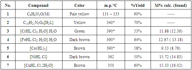
All chemicals were analytical grade and used without any modification. 1HNMR spectra were obtained using a Bruker 300 MHz and d6 –DMSO as a solvent at University of Al Albait, Amman, Jordan. FT-IR spectra were recorded on a Shimadzu spectrophotometer. Elemental analysis for the ligand and metal complexes were carried out using CHNO—elemental analyzer and atomic absorption using Schimadzu model 6809. Electronic spectra using Varian UV–visible spectrophotometer, Molar conductivity of the complexes were measured in DMSO as a solvent in 0.001M solutions using a CON 510 bench conductivity meter with 2-ring stainless steel conductivity electrode (cell constant, K = 1.0), Magnetic susceptibility measurements were carried out using of Curie balance at the Department of Chemistry while Bacterial strains and Mueller Hinton agar were supplied from Department of Biology, College of Science, Mustanseriyah University. Thermal stability (weight changes) of the samples was recorded by Mettler Toledo TGA851 in the temperature up to 600°C at College of Education of Pure Science Ibn-Alhaithum, University of Baghdad, Iraq.
Synthesis of 1-amino-2-quinolone (M)
This ligand (M) was prepared according to literature [11]. Coumarin (1.46g, 0.01 mol) in ethanol (20 mL) and excess of hydrazine hydrate (98%) (5g, 0.1 mol) were stirred under reflux for 12 h., reduce the volume by evaporation, cooled and the separated solid product was filtered and washed with cold ethanol and dried. Recrystallization from ethanol to give pale yellow crystals with yields 90%, m.p. 131 – 133°C. Elemental analysis for C9H8N2O, M.wt. 160; calculated (%) C 67.50, H 5.00 , N 17.50, O 10.00; found (%) C 67.34, H 5.32, N 17.45, O 9.97.
Syntheses of the ligand H2L
2,4 – dihydroxybenzaldehyde (1.12 g, 10 mmol) dissolved in ethanol (20ml) was added to M (1.60 g, 10 mmol) in ethanol (20ml). Few drops of glacial acetic acid was added as catalyst, and then refluxed for 7hrs (Scheme 1). cooled and the formed ligand was collected and recrystallized from ethanol to afford a yellow solid crystals with yield 70%, m.p.278 – 280°C. Elemental analysis for C16H12N2O3, M.wt. 280; calculated (%) C 68.57, H 4.28, N 10.00, O 17.15; found (%) C 69.09, H 5.10, N 9.89, O 17.87.
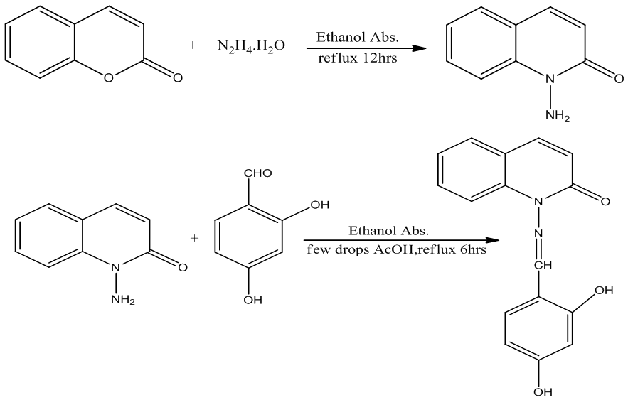
Scheme 1. Synthesis of intermediate M and the ligand (H2L)
Synthesis of complexes: The appropriate quantity of solid ligand (H2L) (0.56 g, 2 mmol) was dissolved in ethanol (20ml). This ligand solution was added dropwise with stirring to 2mmol of an ethanolic solution of; CrCl3. 6H2O, FeCl3, CoCl2., NiCl2. and CuCl2. 4H2O. Adjust the pH to 6.5 – 7.0 and reflux for 2hr to complete the crystal precipitation process, cooled, filtered washed with cold ethanol and dried. Hot ethanol used for recrystalliztion to afford the appropriate precipitate with 53–65% yield, see table 1.
Table 1. Some measured physical data of the ligand and complexes
     
Antimicrobial tests: The synthesized compounds were screened in vitro for their antibacterial activity against two Gram-negative (Escherichia coli and Pseudomonas aeruginous) and two Gram-positive (Streptococcus sp and Staphylococcus aureus) bacterial strains using agar well diffusion method and Primaxin drug as standard. The results of antibacterial studies are presented in Table 3. The tested compounds were dissolved in DMF to get concentrations of 50µg/mL. 0.1 mL from the specific concentration of dissolved tested compounds was carefully placed on incubated agar surface. After incubation for 24 h at 37°C, inhibition of the organisms which evidenced by clear zone surround each disk was measured and used to calculate mean of inhibition zones.

3. Results and Discussion

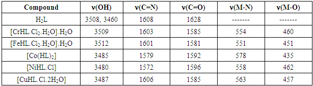
FT – IR spectra: The spectra were recorded in the region 4000–400 cm−1 using KBr disc.
The ligand (H2L) shows two absorption band at 3508 and 3460cm−1 attributed to υ (OH1) and (OH2) groups respectively. Strong band located at 1627 cm-1 is assignable to lacton υ (C=0) of coumarin. The other bands at 1608 cm-1 corresponding to imine (C=N) stretching frequency, while the aromatic ring gave two bands at 3205 and 1450 cm-1 attributed to (υ =CH) and (υ C=C), and the band assigned to aliphatic υ (C–H) appeared at 2986 – 2858 cm−1 [13]. In metal complexes, the disappearance of phenolic υ (OH1) was observed with low shift (5 – 45cm-1) for the position of the amide group (C=N), and lactone group (C=O) bands; this is indicative of formation of coordination bonds with each group. Ligand coordination to the metal centre is supported by the appearance of another two bands at approximately 551– 578 and 435 - 462 cm-1 corresponding to υ (M–N) and υ (M–O) respectively [14, 15] (Table 2). A broad band at approximately 3350 cm-1 and two other weak bands at approximately 750 and 700 cm-1 for CrIII, FeIII and CuII complexes indicate the presence of coordinated water molecule [16].
Table 2. Major Infra – red absorption bands of H2L and complexes (cm-1)
     
Electronic spectra
The ligand and all metal complexes were recorded in a concentration of 10 –3 M using dimethylsulfoxide as a solvent.
Three strong bands recorded for the ligand at 370, 308 and 275nm were attributed to n → π* for (C=O), (C=N) and π →π* for (C=C) groups respectively. Three bands were observed for CrIII complex attributed to U1 =786nm (4A2g→4T2g (F)), U2 = 480nm (4A2g→4T1g (F) and U3 = 385nm (4A2g→ 4T2g (p)), suggesting octahedral geometry, [17–19]. For FeIII complex it seems no d → d absorption bands recorded suggest a high spin octahedral geometry with 6A1g ground energy level [20], and this observation was supported by the magnetic moment value (µeff = 5.93 B.M ) for the complex. CoII complex showed two absorption bands at 399 and 448 nm attributed to [A1g → T1g, A1g → T2g] transitions suggest low spin octahedral complex [21]. The absorption spectra of NiII complex show two peaks, one around 480 nm and the other around 580 nm, assigned to 3T1(F)→3T1(P) and 3T1(F)→3T2(F) transition and when this combined with the calculated magnetic moment of (µeff = 3.48 B.M.) that will agree well with high-spin configuration and indicates the presence of tetrahedral environment [22]. The Cu(II) complex show a magnetic moment (2.14) B.M., the electronic spectrum showed a band at 641 nm which correspond to the transition 2B1g → 2Eg and another strong band at 463 nm correspond to charge transfer. This show that the Cu(II) complex has octahedral geometry [22]. The appearance at lower wavelength could be attributed to distortion geometry [23].
1HNMR spectra: NMR spectra were recorded using DMSO as solvent and TMS was used as an internal standard. Chemical shifts were reported in parts per million (ppm) and coupling constants were given in Hertz (Hz).
Only 1H NMR spectrum for ligands M and L was recorded, due to the paramagnetic properties of the metal complexes (Figures 1 and 2). The spectrum of the ligand (M) showed a multiples signals of aromatic protons appeared at the range 6.70 – 7.50 δppm. A broad peak at 4.1 – 4.4 δ ppm probably assigned to signals of – NH2 proton, while the signal of – C=C-H proton peak were recorded at 3.35 δ ppm. In the spectrum of H2L ligand the signal of – NH2 proton disappeared with the observation of two signals at 11.50 δppm for OH1 and at 10.27 δ ppm for OH2. Signal recorded at 8.27 δ ppm for – CH=N clearly indicates that the magnetic environment is equivalent for such proton. Multiples signals of aromatic protons appeared at the range 6.50 – 7.50 δppm and at 3.35 δ for – C=C – H.
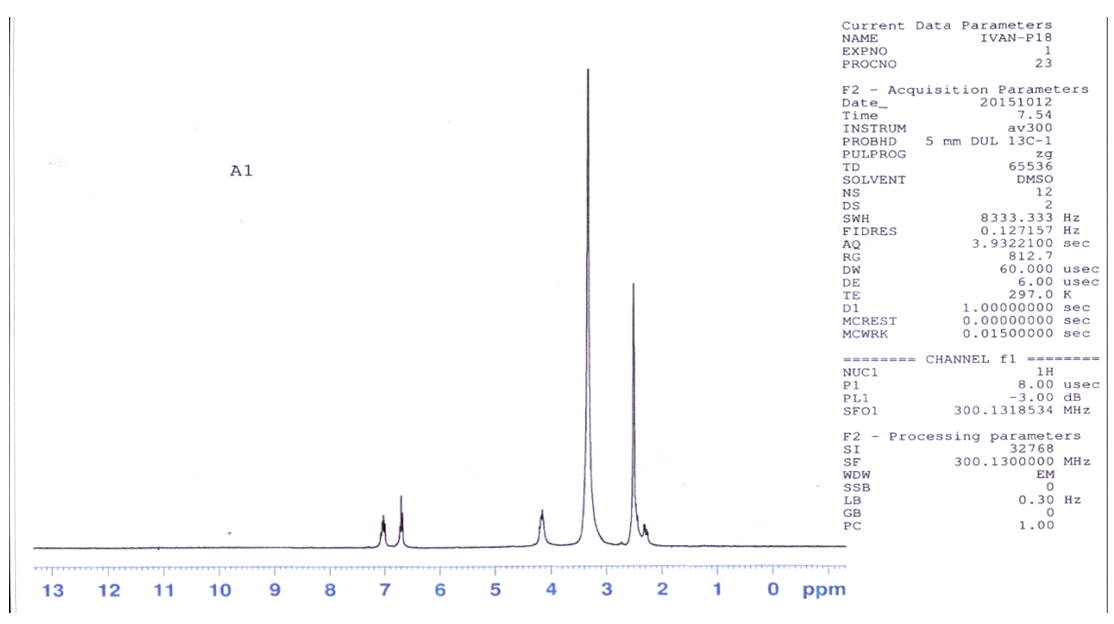
Figure 1. 1HNMR spectrum of M
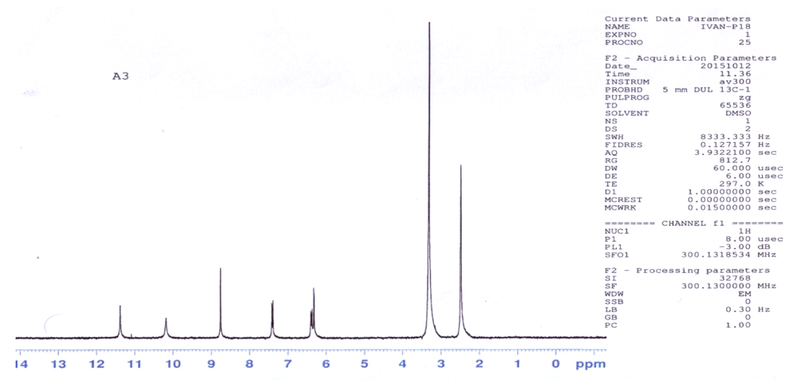
Figure 2. 1HNMR spectrum of the ligand H2L
Molar conductivity measurements
Using DMSO as a solvent with the concentration of 10-3 M, all metal complexes behaved as non-electrolyte which indicates the 1:1 electrolyte type, and these results fit with the geometry of mononuclear complexes [24].
Magnetic measurements
Magnetic susceptibility was measured for all the complexes at room temperature by use of Curie balance. The magnetic moment for the CrIII complex was µeff = 4.15 B.M which is slightly higher than the theoretical one (µeff = 3.89 B.M.) due to the absence of the orbital contribution of octahedral geometry (t2g3eg0) [25]. The same geometry for FeIII and CoII complexes were suggested by the values of µeff = 5.93 B.M (high spin) and µeff = 1.89 B.M. (low spin) respectively. Tetrahedral geometry is suggested for the NiII and CuII complexes by the observed magnetic moment µeff = 3.48 and 2.08 B.M respectively which is higher than the theoretical one because of orbital contribution of the e4t24 and e4t25 configurations.
Thermogravimetric analysis: Thermo gravimetric analyses (weight changes) were performed for complexes in the temperature range from room temperature up to 900°C using helium as inert gas at the heating rate of 20°C/min. The thermogram of complexes exhibits three clear cut stages [Figures 3, 4 and 5]. The first step showed the initial weight loss in the temperature range of 100 - 125°C corresponding to the loss of water molecules (CrIII complex, figure 3). The loss of water molecules in this temperature range indicates that the water molecules are of lattice type [26, 27]. The second step occurred around 200°C due to loss of coordinated water molecules (CrIII and CuII complexes, figures 3 and 5 respectively) [28]. For CoII complex (Figure 4) the second step starts at 360οC and do not show any weight loss below this temperature which indicated that this complex is devoid of coordinated water in the coordination sphere. The anhydrous complexes remain stable up to 340°C then the complexes show rapid degradation presumably due to decomposition of organic constituents of the complex molecules as indicated by the steep fall in the percentage weight loss. The final residue with attaining a constant weight roughly corresponds to metallic oxide [29].
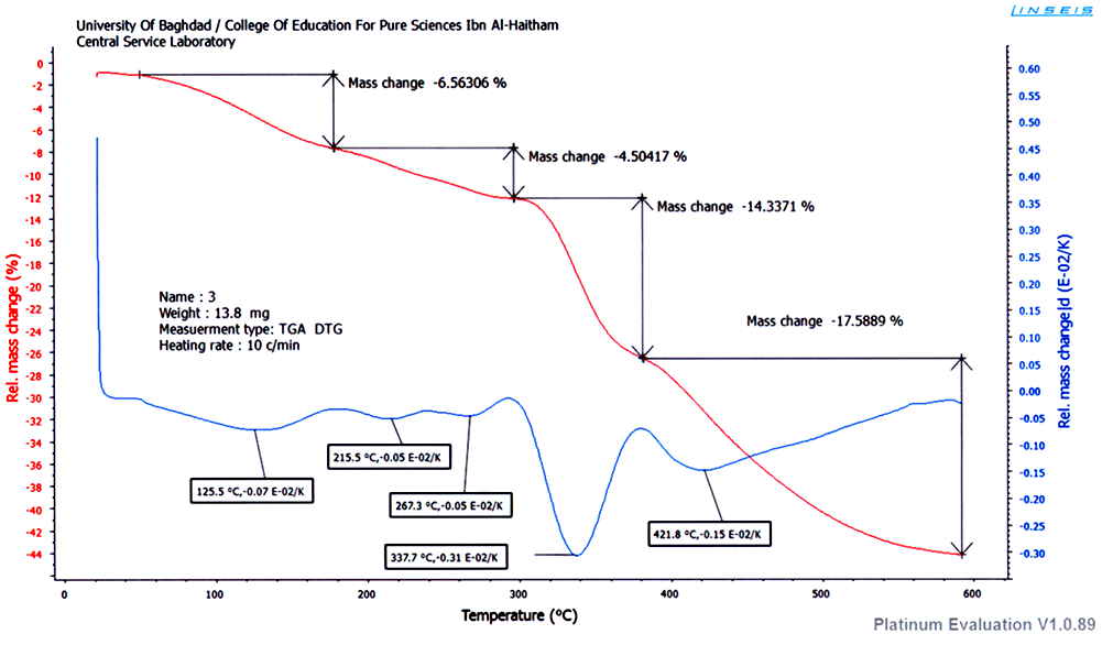
Figure 3. TGA of CrIII complex
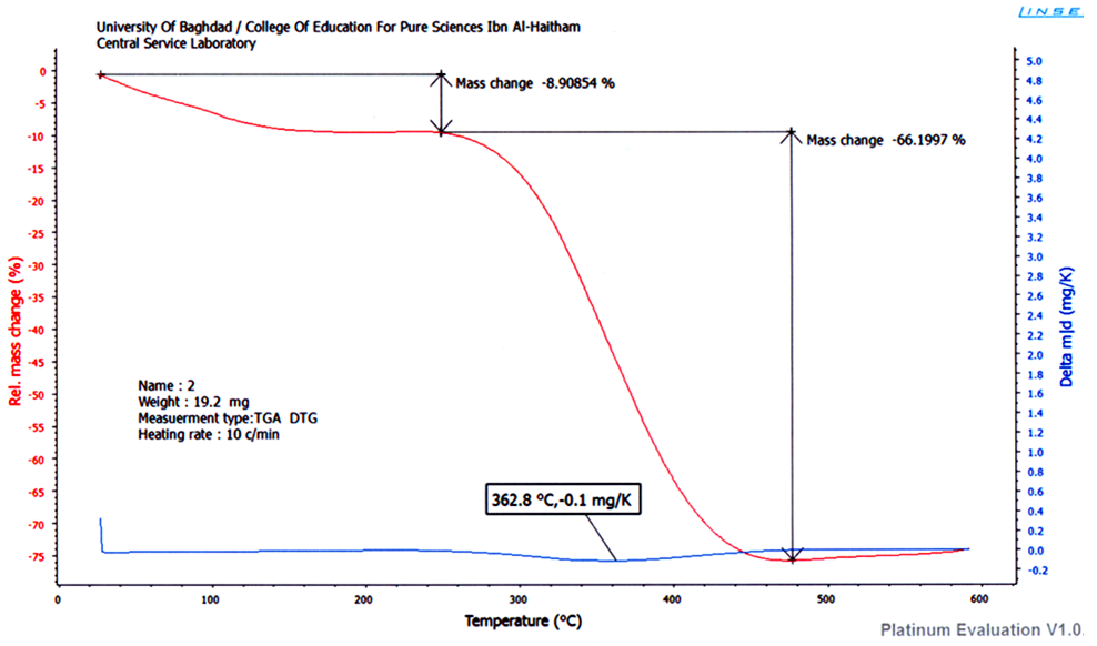
Figure 4. TGA of CoII complex
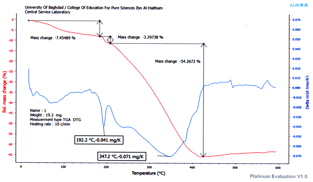
Figure 5. TGA of CuII complex
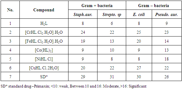
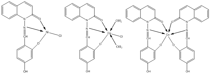
Table 3. Results of antibacterial assay (concentration used 50µg/mL of DMSO)
     
Biological activity study: A comparative study of the ligand and metal complexes indicates that most of the metal complexes exhibit higher antimicrobial activity than that of the free ligand, while CrIII and CuII complexes showed higher activity close to the control; see table 2. Hence complexation increases antimicrobial activity [30]. The higher activity of the metal complexes may be owing to the effect of metal ions on the normal cell membrane. Metal chelates bear polar and nonpolar properties together; this makes them suitable for permeation to the cells and tissues. Changing hydrophilicity and lipophilicity probably leads to bring down the solubility and permeability barriers of cell, which in turn enhances the bioavailability of chemotherapeutics on one hand and potentiality at another [31-33].

4. Conclusions

The condensation reaction of 2,4 – dihydroxybenzaldehyde and N – aminoquinolone- 2 - one affords a new tridentate Schiff base ligand. The CrIII and CuII complexes were found to be the most active against all the tested organisms with MIC close to the standard drug control. The analytical data of the transition metal complexes of the synthesized ligand confirmed a molar ratio (1:1) M:L except CoII complex (1:2) as shown below.

References

[1]  Arzu, G.; Seref, K.; Halil, IU.; Mustafa, B.;(2010) J. Heterocyclic Chem., 47, 1127.
[2]  Indulatha V N., Gopal N. and Jayakar B.; (2011) Int. J. PharmTech Res., 3(4).
[3]  Sarah. B., etal,; (2010) Dalton Transactions, 39, 10854.
[4]  Abhishek K., Jennifer F., Pankaj K. (2014) Int. J. Drug Dev. & Res., 6 (2), 165.
[5]  Park S J., Lee JC. and Lee K I.; (2007) Bull. Korean Chem. Soc., 28(7) 1203.
[6]  Godard A., Fourquez, J. M., Tamion, R., Marsais, F., Queguiner, G.; (1994) Synlett, 235.
[7]  Mohan G. and Rajesh N.; (1992) Indian J. Pharm, 24, 207.
[8]  Kong D., Reibbenspies J., Mao J. and Clearfield A.; (2003) Inorg.Chim .Acta, 342, 158.
[9]  Chellan P, Nasser S, Vivas L, Chibale K, Smith GS (2010) J Org-Met Chem., 695, 2225.
[10]  Khanye SD, Smith GS, Lategan C, Smith PJ, Gut J, Chibale K, Rosenthal PJ (2010) J Inorg Biochem 104,1079.
[11]  Hassan I. A.; (2013) Res. J. Chem. Sci., 3(12), 50.
[12]  AL-Amiery AH, AL-Bayati I H, Radi F.; (2011) British. J.Pharm. Toxi, 2, 5.
[13]  Shakya P. R., Singh A. K. and Rao T. R.; (2012) Mater Sci Eng, C 32, 1906.
[14]  Rao C. N. R. (1963) Chemical Application of Spectroscopy (Academic press, New York)
[15]  Nakamato K.(1986)Infra-red Spectral Inorganic and Coordination Compound, 4th ed.(Wiley, New York).
[16]  Patil S. A., Unk S. N., Badami P. S.; (2013) J. Therm. Anal. Calorim. 111, 1281.
[17]  Lever A. B. P.; (1968) Inorganic Electronic spectroscopy, Elsevier, New York, p. 274.
[18]  Thaker B. T., Lekadia J.; (1996) J. Indian Chem. 35A, 483
[19]  Datto R. L., Syamal A.; (1993) Elemental of Magnetic Chemistry, 2nd edn. (Affiliated East-West Press, New Delhi, p. 101.
[20]  Rao N. R. and Rao P. V.; (1996) Indian J. Chem., 25A, 482.
[21]  Comba P., Hambley W. I., Hifenhans P. and Richens D. T.; (1996) J. Chem. Soc. Dalton Trans, 533.
[22]  Rehman S. U.., Ikram M., Rehman S., Islam N. Z. and Jan N.; (2011) J. Mex. Chem. Soc., 55(3), 164.
[23]  Aputel B.T., Pchaker J.; (1996) J. Indian Chem. 35A, 483.
[24]  Aswale S. R., Mandlik P. R., Aswale S. S. and Aswar A. S.; (2003) Indian J. Chem. , 42, 322.
[25]  Alwan B., Singh H.A., J.; (1986) Indian Chem. Soc. LXIII (12), 106.
[26]  Allan J. R. and Veitch P. M.; (1983) J. Thermal Anal., 27, 3.
[27]  Nikolaev A.V., Logvinenko V. A. and Myachina L.T.; (1969) Thermal Analysis, Academic Pres, New York, 2, P779.
[28]  Bottei R. S. and Quane D.; (1964) J. Inorg. Nucl. Chem., 26, 1919.
[29]  a) Prasad K. S., Kumar L. S., Shekar S. C., Prasad M. and Revanasiddappa H. D.; (2011) Chemical Sciences Journal, 1; b) Jonathan B., Henderson B. L., Kiesz M. D., Whalley A. C., Morris W., Grunder S. and Deng H.; (2013) Chem. Sci., 4(6) 2300.
[30]  Agarwal R. K., Garg R. K., Sindhu S. S.; (2005) J. Iranian Chemical. Society., 2, 203.
[31]  Anjaneyulu, Y.; Rao, P. R.; (1986) Synth. React. Inorg. Met. Org. Chem., 16, 257.
[32]  Dharmaraj, N.; Viswanathamurthi, P.; Natarajan, K.; (2007) Transition Met.Chem, 26, 105.
[33]  D. Rehder; (2003) Inorganic Chemistry Communications; 6, 2003.