International Journal of Biophysics

p-ISSN: 2168-4979    e-ISSN: 2168-4987

2015;  5(1): 1-11

doi:10.5923/j.biophysics.20150501.01

Role of Protein Vibration in Emotion, Attention, Learning and Memory

Brajagopal Majumder

SRC-Tripura, Melarmath, Agartala, Tripura

Correspondence to: Brajagopal Majumder, SRC-Tripura, Melarmath, Agartala, Tripura.

Email:

Copyright © 2015 Scientific & Academic Publishing. All Rights Reserved.

Abstract

A brief account of behavioral traits of an individual particularly with respect to emotion, attention and learning has been discussed. It has been shown that emotion; attention and learning are closely related to each other. Earlier these were used to be discussed from the view point of macro behavioral attitudes like physical changes viz. body language etc. of the individual. With the advancement in molecular biology these are discussed and experimented in the context of micro molecules. The role of protein with its vibration in explaining emotion, attention and learning is explained here briefly. Attempt has been taken to identify different proteins responsible for different characteristics of the behavioral traits of the individual. The results pertaining to learning and retention of the learned events evaluated on the basis of “protein vibration” from the view point of one dimensional Schrodinger Equation are found compatible with those observed by the scholars on undertaking experiment on mice in 1963 and also in subsequent years.

Keywords: Emotion, Attention, Learning, Mental disorders, Bipolar, Depression, Molecular Biology, Protein Vibration

Cite this paper: Brajagopal Majumder, Role of Protein Vibration in Emotion, Attention, Learning and Memory, International Journal of Biophysics , Vol. 5 No. 1, 2015, pp. 1-11. doi: 10.5923/j.biophysics.20150501.01.

1. Introduction

It has been established since decades together that the standing hypothesis of learning are propounded in the form of S-R (stimulus response) connection. S-R hypothesis in turn is concerned with specific regions of the brain which is activated periodically with the sensation created by nerve impulses. And distortion in learning event becomes inevitable if there be any disturbance in the periodic activities of the brain.
It was suggested by Babsky et al. [1] that external stimulations like electric impulses constitute as one of the principal means of central Nervous System (CNS). In response to different impulses induced by external stimulation, it is possible to investigate accurately the transmission pathways of information to various brain structures and localization of sensory system perceiving these stimulations. This constitutes broadly the role of the electrical phenomenon in cerebral cortex of the individual. Since thirties, this has constituted the background of individual behavioral traits particularly with respect to emotion, attention and learning capability of the individual. However, this has now been changed with the advancement in molecular biology and the whole scenario is now looked on the role of micro molecule like protein, lipid etc. It has been established by Kovalyova [2] that when an electric signal gets to a neuron, the structure of the membrane lipid is changed and they pass into a new state with intensifying RNA activity and its constituent protein synthesis. If the cell does not undergo any change, nothing will be recorded. It means it has just reacted to an external irritant. But if the cell passes into a state with some new property, it means that an imprint of the received information is left and a trace of memory has emerged. At this stage, the lipid structure becomes restructured, the nature of the synthesis of constituent protein becomes different and the entire synthesis process of the cell passes into a new state. The re-oriented proteins enter the lipids and stabilize their new structure.
Now the question is – what role is being played by protein in generating emotion and attention in an individual?
Recent investigations on hippocampus, amygdale, thalamus and on frontal cortex with the application of modern technique like PET (Proton Emission Tomography), MRI (Magnetic Resonance Imaging) reveal us the fact that specific regions of the brain are associated with specific activities of the individual. Amygdale is responsible for emotion and craving for learning when the emotion is positive. This in the long run reveals as short memory. On the other hand, thalamus is found to be responsible for sensitivity of pain etc. But the recent achievement in molecular biology paves the way of considering this entire phenomenon in the context of the role of proteins. A brief account on the role of protein in emotion, attention & learning has been discussed in the present paper under the title “Role of micro molecules- Protein”. For example, while serotonin receptor, Dopamine transporter etc. are held responsible for generating emotion- positive or negative whatever it may be, BCMP-1 and other membrane bound proteins etc. are found responsible for learning’s and their retentivity.

2. Back Ground

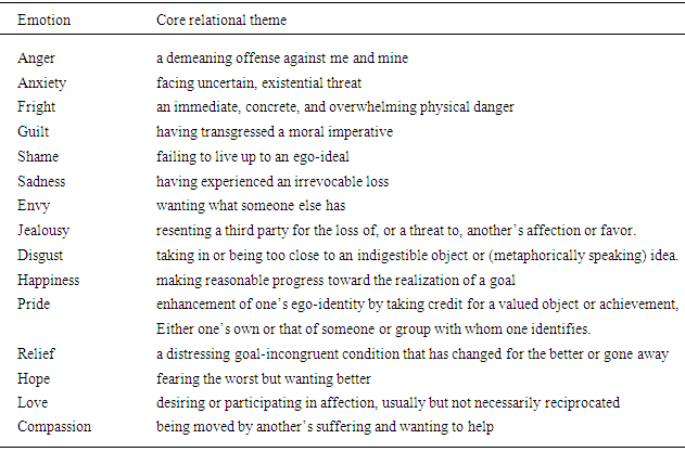
Emotion in broad sense is the mental condition of an individual. It is practically the behavioral outbreak of a person. It depends on both the mental (psychological) and physical status of the individual. There may be mainly a number of causes for generation of emotion in an individual. Usually, mental causes result as physical changes in an individual. These outbreaks may be happened in two categories- positive & negative. This means emotions may be categorized as of positive and negative characteristics.
R.S. Lazarus [3] suggested for 15 different emotions. Among these 15 emotions, there are roughly 9 negative emotions namely anger, fright, anxiety, guilt, shame, sadness, envy, jealousy and disgust. According to Lazarus [3] each is “a product of a different set of troubled condition of living and each involving different harms or Threats”. On the other hand, Lazarus [3] also categorized 7 positive emotions like happiness, pride, relief, love, hope, compassion, and gratitude. He also held that each type of emotion arises from different causes which bond a relationship between a person and the environment in which he lives. As for example- “feeling angry has its own special characteristic and so does for feeling anxious, guilty, ashamed, sad, happy and proud and so on. Though this way of thinking may be complex, yet this enriches the job of understanding and predicting the situation.”
The experiments so far conducted on the effect of emotion are concerned mainly with psychological and physiological aspects of the individuals. From all established experiments it reveals that the individual can recall the experiences generated due to both positive and negative emotions.
Emotion- positive or negative whatever may be, are closely associated with core relational themes. Following Lazarus [3], some of the core relational themes associated with emotions are tabled as,
The relational meaning usually stands for a person’s sense of benefit and harm in a particular person-environment-relationship. To speak of harm and benefit, one has to consider cognitive, motivational and relational aspects of the person-environment situation. Thus, it can be held that emotion is aroused not just by an environmental demand rather with person’s motives and benefits.
However, emotion is always accompanied by stress. There have been two varieties of stress – Eustress and distress. While Eustress is associated with positive emotions like positive feeling and healthy bodily states, distress is associated with negative emotions like negative feelings and disturbed body state. So, it is held that stress is merely a form of activation. Thus, feeling of emotion viz. physical change of an individual is directly proportional to magnitude of mental stress.
This advocates for different appraisal of the personal involvement in an adaptation encounter. From this, one can focus on learning different adaptation ally relevant things as what is happening and what will be the characteristic of the person who is reacting to the happening?
Basing on different experiences of emotions, the scholars are trying to correlate these with the process of learning. However, they are of the opinion that emotions also create attention is an individual which ultimately leads him in learning. Now let us examine the role of attention as preparedness of learning.
Table 1. Emotions and their core relational themes
     
Attention is a complex neural and psychological phenomenon. It originates from different brain structure. It emerges in various forms. The recent developments in attention opened the study of higher cognition to physiological analysis. It also reveals a system of anatomical areas which tends to be basic particularly to the selection of information for foul processing. The importance of attention lies in correlating the mental level of understanding cognitive science with anatomical level of neuroscience. In the context of the role of attention in understanding brain function Sperry [4] held “Control from below upward is retained but is claimed not to furnish the whole story. The full explanation requires that one takes into account new, previously nonexistent, emergent properties, including the mental that inter act causal control from above downward”. This opens a new channel of understanding the human attention system by correlating both the cognitive operations and neuronal activity in an individual. This advocates for more detailed studies of attention from the view point of cognitive neuroscience.
The attention system is concerned with sensory and motor system of an individual. It interacts with other parts of the brain by keeping its own identity aloof from others. It is carried out by a network of anatomical area and hence the area involved in attention usually carries out different functions and the outcome of these functions can be specified in cognitive terms. Hence, it is neither property of a single centre nor a general function of the brain operating as a whole [5].
A good number of experiments conducted on patients with lesions of the thalamus of monkeys with chemical injection into the lateral pulvinar also showed difficulty in covert orienting. All these findings advocated for two important points. While the first one confirms the involvement of specific cognitive operations in anatomical areas, the second one suggest a hypothesis about the circuitry involvement in covert visual attention shifts to spatial locations.
Studies on split brain patients [6] established the fact that two hemispheres of the brain perform different functions. Hemispheric dominance requires to be treated in a more differentiated manner with a view to understanding the cognitive localization. In this respect the experiment of Navon [7] can be cited. Navon formed large letters out of small letters. The use of small and large letters as a method of directing local and global attention results in the allocation of visual channels to spatial frequencies. The experiment conducted by Shulman and Wilson [8] showed that the subjects are relatively more accurate in perception of low spatial frequency and the reverse is observed in case of small letter.
It is observed from a number of studies that patients with right hemisphere is biased towards global processing like low spatial frequencies while local processing (high spatial frequencies) is associated with left hemispheric patients [9]. Examples may be cited as right hemispheric patients may copy small letters but miss the overall form while those with left hemisphere may copy large letter but miscopy the small letters. A number of experiments were conducted by different scholars and observed not only symmetric results but asymmetric results too. Thus, it seems that the hemispheric specializations are neither absolute nor innate. PET studies of the brain reveal that different areas of the brain are found attentive to different forms of activities pertaining to location orienting, visual orienting etc.
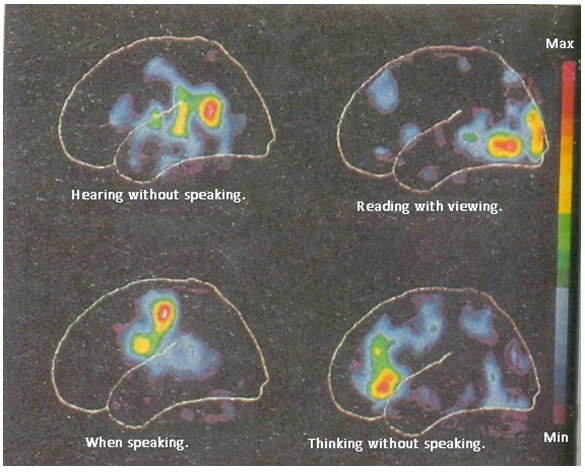
Figure 1. Blood flew with glucose metabolism in different regions of the brain. (Courtesy: J.J. Ghose as depicted in Bengali periodical DESH, 1992,58)
A major aspect of our study of attention is to see how attention can influence the functioning’s of cognitive system. Evidences are available with respect to attention system belonging to both spatial and language orienting from studies of cerebral blood flow during cognitive tasks. Roland [10] identified a lateral superior frontal area found active during cognitive system of language and spatial imaginary tasks. However, these studies do not provide clear evidences with respect to attention system. The study conducted by Peterson et al. [11] concerning midline frontal area including the anterior cingulated gyrus and the supplementary motor area found active during semantic processing of words. On the other hand, Posner et al. [12] observed that the degree of blood flow in the anterior cingulated increases as the number of targets increases. Hence, the anterior cingulated seems to be sensitive to target detection. From PET scanning it is also found that blood circulation also varies with respect to different activities undertaken by the individual. Differentiation of blood flow along with glucose metabolism in different regions of the brain during different tasks undertaken by the individual as observed through PET (positron emission topography) scanning are depicted as
It reveals from different studies of cerebral blood flow and metabolism that the areas of right cerebral hemisphere are responsible for vigilance tasks. However, the areas of right hemisphere do not respond uniformly with respect to other attention demanding activity like semantic tasks, imaginary tasks etc.
In 1978 Posner [13] conducted a study with a view to finding out the relationship between the alert state and other aspects of information processing. It reveals from the study that the alert state produces more rapid responding, without affecting the build-up of information in the sensory or memory system; however, this is accompanied by increase in error. One consistent finding is that alert state depends upon the integrity of the right cerebral hemisphere [14]. It is also verified in clinical observation where patients with right hemisphere lesions often show signs of neglect. This also leads to the notion that all spatial attention is controlled by right hemisphere.
Finally, it can be held that disorders of higher level of cognition like neglect, schizophrenia, closed head injury etc. are said to occur due to defects of attention. However, a combined cognitive and anatomical approach in integrating the physiological and psychological influences in an individual is found viable.
In view of these microscopic (physiological and psychological) approaches, the present paper focuses on the role of different proteins and the mode of their functioning’s in different behavioral traits of an individual. The whole process has been discussed from our hypothetical approaches [15] of “protein vibration” and its reorientation, as and when it is felt necessary.

3. Role of Micro Molecules-Protein

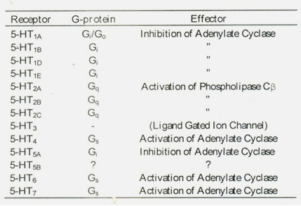
Emotion, Attention and Learning are very much related to each other. Till to date these are viewed by the physiologist, psychologists and educationists from macro behaviorism of the individuals. Different experiments are conducted basing on physical changes occurring in human behavior. Some scholars are also conducting experiments on physiological changes with the help of sophisticated instruments like MIR, PET etc Recently, EEG rhythms of different frequencies are applied to measure the level of learnt events.
Moreover, it is possible today to identify correctly the causes of mental depression, borderline disorder, anxiety disorder, panic disorder (phobia), attention deficit disorder etc. with the help of PET, MRI, FDG etc. Recently, it has been observed through PET and FDG that unipolar depression and bipolar depression are different types of diseases. Differentiation in blood circulation and in glucose metabolic behavior is largely found in different regions of the brain in case of these two types of diseases. Degree of glucose metabolism in basal ganglia of middle lobe and certain regions of frontal lobe increase almost 1000 times in border line disorder persons with respect to normal individuals. On the other hand, it is observed that the metabolic behavior of glucose is found to decrease abnormally in certain regions of the brain of hyperactive children. Though hard, it has become possible to observe the physiological changes in an individual who intends to kill himself with the help of PET. It reveals from PET scanning on similar individual that the number of protein receptors namely serotonin neurotransmitter increases in the individual at the moment of his attempt to kill himself. From these studies scholars now can identify the specific regions of the brain responsible for attention. Thus, from the above studies, it can be held that the causes for emotional changes- positive or negative, attention, learning and subsequently memory traces are practically associated with change in vibration frequencies of certain proteins- most of which are still to explore. However serotonin receptor, Dopamine transporter, noradrenaline transporter, hormone etc. have so far been identified as responsive to certain of emotion- positive or negative. It has been experimentally proved that Dopamine transporter and noradrenaline transporter remain in the cell membrane of the synapses. When a brain stimulant medicine enters the brain, the medicine by organic reactions gets bounded with respective proteins. These in turn generates craving for stimulant medicine by the individual. Thus, it is now an established fact that differentiation in blood metabolism and participation of different proteins etc. are found to be responsible for creation of emotion in an individual.
Recent physiological and biochemical investigation reveal that membrane bound protein play a vital role in storing and also in reproducing the learnt events [15]. So far the mechanism of memory is concerned most of the researcher emphasized on membrane bound proteins. Some of the membrane bound protein with their ID names is listed in.
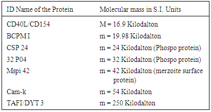
Table 2. ID Name and Molecular weights of some memory related proteins
     
The reason for presumption of these sorts of protein involvement in the process is due to their dependence on lipids while lipids play passive role in organizing memory. Thus, the cell membrane may be considered as double layer of fat like organic lipids with protein ice-bergs floating in it.
The mechanism can better be understood form the simple electrochemical net-working of CNS. With the passage of electrical signals, the neurotransmitter glutamate is released at the axon terminals of one neuron and picked up by the N-methy 1-D-asparatate (NMDA) receptor on the dendrite of another neuron. When an electric signal gets to a neuron, as held by Kovalyova [2] the structure of the membrane lipid is changed and they pass into a new state with intensifying RNA activity and its constituent protein synthesis with reoriented structure. The reoriented proteins enter the lipids and stabilize their new structure. The structural orientation of protein due to external stimuli may be symmetrical to conformational changes under the phenomenon “Steered Molecular Dynamics (SMD)” as described by H. Karcher et al. [16]
Figure 2
C1, C2 represent the structural (conformation) orientation of proteins of proteins under SMD hypothesis proposed by H. Karcher et al.
The left end of both the helixes is held fixed with six hydrogen bonding pairs while the right end of C2 conformation may be due to loose bonding or loss of amino-acid molecules. Thus C2 may be considered as the conformational form of the reoriented protein.
It was established [17] during seventies that increased amount of phosphorylation of synaptosomal protein (C57B1/6J) in mice was subjected to instrumental aversive foot-shock conditioning. Subsequently, number of experimental models emerged out in this area centering on Caenorhabditis elegans, drosophila melanogaster and Aplysia californica. Recently, this is being followed in mammals such as rats, mice, rabbits and primates etc.
The cAMP stimulates protein kinase A (PK-A) which phosphorylates the K+ channels on the membrane resulting in short term memory (STM) or cAMP response to element binding with protein in long term memory (LTM). Along with cAMP, phosphoinositides and diacylglycerol also play a pivotal role in the process of learning and memory formation. Long term memory traces are usually associated with more permanent alteration in the structure and number of synapses. Such an alternation involves new protein synthesis and assemblage of new memory circuits. This means that there occur changes in the electrical activity of specific regions of the brain due to different learning experiences [18]. At this stage acetylcholine and nitric oxide play significant role as neurotransmitters or neuromodulators. The process of learning gets disturbed if recruitment of synaptic vesicle or neurotransmitter is interrupted.
Permanent alteration in the structure and number of synapses involves new protein synthesis and assemblage of new memory circuits resulting changes in electrical activity of specific regions of the brain due to different learning experiences. In his paper [15] the author analytically showed that injections of protein inhibitor in the temporal lobe of mice led to the fact that learning is effective in long-term memory. Studies of long term potentiation (LTP) and long term depression (LTD) in mammalian brains resulted in two distinct phases- the early one (typically lasting 1-3 hours) is dependent of new protein synthesis and the late one (typically lasting more than 8 hours) depends on new protein synthesis [19]. A number of proteins are responsible for memory formation- short term or long term. It was also showed by the author that retentivity of learning events depends on the number of vibration frequencies generated in proteins of large or small molecular weights due to external stimuli or electrostatic force inherit of the proteins.

4. Methodology

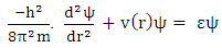
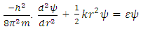
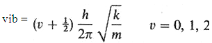
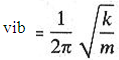
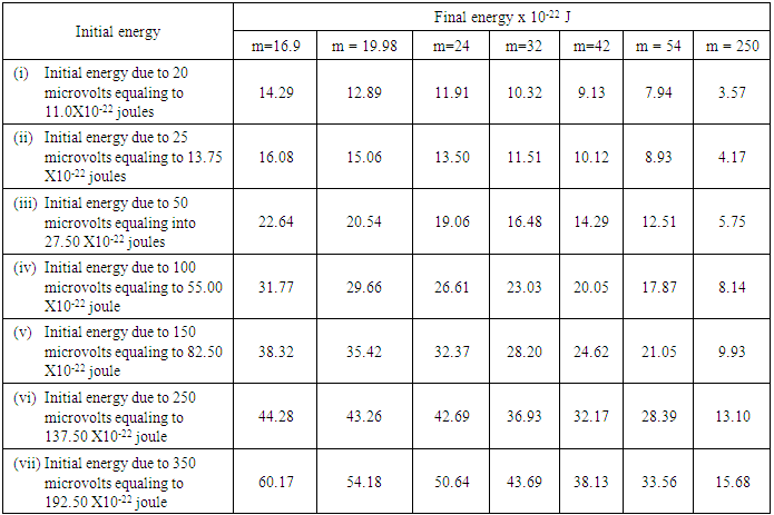
The author in a recent paper [15] showed that the recalling of the learnt event depends on the number of frequencies of vibration in protein.
The problem of protein vibration responsible for memory was considered [15] in the light of the solution of the standard form of Schrodinger Equation
Inserting the potential energy function v = ½ kr2 in one dimensional Schrodinger Equation
(1)
which turns to
(2)
The energies of the allowed vibration states derived from the solutions of the Schrodinger’s Equation.
(3)
While the expression for vibration frequency stands as
(4)
The vibrations occurring in protein depends on molecular weight of the concerning proteins available in particular portions of the brain. Thus, frequent vibrations, as the author [15] held, is created in light proteins of small molecular weight while slow vibrations are created in proteins of comparatively large molecular weight.
The memories emerged due to frequent vibrations may be termed as instant or subconscious memory. As for example, when signal of smell bypasses or erases, the vibration generated in protein will cease. This means the level of newly synthesized protein may go down after its temporary activation. These are diagrammatically described as
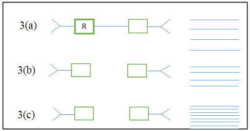
Figure 3. Vibrations of Protein shown in figure 3. (a) represents protein of large molecular weight, 3. (b) represents protein of medium range molecular weight & 3. (c) represents transient protein

5. Results & Discussion

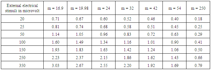
Basing on these approaches the author [15] calculated vibration frequencies of above mentioned proteins as
Table 3. Vibration frequencies (m-1) of some memory related proteins with molecular weights
     
From Table 3 it reveals that frequencies of vibration in protein are in decreasing order with respect to ascending mass of proteins for constant amplitude of external electrical stimuli.
This once again proves our suggestion that vibration frequencies generated in protein are inversely proportional to the molecular weights of proteins.
In support of this protein vibration, the functioning of BCMP1 (Brain Cell Membrane Protein 1) was discussed in his paper [15] by the author. The structure of BCMP1 is represented schematically in the fig as
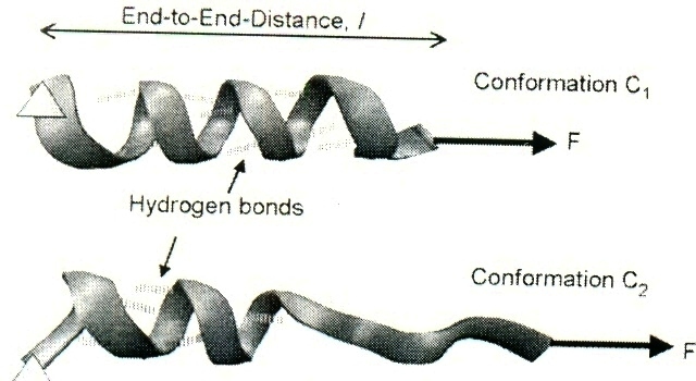
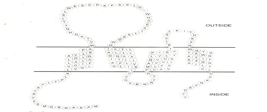
Figure 4. As proposed by Christiane Christophe –Hobertus et al
It is found from the figure (4) that concentration of the amino-acids of the respective protein is bounded to the membrane of the brain cell. The membrane is assumed as the source of energy. When this portion of the protein is activated by external stimuli, vibrations are generated. This means that the cell passes into a new state with the imprint of the received information which results in storing of the learnt events. This in turn may emerge out as memory traces with similar external stimuli.
The author in a paper attempted also to correlate analytically the following experimental findings observed by Louis and Josefa Flexner [19]
i) Learning in effective from day 1 to 3 and are independent of protein synthesis.
ii) Learning lasting for more than 8 (eight) units of time (hour or day) is dependent on new protein synthesis and there should be a critical period for new protein synthesis.
iii) Protein constitutes the molecular basis of memory.
Basing on the role of protein vibration energy in learning and memory it is held by the author [20] that loss of vibration energy means loss of memory. From our mathematical analysis, it is held that learning events initially remain almost constant with respect to t ≥ 3 units of time for frequencies of 0.5 cps to 8 cps of external stimuli irrespective of molecular weights of proteins. However, for t > 8 units of time, reverse found to occur rapidly for ω≥ 3.5 cps with respect to small and middle molecular weights of proteins but in case of protein of large molecular weights, loss of vibration energy is less and hence memory loss is also less. However, in case of lower vibration frequency =0.5 cps of the external stimuli, loss of vibration energy occurs in a consistent rate for t > 8 units of time irrespective of weights of proteins. It has also found to be relevant from the graphical representation of Data. The graphs (Fig-5, Fig-6 and Fig-7) are in decay nature and it found to be compatible with those observed by Ebbinghaus with respect to the amount of retention of learning of unorganized (nonsense) events. The graphs were plotted with decay energy with respect to time for protein of masses m= 19.98, 54,250 KD for external stimuli of 100 microvolt in the form of EEG rhythms for frequency ω= 0.5 to 30 cps. It has also noted that learning with respect to mice may be concerned with one single point but not for multiple issues (organized learning) which are usually concerned with human beings.
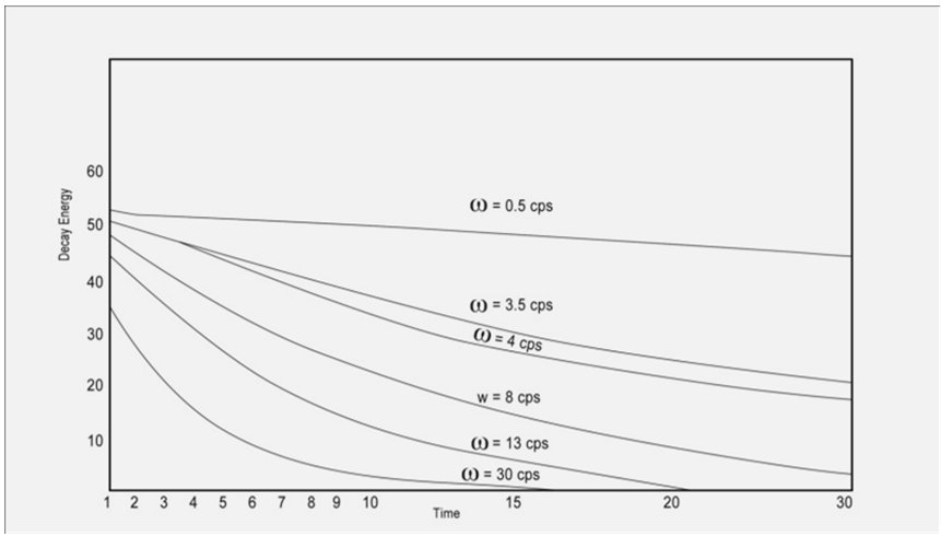
Figure 5. Graphical representation of Decay Energy with Respect to time for protein of mass =19.98kD
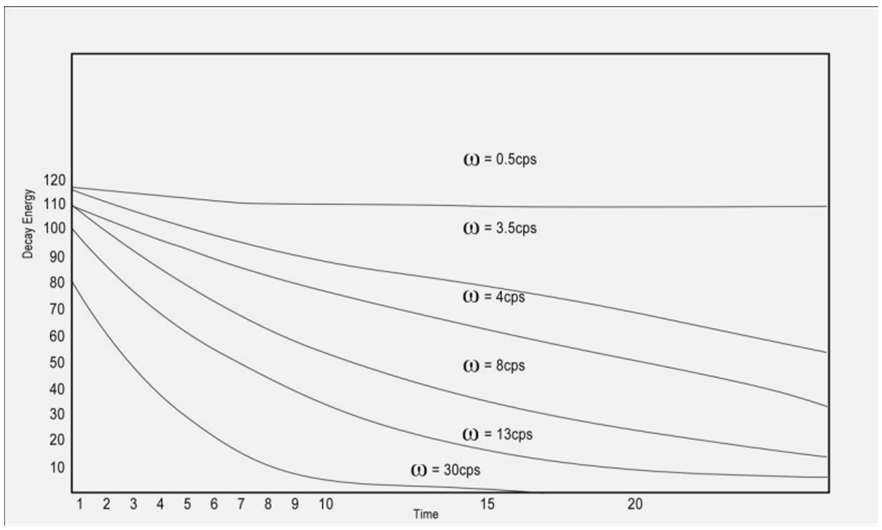
Figure 6. Graphical representation of Decay Energy with respect to time for protein of mass = 54 kD
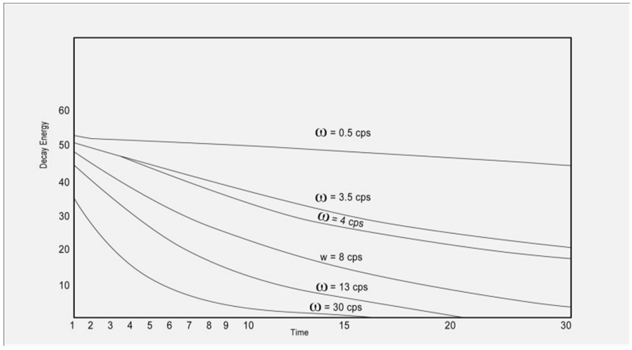
Figure 7. Graphical representation of Decay Energy with respect to time for protein of mass = 250 kD
Thus, it is held that our mathematical analyses on learning and retention of learnt events [20] are in parity with the experimental findings, which read as
i) Learning is effective for day 1 to 3 & is independent of further protein synthesis.
ii) There is a critical time for sustainable- learning.
iii) Learning lasting for more than 8 units of time is dependent of new protein synthesis.
iv) Number of amino acids associated or disassociated from the master protein may be evaluated approximately on the basis of our proposed expression.
v) These amino acid may take part in synthesizing new protein or the disassociated amino acid as a whole, may also act as newly synthesized derivative of the master one.
Similarly, the vibration frequencies of serotonin receptor protein having molecular weights 70.32 KD may be calculated on the same approaches of protein vibration. From the view point of molecular weight, serotonin receptor will also execute vibration similar to 3 (a) as in Fig. 3.
But serotonin receptor has peculiar properties. It may act as a catalyst of generating both negative and positive emotions, stable learning and stable memory. Particularly speaking, “the diversity and specificity of serotonin signaling and function arise from the molecules that receive the signals in various target regions.” These target molecules are protein receptors [21]. There are seven known families of serotonin receptor namely 5-HT1, to 5- HT7. These encompass at least 15 subtypes. Some of the subtypes of mammalian serotonin receptors along with G-proteins primarily coupled to them and the effectors pathway as listed by C.D. Nichols and E. Sanders –Bush [21] are tabled as –
In this context, it may be held that serotonin receptors have got diverse but specific functions in various target regions. As for example LSD (Lysergic acid diethoilamide) bind to a variety of receptors like 5-HT1 2A/2C, 5HT1A, 5-HT6,5-HT7, dopamine D1 and D2 and these receptors take part in different aspects of behavior. However we are here rather interested in receptors for cognitive development of the individual in spite of its involvement in producing hallucinogenic effects. Since we are interested in cognitive development of the individual, we can take the example of 5-TH2A receptor expressed in particular regions of the brains as it is believed to be involved in cognitive process (20). Now it can be held that serotonin receptor protein in measured form acts as a positive catalyst of emotion which helps to get rid of hyperactivity in children and hence to be attentive.
On the other hand, it is also an established fact that depression result from a deficit in brain norepinephire or serotonin or both. Thus for example, it may be held that some depressed patients have reduced concentration of major serotonin metabolites resulting in increase of serotonin receptor protein. The increased serotonin receptors will generate frequent vibrations due to electrostatic forces. As a result, this creates negative emotions which may ultimately tend to commit suicide by an individual.
But it is an established fact that amygdale perform primary role in formation and storage of short term memories. A good number of papers are now available in support of this statement. Research indicates that sensory stimuli reach the lateral nuclei of the basso-lateral complexes of the amygdale where they form associations with the memories of the stimuli. This kind of stimuli association predicted aversive events. This may be treated as fear conditioning and this kind of fear conditioning practically turns to a sustained enhancement of signaling between affected neurons. Several studies show that “patient with bilateral de-generation of the amygdale was told a violent story accompanied by matching picture. Later he was asked to tell the story. It was observed by the researcher that the patient had less collection of story than those without amygdale damage. This proves that amygdale has a strong connection with emotional learning. Lesions of central nucleus in the amygdale have shown to reduce appetitive learning in rats. However, this is not same for all sorts of nuclei within amygdale.
Table 4. Derivatives of Serotonin receptors as proposed by Nicholas, C.D
     
Table 5. Vibration Energy of Proteins at Initial and Final Levels
     
Amygdale also acts as a modulator of memory consolidation. Learning events in amygdale do not turn into long-term memory instantaneous. Rather it is slowly assimilated into long time storage over time by the process of modulation in amygdale. Experiments have shown that mice on administration of stress hormones immediately after they learn something can retain the learnt event even after two days of the 1st learning incident.
A variety of data shows that amygdale has got substantial role in mental states. Many psychological disorders are associated with left or right amygdale. Studies show that children with anxiety disorders tend to have a smaller left amygdale. Left amygdales are linked to social anxiety, obsessive and compulsive disorders, post traumatic stress and also general anxiety. Border line personality disorders showed significantly greater in left amygdale activity. In 2006, researchers observed hyperactivity in amygdale when the individuals are to face threatening or frightening situations. Depressed patients showed exaggerated left amygdale. By and large, amygdale has been found to respond differently in persons with bipolar disorder.
Now the question is- how amygdale gets activated? Recently, it has been established that emotionally arousing information increases amygdale’s activity and that activity correlates with retention. It is an established fact that amygdale neurons show that neural events could promote synaptic plasticity resulting in increased amount of interaction between neocortical storage sites and temporal lobe structure. On the other hand, promoting synaptic plasticity means involvement of neuronal events which is due to the outcome of microscopic chemical changes at the synapse. The changes result in forming new synapse and growing new dendrites along with synthesis of proteins. Thus, it is observed that macro activity of amygdale originates practically from the micro activity concerning with particular protein like serotonin and its derivatives etc.
However, the objective of the present paper focuses on the role of protein vibration basing on which vibration energies of the concerned protein are evaluated. This does not depend on the number of proteins and their derivatives involved in different forms of behavioral traits of the individual like emotion, attention learning and memory traces. In a paper [15] the author evaluated the vibration energies of certain proteins basing on which amount of retained energies may be calculated.
In his proposed approach of electro chemical correlates of bearing and memory, the author held the view that learning and memory are nothing but transformation of energy from one form to another resulting ultimately to chemical energies, which now constitutes the molecular basis of memory.

6. Conclusions

From the above discussion, it is found that emotion, attention, learning and memory traces are correlated not only from macroscopic point of view but also in the context of micro molecules viz. proteins. Different kinds of proteins are associated with the three basic regions of the brain like Amygdale, Hippocampus and Thalamus. They are interconnected with each other though they lie in three different regions of the brain. Interconnection is made through proteins, the mechanism of which lies in the fact that some protein molecules act as adhesive on cell membranes. The main objectives of these proteins are to maintain the communication from cell to cell. These proteins are briefly called CAM (Cell Adhesive Molecules). These are numerous in numbers. Usually, these proteins lie on the outside (membrane) of the cells. These proteins are responsible not only for network functioning between cell to cell but also for looking after the growth and movements of the cells. Thus, it can be concluded that emotional learning, normal learning etc. are closely associated with protein functioning lying particularly in amygdale, hippocampus, and other necessary parts of the brain and the measures of learning outcomes are associated with the level of vibration energies generated due to different external stimuli.

ACKNOWLEDGEMENTS

The author is thankful to Dr. U.C.De, associate Prof. Deptt. Of Chemistry, Tripura University for his helpful discussion. He is also thankful to Mr. Bishnupada Dey, Asst Teacher, Mr Debojit Banik, Mr Rajib Malla and Mr Tanuj Sharma for their co-operation in computer work of the manuscript.

References

[1]  Babsky, E.B, Khodorov, B..I Kosisky, G.I et al.:- Human Phsyology, MIR Publication and Co, Mosco,1875, 2 274.
[2]  Kovalyova, A:- Mapping Memory’s Track, Sci USR ,1990, 4,95-96
[3]  Lazarus, R.S.:- From Psychological Stress to the Emotions: A History of Changing Outlooks, Ann. Rev. Psychol. 1993. 44. 1-21
[4]  Sperry, R. L.:- Psychology’s mentalistparadigm and the religion/science tension. Am. Psychol.1988. 43:607-13.
[5]  (i) Kahneman, D.:- Attention and Effort. Englewood Cliffs, 1973. NJ: Prentice Hall. 246pp. (ii) Mesulam, M. M.:- A cortical networkfor directed attention and unilateralneglect. Ann. NeuroL, 1981. 10:309-25
[6]  Gazzaniga, M. S.:- The Bisected Brain. New York: Appleton, 1970. 171 pp.
[7]  Navon, D.:- Forest before trees: Theprecedence of global features in visual perception. Cog. Psychol, 1977. 9:353-83.
[8]  Shulman, G. L., Wilson, J.:- Spatial fre-quency and selective attention to local andglobal structure. Perception, 1987. 16:89-101.
[9]  (i) Robertson, L., Delis, D. C.:- Part-wholeprocessing in unilateral brain damaged patients: Dysfunction of hierarchicalorganization. Neuropsychology, 1986. 24:363-70. (ii) Sergent, J.:- The cerebral balance of power: Confrontation or cooperation? J.Exp. Psychol. Hum. Percept. Perf, 1982. 8: 253-72.
[10]  Roland, P. E.:- Cortical organization ofvoluntary behavior in man. Hum. Neuro-biol, 1985. 4:155-67.
[11]  Petersen, S. E., Fox, P. T., Miezin, F. M., Modulation of cortical visual responses by Raichle, M. E.:- direction of spa-tial attention measured by PET. Assoc.Res.Vision Ophthal.1988a. p. 22 (Abstr.)
[12]  Posner, M. I.:- Structures and functions of selective attention. In Master Lecturesin Clinical Neuropsychology, ed. T. Boll,B. Bryant, 1988. 173-202 pp:Am. Psych. Assoc. Washington, DC.
[13]  Posner, M. I.:- Chronometric Explor-ations of Mind. Englewood Heights, NJ:Erlbaum, 1978. 271.
[14]  Heilman, K. M., Watson, R. T., Neglect and related disorders. In Clinical Valenstein, E.:- Neuropsychologty, ed. K. M. Heil-man, E. Valenstein, 1985. pp. 243-93. NewYork: Oxford.
[15]  Majumder, B.G.:- Role of Locally Synthesized Protein in Long Term Memory- An Analytical Approach International Journal of Biophysics, 2014: 4(1): 1-8.
[16]  Helene Karcher Seung E. Lee Mohammad A coarse –grained Model for Force –Induced Protein R. Kaazempur-Mofrad and Roger D Kamm.:- Deformation and Kinetics. Biological Journal, 2006 90,2686-2697.
[17]  Perumal, R., Tan, I:- Is there an answer? What proteins are involved in Learning and Memory? IUBMB life, 2007: 59(7): 465-468.
[18]  Martinez JL, Jr. Demick, B.E.:- Longterm potential and Learning Ann. Rev.Psychol, 1996, 47: 173-203.
[19]  Flexner, J.B, Flexner, L.B. Memory in mice as affected by intracerebral and Stellar, E.:- puromycin, Science, 1963, 141, 57-59.
[20]  Majumder, B.G.:- Role of Locally Synthesized Protein in Long Term Memory- An Analytical Approach International Journal of Biophysics, 2014: 4(1): 1-8.
[21]  Nicholas, C.D and Serotonin Receptor Signaling and Hallucinogenic Bush E. Sanders:- Drug Action. The Heffeter Review of Psychedelic Research, 2001, 2, 73-79.