American Journal of Bioinformatics Research

p-ISSN: 2167-6992    e-ISSN: 2167-6976

2012;  2(5): 92-101

doi: 10.5923/j.bioinformatics.20120205.04

Nonlinear Dynamical Systems as Models of Development of Inorganic Cells (iCHELLs) & their Simple Assemblies

Olle Gradoff 1, 2, 3

1Laboratory of Photobionics, Institute of Chemical Physics RAS, Moscow, Russia

2Institute of Mathematical Statistics, Maryland, USA

3SIAM Activity Group of Life Science, Philadelphia, USA

Correspondence to: Olle Gradoff , Laboratory of Photobionics, Institute of Chemical Physics RAS, Moscow, Russia.

Email:

Copyright © 2012 Scientific & Academic Publishing. All Rights Reserved.

Abstract

The rapid development of synthetic biology and biomimetics in recent years leads to the creation of prototypes of inorganic artificial cells with membrane-like properties. In this regard there is an obvious need in the development of a universal formalized mathematical description of morphology, dynamics and cooperative behavior, which results in the formation of supracellular assemblies, applicable to both organic and inorganic cells. For this purpose we propose to use invariant sets of nonlinear dynamical systems, which in some variations are isomorphic to biological cells or can be considered as a good morphological metaphor of their morphogenesis and assotiation. This paper compares graphical visualization of invariant sets of nonlinear dynamical systems with the results of experiments on the chemical synthesis of biomimetic structures. A number of maps is considered: Poincare map, Henon map, Chirikov and Gumovski-Mirr maps, systems by Kepler, Volterra, Dyufing and Henon-Heiles. The possibility of approximation of the experimentally observed structures by those mathematical models is shown. The given results of computer simulations are applicable to inorganic cells (iCHELLs), obtained recently in the University of Glasgow, but the proposed method was originally developed for functionally identical inorganic cells, obtained by the author according to the Russian technique, proposed in the early 2000s. The micrographs of such inorganic cells and their assemblies are given below in comparison with the theoretical model.

Keywords: Artificial Cells, Inorganic Cells, Nonlinear Dynamical Systems, Morphogenesis, iCHELLs, Biomimetics, Cellular Adhesion, Cell Colonies, Philopodial locomotoion

1. Introduction

With the development of biomimetic technology much attention has been paid to the creation of a full structural and functional analogue of a biological cell (Artificial Cell, or ACell). Originally attempts were made either to assembly primitive semi-synthetic vesicular structures, capable of simulating a number of functions of a biological cell due to the identity of chemical composition[1-3], or to create new artificial species by transplantation of a synthetic genome into bacterial cells[4-5]. However the works of S. Rasmussen demonstrate the ability to create completely synthetic complex biomimetic systems via self-assembly[6].
The possibility of creating inorganic artificial cells through self-assembly was first shown by A. Muller[7-8]. Later the concept of self-assembly of inorganic chemical cells (iCHELLs) combined with an evolutionary approach to biomimetics was developed by L. Cronin and his group[9].
To date the most promising area is the combination of evolutionary biomimetics with the works on protocell synthesis[10-11], as well as the convergence of Soft- и Dump ALife, allowing to simulate complex biomimetic structures with their subsequent chemical synthesis[12].
Simulation of the formation of biomimetic structures, including iCHELLs, using nonlinear dynamical systems has not yet been performed. However, seeing nonlinear reaction-diffusion nature of morphogenetic processes[13], this approach is extremely useful and can be considered as a key step towards the convergence of Soft- and Dump ALife.
This paper presents only the methods for simulation and approximation of the formation of artificial cells and their assemblies in terms of the theory of nonlinear dynamical systems, without the emphasis on biomimetic properties of and methods of self-assembly of iCHELLs, described in another paper.

2. Simulation of Synthetic Protoplast Formation

The given results were performed using a specialized program WINSET, simulating invariant sets of dynamical systems (for example see[14-20]). Visualized sets were imported from the relevant program slides. All variables were taken as spatial coordinates.
Simulation of the protoplast form was produced by volume-preserving map
(1)
and a Henon-Heiles system
(2)
Fig. 1a shows the trajectory of the volume-preserving map, and Fig. 1b - micrograph of an "artificial cell" - vesicular system, which includes osmotic fiber, membrane components and the dispersion medium, obtained by the method[21].
Figure 1. a - computer simulation model, b - an “artificial cell”
One can observe a topological similarity between fibrillar components in the cellular micro-object and the corresponding substructures in a computational model. In accordance with the osmotic laws, protoplast has the form of spheroplast, whereas the computational model can be any shape other than spherical (depending on the formalism applied).
Fig. 2a shows the Poincare map of the system
(3)
at p1 = 1, p2= - 1, p3 = - 0.1, p4 = 1. This map, close to the Henon-Heiles system, also has a drop-shaped structure with singularities in the centre of "drops". Fig. 2b gives the Poincare map of Henon-Heiles-type system at p1 = 1, p2 = 1, p3= 1, p4 = 0.5, p5 = 0, p6 = 0, h = 1/6, where h - the energy integral. Such structures occur as a result of morphological aberrations in "artificial cells" associated with their adhesion to the substrate. In such cases it is due to a violation of the hydrophobic surface properties. Drop-shaped protoplasts experiencing strain when moving on a surface of the substrate - quartz glass with a medium, are shown in Fig. 3. Obviously, such structures can be approximated by the graphs given in Fig. 2a, 2b.
Figure 2. Poincare maps of a Henon-Heiles-type systems and its analogue
Figure 3. The droplet-type structures of “artificial cells” in the medium
Volterra system
(4)
adequately describes the shape of the protoplast projections at the two-dimensional plane with relatively small perturbations from the environment. Fig. 4a shows the phase plane of the Volterra system when p1= 2, p2= 0. Fig. 4b shows the sectional map of the gradient of a slightly deformed "artificial cell" obtained by the method of gradient mapping. It is obvious that at small axial deformation structure does not require special amendments and can be described by the Volterra model. The frequency of the Volterra system allows to realize the structures with a stratified growth. For vesicular structures that indicates the possibility of a stratified growth by increasing stratification of membranes, similar to such a process in liposomes.

3. Simulation of Morphogenesis of Colonial Associates

Systems of biological cells or “artificial cells” organized in the structures like streptococcal chains may be regarded as periodic oscillatory structures of the pendulum nature
(5)
or as visualizations of Poincare maps for the motion equation of the body around a single fixed point in the variables of Euler-Poisson
(6)
(7)
Here M - the angular momentum vector, - unit vector vertical, r0 - radius vector of the center of mass, - body weight, I = diag (I1, I2, I3 - inertia tensor, .
Figure 4. a - Volterra phase plane, b - an “artificial cell”
In this case, they acquire only a geometrical meaning, when the actual mechanical formulation of the problem does not matter. Therefore, interpretation can be done without relying on their physical significance, which occurred initially. Fig. 5a shows a phase portrait of a polyharmonic pendulum at p1 = 0.5, p2 = – 1, p3 = 0, and Fig. 5b - the structure of the resonances in this system at p1 = 0.2, p2 = – 1, p3 = 0.1.
Fig. 5c is the Poincare map for the system of Euler-Poisson at H = 50, Н = 5, 
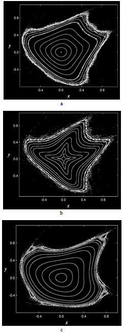
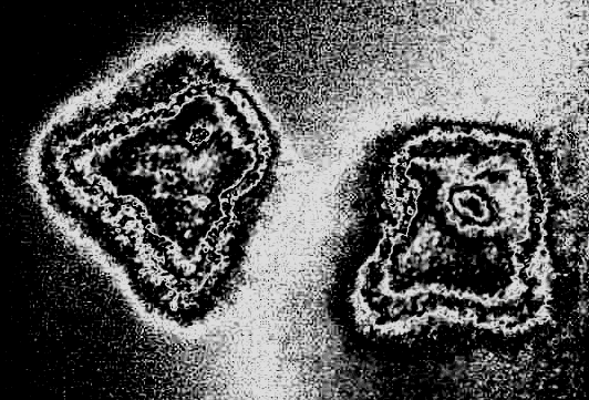
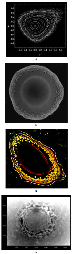
= 0.5, I = diag (2,3,1), r0 = (1, 1, 0). This map, as well as the above equation of a polyharmonic pendulum, can approximate the shape of the cell colonies shown in Fig. 5c. Comparing the system of resonances (5b) and the phase portrait of polyharmonic pendulum (5a), one can conclude that the colony formation is due to cell division. The structure reminiscent of the Cassini ovals or their special case – lemniscate of Bernoulli (Fig. 5a) is a structure-approximant for the fission process of artificial cells. This finding is under a strong mathematical foundation.
Figure 5. Simulation of colonial forms of association. a-c - elementary mathematical modelling; d - artificial cell's colonial associations

4. Philopodial Locomotion and Morphogenesis of Artificial Cells Models

Many “artificial cells” obtained on the basis of polyelectrolyte or electrically-activated media (see, for example,[22]), tend to move in space. Locomotor activity of "artificial cells" in most cases is carried out through the formation of pseudopodia. The emergence and changes of the pseudopodia forms can be described using the Hénon map:
(8)
and visualized in the dynamics through the behaviour of invariant curves of the map T in the variation of the parameter p in a limited range.
Fig. 6 a-c show the invariant curves of the Henon map with decreasing of the parameter p, as Fig. 7 shows map of the gradient for the image of "artificial cells” with pseudopodia. One can see that the "cell" on the left (in Fig. 9) reveals the form shown in Fig. 8 in the top row, and the "cell" on the right - the transformation tendency of the lower row.
In more complex cases, the Hénon map is able to simulate the dynamics of multicellular systems and systems of multiple artificial cells. Consider the map
(9)
When p ≥ 5.6 (i.e. already beyond the formation of pseudopodia by individual cells) it forms the structures with nested contours, which are comparable with the results of the morphogenesis modelling in the systems of "artificial cells" under the influence of environmental factors (such as microwave irradiation, changes in temperature of the carrier).
Fig. 8a shows the invariant curves of the Hénon map for p = 5.6. One can observe the internal structure of the curves and the distinct central organization, little dependent on the external contour deformities. Fig. 8 b shows the phase-contrast micrograph of the product of induced "artificial cells" association into an undifferentiated multicellular structure. A central (matrix) contour and the peripheral single cells are clearly visible. A more highly organized sample is given in Fig. 8 c: it shows the results of contour visualization of a synthetic "multicellular" structure obtained in the same way, but at higher specific concentration of cells - products of precipitation. Visualization is made via the method of Kirsch-Sobel-Prewitt in RGB-tone mode.
As in the previous micrographs, one can observe a clear central contour, independent of the membrane deformations (in this case they are stronger than in Fig. 8 b), “artificial cells”, located on the edges of the structure, and a diffuse contour of the membrane, as in Fig. 8 a. Fig. 8 d shows an interference micrograph of a cellular substructure, which is the basis of multicellular products self-organization. Thus, the Hénon map allows to visualize the morphogenesis of such structures and to reconstruct the evolution processes of primitive Metazoa and their predecessors.
Figure 6. Locomotor activity of “artificial cells” and the appearance of pseudopodia (dynamic models based on the Henon map)
Figure 7. “Artificial cells” with pseudopodia
Figure 8. . a - the Henon map, р=5.6; b - “multicellular" structure; c - real inorganic “multicellular structure”, d - cellular substructure (interference micrograph)

5. Kepler System & Morphogenetic Errors

In the case of incorrect implementation of the mathematical problem, the structure of morphogenesis will be distorted. Failure in management of the morphogenesis can be considered as a result of incorrect analytical implementation of the problem. For example, it was previously shown that under certain conditions associated with the introduction of irrational values into the model, programmed using numerical methods, there are additional contours, whose existence doesn’t possess any physical content. However, for some models this effect is a direct consequence of the problem and it can be analytically identified in the formalism. Consider a Kepler system, which particularly describes the motion of particles and micro-objects in a gravitational field. Strictly speaking, it is also valid for biological microstructure. It overcomes the resistance of a dense incompressible medium during morphogenesis and cytokinesis during growth in condensed medium.
Figure 9. a - the result of incorrect computer simulation of cytodieresis, b - the structure of resonances in the Kepler system at p1= 1, p2= 1, p3= – 0.01, p4= 1
Formalization of the problem may have the form:
(10)
or
(11)
Fig. 10 provides the resonances structure in the Kepler system at p1 =1, p2 = 1, p3 = – 0.01, p4 = 1. There is a certain affinity between Fig. 10 and a defect of cytodieresis shown in Fig. 9. Typically, a cell moving in a condensed medium, undergoes elastic deformation and generates a "wakefield" as shown in Fig. 9 a. In Fig. 9 b it is supplemented by the formation of resonance structures in the plume area.
Figure 10. A tissue model based on the Chirikov map and its real analogue

6. Visualization and Modelling of Histological Structures

The transition to multicellular structures involves the emergence of tissue organization[23]. If the mathematical mechanism of transition between the models of Protozoa and Metazoa really exists, there must exist a formally defined mechanism for transition from a single cell to a tissue. This transition can be accomplished by the Chirikov map, in which the degenerate fixed periodic points arise without degenerate resonances. Consider the following map:
(12)
where n = 0, 1, 2 etc. In the simplest case the Chirikov map, obtained from the study of stability of the charged particles motion in magnetic traps has the form
(13)
The parameter K determines the width of ergodic layer, separating the captured particles from the migrating ones. In the case of biological models – external particles from the components of cellular organization.
Figure 10. A tissue model based on the Chirikov map and its real analogue
Fig. 10 a gives the trajectories of this map at p1 = 0.4, p2 = 2, p3 = 0. Next, Fig. 10 b shows the contour image processing of an artificial tissue unit, considered earlier. (Green indicates the cell clusters).

7. Prospects of Embryological Simulation

Figure 11 a. Natural gastrulation in biological embryogenesis
Figure 11 b. Simulation of gastrulation on the basis of Duffing equations
Successful modelling of cytological and histological structures by means of regular and chaotic dynamics, both in theoretical and experimental forms, gives the prerequisites for the development of these methods in the future. Mathematical theory outstrips the development of the experiment. Therefore it is useful to analyse the potential opportunities arising from the development of the theory. Particularly the transition from morphogenesis to histogenesis is hierarchically justified. Despite the specific nature of morphogenesis in model chemical systems and their inconsistency to biological objects that implement this function completely, it makes sense to point to the invariant sets of biomorphic dynamical systems.
In this aspect the most advanced is the mathematical apparatus based on the Duffing equations:
where - 
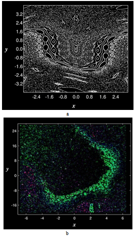
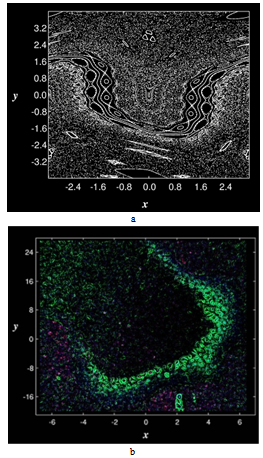
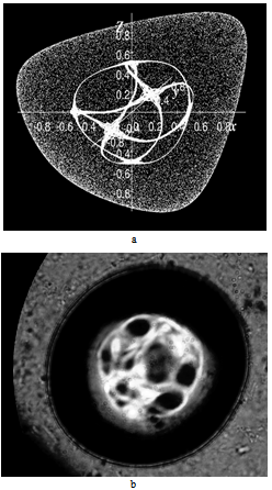
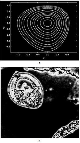
parameters and ε - a small parameter. These parameters theoretically lead to a specific type of invariant curves behaviour, which, in terms of morphology, forms the structures similar to embryonic sections in the early stages of morphogenesis - in particular, at the stage of gastrulation (Fig. 11 a, b).

8. Discussion

This paper compares graphical visualization of invariant sets of nonlinear dynamical systems with the results of experiments on the physico-chemical synthesis of biomimetic structures. Since we simulate the deformation, locomotion and morphogenesis of cellular structures, the results should be compared with biological prototypes despite the fact that in this paper two-dimensional projections are mostly considered.
a) Simulation of synthetic protoplast form. Initially the cytoplasmic structure of artificial cells was simulated. Inorganic cells were obtained as giant vesicles by a method, similar to that described in[24]. The vesicle size was limited by reaction-diffusion mechanisms due to semipermeable properties of inorganic membranous structures. This allowed to simulate the growth of biological cells (on the control of cell growth see[25]) and morphogenesis of cell organelles[26], which is also limited by similar reaction-diffusion processes associated with cell membranes. In particular fig. 1 apparently shows the analogue of cytoskeleton, which is the basis of cell differentiation and development[27] together with cell motility[28]. This implies the possibility of obtaining of morphologically differentiated iCHELLs with a primitive spherical symmetry. The exact morphological analogue of intercellular structures, shown in fig. 1, are known to be found in protozoa[29].
Fig. 3 shows artificial cells adhered to the surface[30], which undergo deformation according to hydrodynamic laws, which are the same for biological cells and porous capsules[31, 32]. This is consistent with the kinetic theory of living patterns formation[33], as well as the modern concepts of cell differentiation on different surfaces[34] and interfacial interactions in biological structures[35]. From the standpoint of biomechanics[36], such behaviour can become a basis of the changes in the shape and direction of growth of cellular structures, similar to those shown in Fig. 3.
b) Computer simulation of colonial associated forms. The adhesion to the surface in physico-chemical parameters is not much different from the cell adhesion, except for the molecular specificity[30]. Therefore in the presence of specific binding between iCHELLs, which is inevitable due to the similarity in the composition and structure of their membranes, the formation of colonies and associated structures is rather possible. These are the same forces that underlie morphogenesis of organs from different cells[37] and correspond to the biophysical concepts of embryogenesis[38]. As it was noted in earlier studies, the development of cells in artificial media is characterized by another phenomenology of colony formation on the surface[39]. Fig. 5d shows inorganic cellular adhesion, which leads to the colony formation of the artificial analogues of aggregated erythrocytes[40]. This phenomenon is based on the rheological similarity of the surface properties between iCHELLs and biological surfactants[41].
c) Philopodial locomotion and simulation of morphogenesis of artificial cells. The above statement implies the mechanical possibility of pseudopodia formation. This statement does not contradict the above mentioned possibility of the formation of cellular assemblies[42] and elementary interactions between the ameboid cells, according to protozoology[43]. Fig.6 shows the results of simulation and fig. 7 - the micrographs of the obtained iCHELLs with pseudopodia in motion with the observed sol-gel transitions at the edges of pseudopodia; fig. 8 illustrates the formation of "multicellular" structures with a common membrane from individual iCHELLs. This can be considered either as an analogue of compartmentalisation[26], depending on the scale, that is the size of the structures, based on vesicles, or as the emergence of elementary multicellular structures, characteristic of invertebrate embryogenesis and ontogenesis[44] and their occurrence in phylogeny[45]. It should be noted that at present moment it is impossible to compare specific structures as microreactors with the functions performed by them. However the simulation of organelles, carried out by WINSET, as well as the experimentally obtained iCHELLs with such functions, can be considered as a metaphor of biological properties of the prototype[46], but not as a simple morphological[47] or an abstract modelling of morphogenesis[48] and the resulting structures. Making visualization of invariant sets of nonlinear dynamic systems, it is closer to the dynamic modelling in biology[49]. However, it does not consider the genetic and molecular biological basis of morphogenesis[50], operating, as[51], only with the second part of morphogenesis - the shape formation without reducing it to vulgar hypothetical models like[52, 53]. Therefore, motility and other biomimetic properties of iCHELLs are considered here not as a phenomenon, but as a result of the similarity of mechanisms underlying the properties of biomolecular medium and colloidal soft matter. Chaotic motion is generally characteristic of nonlinear systems[54], while random walks are inherent in biological objects[55], so if we consider iCHELLs as dynamic models, the simulation of their locomotion will become possible.
d) Visualization and modelling of histological structures. It's obvious that using the elements of a cellular type, generated by WINSET, it is possible to create models of multicellular structures, such as tissues, as it is done in many approaches to biomodel pattern formation (see, e.g.[56, 57]). We have implemented the above approach using WINSET and iCHELLs (see fig. 10). From the standpoint of membrane mimetics[58], it is associated with the ability to transport[59] - transmembrane transport between iCHELLs membranes, similar to biological transport[60, 61]. This is due to cell adhesion, because such adhesion and cell contacts are well known determinants of morphogenesis[62].
e) Prospects of embryological simulation. In this regard, there is a possibility of morphogenesis modelling with both WINSET and iCHELLs (the latter is still in the boundless future). Since multicellular structures based on iCHELLs are rather stable (for example, see fig. 8d), they satisfy the requirements for potential participants in embryogenesis. There are parallels between the results of a simplified computer simulation of histogenesis (fig. 10 а) and gastrulation processes (fig. 11 b) with WINSET, the formation of distorted tissues[63], modelled on iCHELLs (fig. 10 b), and a real optical micrograph of the gastrula[64] (fig. 11 а).
The author did not aim to review the current state of the problems of morphogenesis because today there is a lot of monographic literature on this subject cited in the review. In conclusion we would like to note the paper, which confirms the membranological/membranomimetic consequences of our modelling in the membranopaty simulation[65]

References

[1]  Noireaux V., Libchaber A. A. Vesicle bioreactor as a step toward an artificial cell assembly, PNAS, Vol. 101, pp. 17669-17674 (2004).
[2]  Noireaux V., Bar-Ziv R., Godefroy J., Salman H., Libchaber A. Toward an artificial cell based on gene expression in vesicles. Phys. Biol., Vol. 2, No 3, pp. 1-8 (2005).
[3]  Murtas G., Construction of a Semi-Synthetic Minimal Cell: A Model for Early Living Cells. Origins of Life and Evolution of Biospheres, Vol. 37, No 4-5, pp. 419-422 (2007).
[4]  Lartigue C., Glass J. I., et al. Genome Transplantation in Bacteria: Changing One Species to another. Science, Vol. 317, pp. 632-638 (2007).
[5]  Gibson D.G., Glass J.I., et al. Creation of a Bacterial Cell Controlled by a Chemically Synthesized Genome. Science, Vol. 329, pp. 52-56 (2010).
[6]  Rasmussen S., Chen L., Deamer D., Krakauer D., Packard N., Stadler P., Bedau M. Transitions from nonliving to living matter. Science, Vol. 303, pp. 963-965 (2004).
[7]  Müller A., Roy S. Multifunctional Metal Oxide Based Nanoobjects: Spherical Porous Capsules/Artificial Cells and Wheel-Shaped Species with Unprecedented Materials Properties. J. Mater. Chem., Vol. 15, p. 4673-4677 (2005).
[8]  Müller A., Toma L., et al. Reactions inside a porous nanocapsule/artificial cell: encapsulates’ structuring directed by internal surface deprotonations. Chem. Commun., Vol. 32, pp. 3396–3398 (2006).
[9]  Cooper G.J.T., Kitson P.J., Winter R.S., Zagnoni M., Long D-L., Cronin L. Modular Redox-Active Inorganic Chemical Cells: iCHELLs. Angew. Chem. Int. Ed., Vol. 50, pp. 10373-10376 (2011).
[10]  Mansy S.S., Szostak J.W. Reconstructing the Emergence of Cellular Life through the Synthesis of Model Protocells. Cold Spring Harb. Symp. Quant. Biol., Vol. 74, pp. 47-54 (2009).
[11]  Mansy S.S., Schrum J.P., Krishnamurthy M., Tobé S., Treco D.A., Szostak J.W. Template-directed synthesis of a genetic polymer in a model protocell. Nature, 2008, Vol. 454, pp. 122-125.
[12]  Deamer D. On the origin of systems. Systems biology, synthetic biology and the origin of life. EMBO Rep., No. 10(S1), pp. S1-S4 (2010)
[13]  Turing A. M. The Chemical Basis of Morphogenesis. Phil. Trans. R. Soc. B, Vol. 237, No 641, pp. 37-72 (1952).
[14]  Haddad W.M., Chellaboina V.S., Hui Q. Nonnegative and Compartmental Dynamical Systems, 624 p., Princeton University Press, 2010.
[15]  Haddad W.M., Nersesov S.G. Stability and Control of Large-Scale Dynamical Systems: A Vector Dissipative Systems Approach, 392p., Princeton University Press, 2011.
[16]  Smale S., Hirsch M.W., Devaney R.L. Differential Equations, Dynamical Systems, and an Introduction to Chaos, Second Edition, 425 p., Academic Press, 2003.
[17]  Strogatz S.H. Nonlinear Dynamics And Chaos: With Applications To Physics, Biology, Chemistry, And Engineering, 512 p., Westview Press, 2001.
[18]  Wiggins S. Introduction to Applied Nonlinear Dynamical Systems and Chaos, 808 p., Springer, 2010.
[19]  Feudel U., Kuznetsov S., Pikovsky A. Strange Nonchaotic Attractors: Dynamics Between Order And Chaos in Quasiperiodically Forced Systems, 228 p., World Scientific Pub Co Inc, 2006.
[20]  Tel T.., Lai Y.C. Transient Chaos: Complex Dynamics on Finite Time Scales, 512 p., Springer, 2011.
[21]  Cooper G.J.T., Kitson P.J., Winter R., Zagnoni M., Long D.-L., Cronin L. Modular Redox-Active Inorganic Chemical Cells: iCHELLs. Angew. Chem. Int. Ed., Vol. 50, Issue 44, pp. 10373–10376 (2011).
[22]  Toyota T., Maru N., Hanczyc M.M., Ikegami T., Sugawara T. Self-Propelled Oil Droplets Consuming “Fuel” Surfactant. J. Am. Chem. Soc., 2009, 131 (14), pp. 5012–5013.
[23]  Kaushal M., Singh A., Singh B. Adaptive Thresholding for Edge Detection in Gray Scale Images. International Journal of Engineering Science and Technology, Vol. 2(6), pp. 2077-2082 (2010).
[24]  Giant Vesicles: Perspectives in Supramolecular Chemistry (Ed. by P.L. Luisi, P. Walde), 426 p., Wiley-Interscience, 2000.
[25]  Cell Growth: Control of Cell Size (Ed. by M.N. Hall, R. Martin, G. Thomas), 652 p., Cold Spring Harbor Laboratory Press, 2004.
[26]  Mullins C. The Biogenesis of Cellular Organelles, 192 p., Springer, 2010.
[27]  Plant Cytoskeleton in Cell Differentiation and Development (Ed. by P. Hussey), 325 p., Blackwell, 2004.
[28]  Preston T.M., King C.A., Hyams J.S. Cytoskeleton and Cell Motility, 202 p., Chapman & Hall, 1990.
[29]  The Cytoskeleton of Flagellate and Ciliate Protists (Ed. by M. Melkonian, R.A. Andersen, E. Schnepf), 174 p., Springer, 2001.
[30]  Kendall K., Kendall M., Rehfeldt F. Adhesion of Cells, Viruses and Nanoparticles, 297 p., Springer, 2010.
[31]  Pozrikidis C. Modeling and Simulation of Capsules and Biological Cells, 344 p., Chapman and Hall, 2003.
[32]  Pozrikidis C. Computational Hydrodynamics of Capsules and Biological Cells, 327 p., CRC Press, 2010.
[33]  Harrison L.G. Kinetic Theory of Living Pattern, 376 p., Cambridge University Press, 2005.
[34]  Muramatsu T. Cell Surface and Differentiation,172 p., Springer, 1990.
[35]  Interfacial Phenomena in Biological Systems (Ed. by M. Bender), 448 p., CRC Press, 1991
[36]  Fung Y.C. Biomechanics: Motion, Flow, Stress, and Growth, 607 p., Springer, 1990.
[37]  Trinkaus J.P. Cells into Organs: The Forces That Shape the Embryo, 560 p., Prentice Hall College, 1984.
[38]  Forgacs G., Newman S.A. Biological Physics of the Developing Embryo, 346 p., Cambridge University Press, 2005.
[39]  Willmer E.N. Tissue culture: The growth and differentiation of normal tissues in artificial media, 126 p., Methuen & Co., 1935.
[40]  Gulati G., Caro J. Blood Cells An Atlas of Morphology with Clinical Relevance, 236 p., American Society for Clinical Pathology, 2007.
[41]  Biosurfactants: From Genes to Applications (Ed. by G. Soberon-Chavez), 223 p., Springer, 2010.
[42]  Kessin D.H. Dictyostelium: Evolution, Cell Biology, and the Development of Multicellularity, 308 p., Cambridge University Press, 2001.
[43]  Bonner J.T. The Social Amoebae: The Biology of Cellular Slime Molds, 156 p., Princeton University Press, 2008.
[44]  Sawyer R.H., Showman R.M. The Cellular and Molecular Biology of Invertebrate Development, 339 p., University of South Carolina Press, 1985.
[45]  Minelli A. Forms of Becoming: The Evolutionary Biology of Development, 242 p., Princeton University Press, 2009.
[46]  Keller E.F. Making Sense of Life: Explaining Biological Development with Models, Metaphors, and Machines, 400 p., Harvard University Press, 2003.
[47]  Sharpe J., Lumsden C.J., Woolridge N. In Silico: 3D Animation and Simulation of Cell Biology with Maya and MEL, 656 p., Morgan Kaufmann, 2008.
[48]  Deutsch A., Dormann S. Cellular Automaton Modeling of Biological Pattern Formation, 300 p., Birkhäuser Boston, 2004.
[49]  Ellner S.P., Guckenheimer J. Dynamic Models in Biology, 352 p., Princeton University Press, 2006.
[50]  Davies J. Mechanisms of Morphogenesis, 385 p., Academic Press, 2005.
[51]  Stewart I. Life's Other Secret: The New Mathematics of the Living World, 285 p., Wiley, 1999.
[52]  Pivar S. On the Origin of Form: Evolution by Self-Organization, 208 p., North Atlantic Books, 2009.
[53]  Pollack G.H. Cells, Gels and the Engines of Life, 320 p., Ebner & Sons, 2001.
[54]  Szemplinska-Stupnicka W., Iooss G., Moon F.C. Chaotic Motions in Nonlinear Dynamical Systems, 198 p., Springer, 1988.
[55]  Berg H.C. Random Walks in Biology, 164 p., Princeton University Press, 1993.
[56]  Mathematical Models for Biological Pattern Formation (Ed. by P.K. Maini, H.G. Othmer), 327 p., Springer, 2000.
[57]  Harrison L.G. The Shaping of Life: The Generation of Biological Pattern, 272 p., Cambridge University Press, 2011
[58]  Fendler J.H. Membrane Mimetic Chemistry: Characterizations and Applications of Micelles, Microemulsions, Monolayers, Bilayers, Vesicles, Host-Guest Systems, and Polyions, 522 p., John Wiley & Sons, 1982.
[59]  Perthame B. Transport Equations in Biology, 206 p., Birkhäuser Basel, 2006.
[60]  Cell-Cell Channels (Ed. by F. Baluka, D. Volkmann, P.W. Barlow), 339 p., Springer, 2006
[61]  Cell-Cell Junctions (Ed. by J.W. Nelson, E. Fuchs), 443 p., Cold Spring Harbor Laboratory Press, 2010
[62]  Edelman G.M., Thiery J.-P. The Cell in Contact: Adhesions and Junctions as Morphogenetic Determinants, 516 p., Wiley, 1986
[63]  Thom R. Structural Stability And Morphogenesis, 400 p., Westview Press, 1994.
[64]  Slack J.M. From Egg to Embryo: Regional Specification in Early Development, 352 p., Cambridge University Press, 1991.
[65]  Gradov O., Gradova M., Rybakov S., Choon S.J. Inorganic biomimetics for clinical hematology. Medical and Health Science Journal, Vol. 2, pp. 10 -17 (2010).