
-
Paper Information
- Paper Submission
-
Journal Information
- About This Journal
- Editorial Board
- Current Issue
- Archive
- Author Guidelines
- Contact Us
Advances in Life Sciences
p-ISSN: 2163-1387 e-ISSN: 2163-1395
2026; 14(1): 1-11
doi:10.5923/j.als.20261401.01
Received: Jun. 8, 2026; Accepted: Jun. 29, 2026; Published: Jul. 10, 2026

Assessment of Liver Function, Kidney Function, and Lipid Metabolism in Fasciola-Infected Male Cattle Slaughtered at the Main Abattoirs of Buea, Cameroon
 Abstract
 Reference
 Full-Text PDF
 Full-text HTML
Mbafor Fidelia Lem1, Jubin Osei-Mensah2, 3, Ngeh Frankline Konfor1, Archille Paguem1, Naihibu Musa Ndemsah1, Arrey Oben Ebob Ashu4, Tiencheu Bernard4, Manchang Tanyi Kingsley1
1Department of Veterinary Medicine, Faculty of Agriculture and Veterinary Medicine, University of Buea, Cameroon
2Department of Pathobiology, School of Veterinary Medicine, KNUST, Kumasi, Ghana
3Kumasi Centre for Collaborative Research in Tropical Medicine, KNUST, Kumasi, Ghana
4Department of Biochemistry and Molecular Biology, Faculty of Science, University of Buea, Buea, Cameroon
Correspondence to: Mbafor Fidelia Lem, Department of Veterinary Medicine, Faculty of Agriculture and Veterinary Medicine, University of Buea, Cameroon.
| Email: |  |
Copyright © 2026 The Author(s). Published by Scientific & Academic Publishing.
This work is licensed under the Creative Commons Attribution International License (CC BY).
http://creativecommons.org/licenses/by/4.0/

Fascioliasis, caused by Fasciola hepatica, is one of the most important parasitic diseases of ruminants worldwide and a neglected zoonotic disease of public health significance. The infection causes substantial economic losses through reduced productivity, organ condemnation, and impairment of physiological functions, particularly those involving the liver, kidneys, and lipid metabolism. Despite its importance, limited information exists on the prevalence and biochemical effects of fascioliasis among cattle slaughtered in Fako Division, Cameroon. This study aimed to determine the prevalence of Fasciola hepatica infection and assess its effects on liver function, kidney function, and lipid metabolism in cattle slaughtered in the municipal abattoirs of Buea, Tiko, and Limbe in Fako Division, Cameroon. A cross-sectional study was conducted from September 2024 to February 2025 involving 565 cattle slaughtered in the municipal abattoirs of Buea, Tiko, and Limbe. Fecal samples were collected post-slaughter and examined using the sedimentation technique for the detection of F. hepatica eggs. Blood samples were collected for biochemical analyses including liver enzymes (ALT, AST, and ALP), kidney function indicators (uric acid and creatinine), protein profile (albumin, total protein, and globulin), and lipid profile parameters (total cholesterol, triglycerides, HDL, and LDL). Data were analyzed using descriptive statistics and Pearson’s Chi-square test at a significance level of P < 0.05. Of the 565 cattle examined, 342 were positive for F. hepatica, resulting in an overall prevalence of 60.5%. Infection prevalence was 60.4% (336/556) in males and 66.7% (6/9) in females, with no significant association between sex and infection (χ² = 1.44, P = 0.740). Young cattle showed a higher prevalence (74.1%; 53/85) than adults (60.2%; 289/480), although the difference was not statistically significant (χ² = 0.603, P = 0.740). A significant difference was observed among subdivisions (χ² = 7.427, P = 0.024), with Buea recording the highest prevalence (62.4%), followed by Tiko (59.8%) and Limbe (56.5%). Biochemical analyses revealed elevated liver enzyme activities, particularly AST, which reached 375.63 ± 103.13 U/L in Limbe. Mean ALT concentrations ranged from 39.68 ± 9.03 to 48.64 ± 18.14 U/L, while ALP values ranged from 31.84 ± 12.88 to 56.47 ± 12.02 U/L. Uric acid concentrations ranged from 2.48 ± 1.34 to 4.23 ± 1.90 mg/dL and creatinine concentrations from 0.72 ± 0.08 to 0.99 ± 0.72 mg/dL. Lipid profile analysis showed cholesterol concentrations ranging from 56.68 ± 13.03 to 116.82 ± 30.89 mg/dL and triglyceride concentrations ranging from 26.63 ± 4.43 to 41.74 ± 17.43 mg/dL. Albumin concentrations ranged from 2.40 ± 0.33 to 3.09 ± 0.59 g/dL, whereas total protein and globulin concentrations were elevated across study sites. Fasciola hepatica infection was highly prevalent among cattle slaughtered in Fako Division and was associated with significant alterations in biochemical indicators of liver function, kidney function, protein metabolism, and lipid metabolism. These findings highlight the need for strengthened fascioliasis control strategies, routine surveillance, and improved livestock management practices.
Keywords: Fasciola hepatica, Fascioliasis, Prevalence, Liver function, Kidney function, Lipid profile, Cattle, Fako Division, Cameroon
Cite this paper: Mbafor Fidelia Lem, Jubin Osei-Mensah, Ngeh Frankline Konfor, Archille Paguem, Naihibu Musa Ndemsah, Arrey Oben Ebob Ashu, Tiencheu Bernard, Manchang Tanyi Kingsley, Assessment of Liver Function, Kidney Function, and Lipid Metabolism in Fasciola-Infected Male Cattle Slaughtered at the Main Abattoirs of Buea, Cameroon, Advances in Life Sciences, Vol. 14 No. 1, 2026, pp. 1-11. doi: 10.5923/j.als.20261401.01.
Article Outline
1. Introduction
- Fasciolosis is a major parasitic disease of livestock caused by trematodes of the genus Fasciola, principally Fasciola hepatica and Fasciola gigantica, which infect a wide range of domestic and wild ruminants worldwide [1]. The disease is recognized as one of the most important helminth infections affecting cattle because of its significant impact on animal health, productivity, and economic returns to livestock farmers [2]. Globally, fasciolosis affects more than 600 million domestic animals and causes annual economic losses estimated at several billion US dollars through reduced meat production, decreased milk yield, liver condemnation, poor growth performance, reduced fertility, and increased susceptibility to secondary infections [3]. In tropical and subtropical regions, favorable climatic conditions support the survival and multiplication of intermediate host snails, thereby increasing transmission rates among grazing livestock populations [1]. The disease has become an important veterinary and public health concern because it is classified as a neglected tropical disease and has zoonotic significance in many parts of the world [4].In Africa, fasciolosis remains highly prevalent due to extensive livestock management systems, abundant rainfall, and the widespread distribution of lymnaeid snail intermediate hosts [5]. Several studies conducted in sub-Saharan Africa have reported prevalence rates ranging from 20% to over 70% depending on geographical location, season, husbandry practices, and diagnostic methods employed [6,7]. In Cameroon, livestock production contributes significantly to food security and household income; however, parasitic diseases such as fasciolosis continue to undermine productivity and profitability within the cattle sector [8]. The humid ecological conditions of the South West Region, particularly within Fako Division, provide suitable habitats for the development of intermediate host snails and the maintenance of transmission cycles throughout much of the year [9].The liver is the primary target organ affected by Fasciola hepatica infection. During migration through the hepatic parenchyma, juvenile flukes induce mechanical destruction of liver tissues, hemorrhage, necrosis, and inflammatory reactions, while adult parasites residing in the bile ducts cause cholangitis, hyperplasia, fibrosis, and biliary obstruction [2]. These pathological alterations can result in significant changes in serum biochemical parameters commonly used to assess liver function. Elevated activities of liver enzymes such as alanine aminotransferase (ALT), aspartate aminotransferase (AST), alkaline phosphatase (ALP), and gamma-glutamyl transferase (GGT) have been reported in Fasciola-infected cattle, reflecting hepatocellular damage and biliary dysfunction [10,11]. Consequently, biochemical evaluation of liver function serves as an important tool for assessing the physiological consequences of fasciolosis in infected animals [12].Although the liver is the principal organ affected, increasing evidence suggests that fasciolosis may also influence kidney function through systemic inflammatory responses, oxidative stress, altered protein metabolism, and circulating toxic metabolites associated with chronic parasitic infection [13]. Renal function indicators such as serum urea, creatinine, and uric acid concentrations may become altered in infected animals, reflecting changes in glomerular filtration and metabolic homeostasis [14]. Studies have demonstrated significant differences in kidney biochemical markers between infected and non-infected cattle, suggesting that the disease exerts effects beyond the hepatobiliary system [11]. Therefore, assessment of renal function provides valuable information regarding the systemic impact of fasciolosis on animal health and physiological status.Lipid metabolism is another important physiological process that may be affected by Fasciola hepatica infection. The liver plays a central role in lipid synthesis, transport, storage, and metabolism; therefore, hepatic damage caused by fasciolosis can lead to disturbances in lipid profiles [14]. Previous studies have reported alterations in serum concentrations of total cholesterol, triglycerides, high-density lipoproteins (HDL), and low-density lipoproteins (LDL) in cattle infected with liver flukes [15]. These alterations may result from impaired hepatic function, reduced feed utilization efficiency, inflammatory responses, and increased metabolic demands associated with chronic parasitic infection [10]. Evaluation of lipid profiles may therefore provide useful insights into the metabolic consequences of fasciolosis and its effects on animal productivity.Age is recognized as an important epidemiological factor influencing the prevalence and severity of fasciolosis in cattle populations. Older cattle are generally exposed to infective stages for longer periods and may therefore accumulate higher parasite burdens compared with younger animals [16]. Several studies have demonstrated significant associations between age and fasciolosis prevalence, emphasizing the importance of considering age when evaluating disease epidemiology and its physiological consequences [7,6]. Understanding age-related patterns of infection is therefore essential for developing targeted control and management strategies.Abattoir-based studies are particularly valuable in regions where routine farm-level disease monitoring is limited, as they provide reliable estimates of disease prevalence and associated pathological changes [17]. The municipal abattoirs of Buea, Tiko, and Limbe serve as major slaughter facilities within Fako Division and receive cattle from various parts of Cameroon, making them suitable locations for assessing the epidemiological and biochemical impacts of fasciolosis.Despite the economic and veterinary importance of fasciolosis, limited information exists regarding the effects of Fasciola hepatica infection on liver function, kidney function, and lipid metabolism among slaughtered cattle in Fako Division, Cameroon. Furthermore, few studies have simultaneously evaluated the relationship between infection status, biochemical alterations, and age-related differences within cattle populations in the region. Such information is necessary for understanding the broader health implications of fasciolosis and for informing effective disease control programs.Therefore, this study aimed to assess the prevalence of Fasciola hepatica infection and evaluate its effects on liver function, kidney function, and lipid metabolism among male cattle slaughtered in the municipal abattoirs of Buea, Tiko, and Limbe in Fako Division, South West Region of Cameroon, while taking into account the age of the cattle. The findings of this study are expected to contribute to the understanding of the epidemiology and pathophysiological effects of bovine fasciolosis and provide evidence for improved disease surveillance, prevention, and control strategies in Cameroon.
2. Materials and Methods
2.1. Study Period and Area
- This study was conducted between September 2024 and February 2025 in Fako Division, located in the South West Region of Cameroon. Geographically, Fako Division lies between latitudes 4°0′ and 4°0.5′ North and longitudes 9°10′ and 9°13′ East. The division comprises seven subdivisions: Limbe I, Limbe II, Limbe III, Tiko, Buea, Muyuka, and Idenau. Covering an area of approximately 2,093 km², it includes elevations reaching up to 2,833 m above sea level.Fako Division experiences a tropical climate characterized by rainforest, savannah, and mountainous ecosystems, which provide favorable conditions for livestock production. The area has two distinct seasons: a rainy season extending from March to October and a dry season from November to February. Average temperatures range from 20°C to 28°C, while annual rainfall varies between 4,000 mm in lowland areas and 10,000 mm at higher altitudes. Relative humidity ranges from 75% to 85%. According to recent demographic data, the division had an estimated population of 466,412 inhabitants in 2021, with major urban centers including Buea, Limbe, and Tiko [18].
2.2. Study Animals
- The study population consisted of both male and female cattle slaughtered at three municipal abattoirs within Fako Division. These animals were primarily sourced from the Adamawa and North Regions of Cameroon and supplied for local meat consumption. The cattle were reared under an extensive production system, allowing free grazing on natural pastures. Animals of different local breeds and age categories were included in the study.For analytical purposes, cattle were classified into two age groups based on dentition: young animals (<3 years) and adults (>5 years). This classification was used to evaluate the occurrence of bovine fascioliasis across different age categories.
2.3. Study Design
- A cross-sectional study design was employed from September 2024 to February 2025 in three municipal abattoirs located within Fako Division. The objectives were to determine the prevalence of bovine fascioliasis and estimate the direct economic losses associated with liver condemnation during routine meat inspection.The selected abattoirs were located in Buea, Limbe I, and Tiko subdivisions. Buea serves as the administrative headquarters of the South West Region, while Limbe and Tiko are major coastal and tourism centers. Owing to their large human populations and commercial activities, these municipalities record relatively high cattle slaughter rates, making them suitable sites for epidemiological investigations.
2.4. Sampling Method and Sample Size Determination
- A simple random sampling technique was employed during the abattoir survey to select study animals for examination. The required sample size was determined using the formula described by Thrusfield [19], assuming an expected prevalence of 50%, a desired absolute precision of 5%, and a confidence level of 95%.The sample size was calculated according to the following formula:
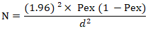 Where: N= Required sample size; Pex = Expected prevalence; d= Desired absolute precision = 5% = 0.05; 1.96= Constant for 95% confidence interval.For the cattle examine for Fascioliasis, the sample size (N) was calculated as follows:
  Given N = 268, Thus, 565 fecal samples were collected and livers inspected to maximized precision.
2.5. Sample Collection
2.5.1. Fecal Sample Collection
- To determine the parasitic burden of Fasciola spp. eggs in cattle, fecal samples were collected immediately after slaughter directly from the rectum of each animal using sterile disposable gloves. Approximately 10–20 g of fecal material was placed into sterile, labeled containers indicating the date of collection, sex, age, breed, and species of the animal. Age determination was performed based on dentition, considering the eruption of permanent teeth, and animals were subsequently classified as young or adult following the criteria described by Muylle [20].Samples were preserved in a cooler maintained at approximately 4°C and transported to the Veterinary Parasitology Laboratory, Faculty of Agriculture and Veterinary Medicine, University of Buea, for analysis within 24 hours of collection. When immediate examination was not feasible, samples were refrigerated at 3–5°C for a maximum period of 48 hours before processing [21].
2.5.2. Blood Sample Collection and Serum Preparation
- Blood samples were collected exclusively from male cattle, as female animals were generally retained for breeding and reproductive purposes and were therefore rarely slaughtered. During exsanguination, approximately 10 mL of blood was aseptically collected from each selected animal into plain vacutainer tubes without anticoagulant.The samples were transported in an ice-packed cooler to the Veterinary Clinical Pathology Laboratory of the Faculty of Agriculture and Veterinary Medicine, University of Buea. Blood samples were allowed to clot at room temperature for 30 minutes and subsequently centrifuged at 3,000 rpm for 10 minutes. The resulting serum was carefully separated, transferred into sterile cryovials, and stored at −20°C until biochemical analyses were conducted [14].
2.6. Analysis of Collected Specimens
2.6.1. Fecal Examination and Liver Inspection
- Fecal samples obtained from both male and female cattle were processed using the sedimentation technique described by Opio et al. [22]. Each sample was analyzed in triplicate, and the resulting sediments were combined in a Petri dish, stained with 1% methylene blue, and examined microscopically under 10× magnification for the detection of trematode eggs [23]. To improve analytical reliability, each sample was evaluated twice, and the mean value of the two observations was used for subsequent analysis.Post-mortem liver examination was performed immediately after slaughter and evisceration. Each liver was visually inspected, palpated, and incised to detect the presence of adult flukes, migratory tracts, or pathological lesions associated with fascioliasis, following established meat inspection procedures [24].
2.6.2. Determination of Liver Function Parameters (ALT, AST, and ALP)
- Serum alanine aminotransferase (ALT) and aspartate aminotransferase (AST) activities were determined using the kinetic enzymatic method recommended by the International Federation of Clinical Chemistry (IFCC) with commercially available Chronolab diagnostic kits (Spain). The assays are based on the enzymatic conversion of alanine and aspartate into pyruvate and oxaloacetate, respectively, followed by spectrophotometric monitoring of nicotinamide adenine dinucleotide (NADH) oxidation at 340 nm [25].Serum alkaline phosphatase (ALP) activity was measured using a colorimetric kinetic assay employing p-nitrophenyl phosphate as substrate. Under alkaline conditions, ALP hydrolyzes p-nitrophenyl phosphate to p-nitrophenol, and the resulting product was quantified spectrophotometrically at 405 nm [26].
2.6.3. Determination of Kidney Function Parameters
- Serum creatinine concentration was determined using the modified Jaffe colorimetric method, whereby creatinine reacts with picric acid in an alkaline medium to form a colored complex measurable at 520 nm [25].Serum uric acid concentration was measured using the enzymatic uricase-peroxidase method. In this assay, uric acid is oxidized by uricase to produce allantoin and hydrogen peroxide. The hydrogen peroxide subsequently reacts with chromogenic substrates in the presence of peroxidase to form a colored compound, the absorbance of which was measured at 546 nm [26].
2.6.4. Determination of Serum Albumin and Globulin Concentrations
- Serum albumin concentration was determined using the bromocresol green (BCG) colorimetric method. Under acidic conditions, albumin binds specifically to bromocresol green to form a green-colored complex, which was quantified spectrophotometrically at 630 nm [27].Total serum protein concentration was measured using the Biuret method, based on the formation of a violet-colored complex between peptide bonds and cupric ions in an alkaline solution. Absorbance was read at 540 nm [28].Serum globulin concentration was calculated indirectly by subtracting albumin concentration from total protein concentration according to the following equation:Globulin (g/dL) = Total Protein (g/dL) − Albumin (g/dL)[14].
2.7. Determination of Lipid Profile Parameters
- Serum total cholesterol concentration was determined using the cholesterol oxidase-peroxidase (CHOD–PAP) enzymatic method. Cholesterol esters were hydrolyzed by cholesterol esterase, and the liberated cholesterol was subsequently oxidized by cholesterol oxidase, generating hydrogen peroxide that reacted with a chromogenic substrate to form a colored compound measured at 500 nm [29].Serum triglyceride concentration was determined using the glycerol phosphate oxidase-peroxidase (GPO–PAP) enzymatic method. Triglycerides were hydrolyzed by lipase to glycerol and fatty acids, followed by enzymatic reactions producing a colored end product measured spectrophotometrically at 500 nm [30].High-density lipoprotein cholesterol (HDL-C) concentration was measured following selective precipitation of low-density and very-low-density lipoproteins, after which cholesterol in the supernatant was quantified using the CHOD–PAP method [31].Low-density lipoprotein cholesterol (LDL-C) concentration was estimated using the Friedewald equation:LDL-C (mg/dL) = Total Cholesterol − HDL-C − (Triglycerides/5)This calculation was applied only when serum triglyceride concentrations were below 400 mg/dL [32].
2.8. Statistical Analysis
- Data generated from the study were coded and entered into Microsoft Excel before being exported to IBM SPSS Statistics version 27.0.1 for analysis. Descriptive statistics were used to summarize the data, while Pearson’s Chi-square (χ²) test was employed to assess associations between the prevalence of bovine fascioliasis and potential risk factors. Statistical significance was established at a probability level of P < 0.05.
2.9. Ethical Clearance
- Ethical approval for this study was obtained from the Refotde Institutional Animal Ethics Committee (RIAEC) under certificate reference number 014/RIAE/2024, issued on 14 November 2024.
3. Results and Discussion
3.1. Results
3.1.1. Prevalence of Fasciola Hepatica Infection According to Sex, Age, and Subdivision
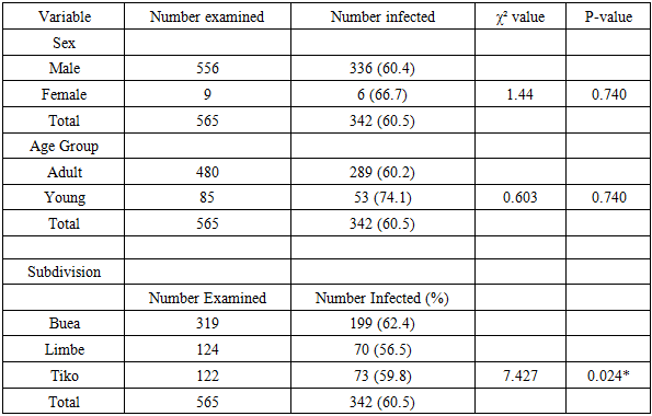
- Table 1 present the prevalence of bovine fasciola in the three municipalities. A total of 565 cattle slaughtered in the municipal abattoirs of Buea, Tiko, and Limbe were examined for Fasciola hepatica infection. Of these, 342 animals were positive, giving an overall prevalence of 60.5%.
|
3.2. Serum Biochemical Parameters of Slaughtered Cattle
3.2.1. Kidney and Liver Functions
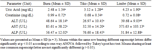
- The biochemical parameters measured in cattle infected with Fasciola hepatica revealed variations in liver and kidney function indices across the three abattoirs (Table 2). in Buea, Tiko, and Limbe. Serum uric acid concentration was highest in cattle slaughtered in Limbe (4.23 ± 1.90 mg/dL) compared to Buea (2.48 ± 1.34 mg/dL), while values in Tiko ranged from 0.30 to 15.10 mg/dL, indicating considerable variation among animals. Elevated uric acid levels may suggest altered renal excretion or increased tissue catabolism associated with chronic fasciolosis.
|
3.2.2. Serum Protein Profile of Male Cattle Infected with Fasciola Hepatica
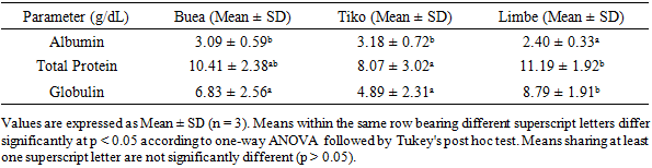
- The serum protein profile of cattle infected with Fasciola hepatica varied among the three abattoirs (Table 3). Albumin concentrations ranged from 2.40 ± 0.33 g/dL in Limbe to 3.09 ± 0.59 g/dL in Buea. Total protein concentrations were 10.41 ± 2.38 g/dL and 11.19 ± 1.92 g/dL in Buea and Limbe, respectively, while globulin concentrations ranged from 6.83 ± 2.56 g/dL to 8.79 ± 1.91 g/dL.
|
3.2.3. Serum Lipid Profile of Male Cattle Infected with Fasciola hepatica Slaughtered in Buea, Tiko and Limbe Abattoirs
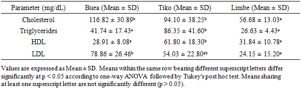
- The lipid profile of cattle infected with Fasciola hepatica varied among the three abattoirs (Table 4). Total cholesterol concentration was significantly higher in cattle slaughtered in Buea (116.82 ± 30.89 mg/dL) and Tiko (94.10 ± 38.25 mg/dL) than in Limbe (56.68 ± 13.03 mg/dL) (p < 0.05).
|
3.3. Effect of Age on Lipid Profile, Liver Function, Kidney Function and Protein Profile of Fasciola hepatica-Infected Cattle
3.3.1. Effect of Age on Lipid Profile, Liver Function, Kidney Function and Protein Profile of Fasciola hepatica-Infected Cattle
- The biochemical parameters of Fasciola hepatica-infected cattle from Buea varied with age (Table 5). Total cholesterol concentrations increased significantly (p < 0.05) with age, with the highest value recorded in 7-year-old cattle (162.20 ± 10.00 mg/dL) and the lowest in 5-year-old cattle (108.63 ± 26.00 mg/dL). This increase may suggest age-related alterations in lipid metabolism and prolonged exposure to infection.
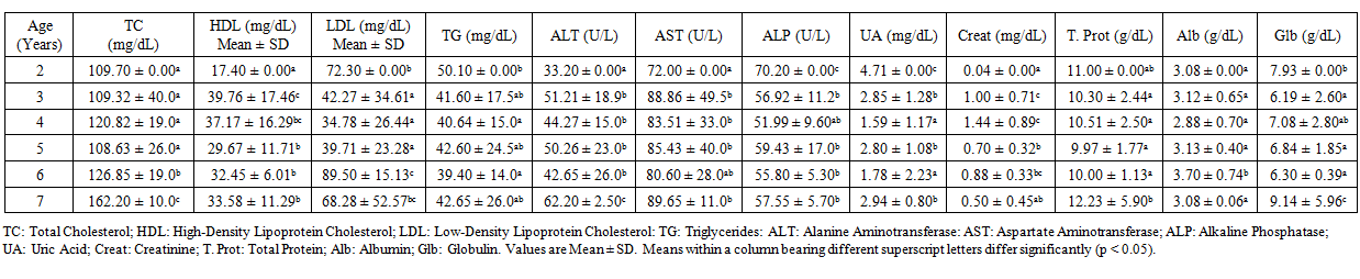 | Table 5. Effect of Age on Lipid Profile, Liver Function, Kidney Function and Protein Profile of Fasciola hepatica-Infected Cattle |
3.3.2. Effect of Age on Lipid Profile, Liver Function, Kidney Function and Protein Profile of Fasciola hepatica-Infected Cattle in Limbe
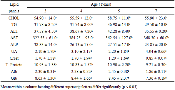
- Significant age-related differences were observed in Limbe cattle (Table 6). AST activity was markedly elevated in all age groups, with the highest value recorded in 4-year-old cattle (384.25 ± 93.00 U/L). ALT activity also increased significantly in 5-year-old cattle (42.28 ± 8.40 U/L) compared with younger animals.
|
3.3.3. Effect of Age on Lipid Profile, Liver Function, Kidney Function and Protein Profile of Fasciola hepatica-Infected Cattle in Tiko
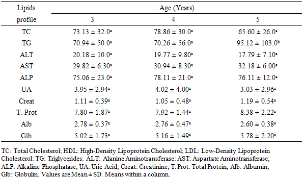
- Effect of Age on Lipid Profile, Liver Function, Kidney Function and Protein Profile of Fasciola hepatica-Infected Cattle in Tiko is presented on table 7. The biochemical parameters of infected cattle from Tiko showed comparatively less variation with age than those observed in Buea and Limbe. Cholesterol concentrations ranged from 65.60 ± 26.00 mg/dL in 5-year-old cattle to 78.86 ± 30.00 mg/dL in 4-year-old cattle, with no significant differences among age groups.
|
4. Discussion
4.1. Prevalence of Fasciola hepatica Infection
- The overall prevalence of 60.5% recorded in this study indicates that bovine fascioliasis remains highly endemic in Fako Division. This prevalence is comparable to reports from other tropical and subtropical regions where environmental conditions favor the survival of lymnaeid snails, the intermediate hosts of Fasciola species [1]. The high prevalence observed may be attributed to extensive grazing systems, poor pasture management, and continuous exposure of cattle to contaminated water bodies and marshy grazing areas [16].Young cattle exhibited a higher prevalence (74.1%) than adults (60.2%), although the difference was not statistically significant. This may be due to the relatively immature immune system of younger animals, making them more susceptible to infection compared with adults that may have acquired partial immunity following repeated exposure [3,5].Variation in prevalence among subdivisions suggests that environmental conditions influence transmission dynamics. The higher prevalence in Buea may be associated with its humid climate, high rainfall, and abundant waterlogged grazing areas that favor snail proliferation and parasite transmission [2].
4.2. Liver and Kidney Function Parameters
- Biochemical analysis revealed significant alterations in liver and kidney function parameters among infected cattle, consistent with the pathological effects of fascioliasis. The migration of immature flukes through hepatic tissues causes cellular damage, inflammation, and fibrosis, leading to leakage of intracellular enzymes into circulation [33,34].Serum ALT and AST activities were elevated, indicating hepatocellular injury, while ALP increases reflected cholestasis and biliary obstruction associated with adult flukes in the bile ducts [35,36].Creatinine and uric acid concentrations were also altered, suggesting possible secondary effects on renal function and systemic metabolism. Although the kidney is not the primary target of F. hepatica, chronic infection may induce metabolic stress, oxidative damage, and inflammatory responses that indirectly affect renal biomarkers [11,13].
4.3. Protein and Lipid Profile
- Serum albumin concentrations were reduced, particularly in cattle from Limbe, indicating impaired hepatic protein synthesis. This may result from liver tissue damage, reduced synthetic capacity, and chronic inflammatory processes. In contrast, increased globulin levels reflect prolonged antigenic stimulation and enhanced immunoglobulin production during chronic infection [14,35].Total protein concentrations remained relatively elevated due to compensatory globulin production, a typical response to chronic parasitic infection. The combined pattern of hypoalbuminemia and hyperglobulinemia suggests chronic hepatic dysfunction and sustained immune activation [33,36].Alterations in lipid profile parameters further indicate disruption of hepatic lipid metabolism. Total cholesterol was significantly reduced in Limbe cattle, reflecting impaired hepatic synthesis and altered lipoprotein metabolism. Similar hypocholesterolemia has been widely reported in fasciolosis due to liver dysfunction and parasite utilization of host lipids [14,15].Changes in HDL, LDL, and triglycerides suggest disturbances in lipid transport and energy metabolism, with variations likely influenced by infection severity, nutritional status, and liver function [37,36].
4.4. Effect of Age on Biochemical Parameters (All Municipalities)
- Age-related variations indicate that older cattle generally exhibit more pronounced biochemical alterations, suggesting cumulative effects of chronic infection. Increased AST, creatinine, and lipid abnormalities in older animals reflect progressive hepatic damage and metabolic stress associated with prolonged exposure to F. hepatica [38].Lower albumin and higher globulin levels in older cattle indicate reduced hepatic synthetic function and sustained immune stimulation. Similarly, alterations in lipid metabolism, particularly HDL and LDL variations, suggest that liver function deteriorates progressively with age and infection duration [35,34].
4.5. Effect of Age per Municipality
4.5.1. Buea
- In Buea, age-related increases in cholesterol and liver enzymes suggest progressive hepatic dysfunction with advancing age. Elevated ALT and AST levels in older cattle indicate ongoing hepatocellular damage. Increased globulin and total protein levels further reflect chronic immune stimulation due to persistent infection [39,33].
4.5.2. Limbe
- Cattle from Limbe showed the most severe biochemical alterations, particularly elevated AST levels across all age groups, indicating marked hepatocellular injury. The decline in albumin with age suggests worsening hepatic synthetic function, while elevated globulin levels indicate chronic immune activation. Reduced cholesterol concentrations further confirm impaired lipid metabolism associated with advanced liver damage [38,36].
4.5.3. Tiko
- In Tiko, relatively stable biochemical parameters across age groups suggest lower infection intensity or less severe hepatic involvement. Liver enzyme activities remained comparatively stable, indicating reduced hepatocellular damage. Protein and lipid profiles showed minimal variation, suggesting a more balanced physiological response to infection or better adaptation to parasitic exposure [40].
5. Conclusions
- The present study demonstrates that bovine fascioliasis is highly endemic in Fako Division, with a prevalence of 60.5%. Infection is associated with significant alterations in liver, kidney, protein, and lipid biochemical parameters, reflecting substantial physiological and metabolic disturbances in affected cattle.Overall, the findings highlight the negative impact of Fasciola hepatica infection on cattle health, productivity, and metabolic homeostasis, underscoring the need for effective control strategies to reduce infection prevalence and associated economic losses.
Funding
- This research did not receive any specific grant from funding agencies in the public, commercial, or not-for-profit sectors.
Data Availability Statement
- The authors declared that all related data are included in the article.
Declarations
- Competing interests: The authors declare no competing interests.Ethical Approval: Ethical approval for this study was obtained from the Refotde Institutional Animal Ethics Committee (RIAEC) under certificate reference number 014/RIAE/2024, issued on 14 November 2024.