Advances in Life Sciences

p-ISSN: 2163-1387    e-ISSN: 2163-1395

2014;  4(2): 94-99

doi:10.5923/j.als.20140402.10

Investigation of the Pattern of Antibiotic Resistance and Frequency of the AmpC ß-lactamase Gene in Pseudomonas Aeruginosa Isolated from Clinical and Environmental Samples in Sanandaj Hospitals

Shadi Rahimi1, Bahareh Rahimian Zarif2

1Department of biology, Kurdistan science and research branch, Islamic Azad University, Sanandaj, Iran

2Department of biology, Sanandaj branch, Islamic Azad University, Sanandaj, Iran

Correspondence to: Bahareh Rahimian Zarif, Department of biology, Sanandaj branch, Islamic Azad University, Sanandaj, Iran.

Email:

Copyright © 2014 Scientific & Academic Publishing. All Rights Reserved.

Abstract

Background:Pseudomonas aeruginosa is a very important pathogen with intrinsic resistance to different antimicrobial agents. In Pseudomonas aeruginosa the resistance mechanisms result from different ways like changes in gene expression for example by the Mex drug effluxpumps, the AmpC ß-lactamase and the carbapenem-specific porin OprD. AmpC is classification in group I and class C ß-lactamase that present in P. aeruginosa and causing resistance to penicillins and most cephalosporins. Now, bacterial resistance to antibiotics is a major and important problem in hospital and community in the world. Materials and Methods:100 P. aeruginosa were examination that isolates from different specimens in all of the Hospitals in Sanandaj and the susceptibility of the isolates to 10 antimicrobial agents was determined by the disc diffusion method on Mueller–Hinton agar plates. ESBL-producing strains were confirmed using double-disk diffusion test. Using CTAB method DNA was extracted and PCR assay was performed for detection of ampC gene. Results: Among all P. aeruginosa isolates, the highest resistance was seen for Ceftazidime and Ofloxacin (30%) and the least resistance was seen for Amikacin (11%). 11 isolates (11 %) of them were multiple drugs resistant. 13% (13 isolates) of them were found to be ESBL-producing strains in phenotypic tests and11 of them (85%) carried AmpC genes from different families so they shows polymorphism. Discussion: This study emphasizes the high prevalence of MDRP. aeruginosa in clinical and environmental specimens isolated from this hospitals. It is important to reduce these pathogens in hospitals and preventing of more resistance in this bacteria by using a suitable treatment protocol based on the antibiogram pattern of the isolates.

Keywords: Pseudomonas aeruginosa, Antibiotic resistance, Multiple drug resistance (MDR), ß-lactamase, AmpC

Cite this paper: Shadi Rahimi, Bahareh Rahimian Zarif, Investigation of the Pattern of Antibiotic Resistance and Frequency of the AmpC ß-lactamase Gene in Pseudomonas Aeruginosa Isolated from Clinical and Environmental Samples in Sanandaj Hospitals, Advances in Life Sciences, Vol. 4 No. 2, 2014, pp. 94-99. doi: 10.5923/j.als.20140402.10.

1. Background

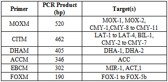
Pseudomonas aeruginosa is an opportunistic human pathogen that have second rank among Gram-negative hospital acquiring pathogens. The ability of P. aeruginosa to survive on minimal nutritional mediums and to tolerate different physical conditions make it adapted to hospital environment, so they are formidable nosocomial pathogens and one of the leading cause of nosocomial infections [1, 2, 3, and 4]. P. aeruginosa characterized by an innate resistance tomultiple antimicrobial agents that involving different enzymic and mutational mechanisms. These mechanisms are often present concurrently that conferring combined resistance in many strains [5]. Multidrug-resistant strains of P. aeruginosa are often isolated from patients with weak immune system [6]. MDR-isolates (multidrug resistant) were defined as those resistant toat least three or more classes of antibiotics [7]. Combination of different mechanisms is usually the cause of resistance to multiple antibiotics [8, 9, and 10]. The main mechanism for resistance to antibiotics in gram-negative bacteriais the synthesis of ß-lactamase [11, 12, 13, and 14]. They cleavage the amide bond in the ß-lactam ring that inactivating these antimicrobials. ß-lactamase enzymes are concentrated into the priplasmic space so ß-lactamase-mediated resistance is very efficient especially in Gram-negative bacilli, because they destroying ß-lactams before they can reach the penicillin-binding protein (PBP) targets in the cytoplasmic membrane, so they stay alive [15, 16, and 17]. Resistance appeared in organisms that could by mutation or some ß-lactam inducers, overproduce their chromosomal AmpC ß-lactamase because usually this enzymes are expressed at very low levels [18, 19, and 20]. AmpC ß-lactamase, the chromosomal encoded cephalosporinases, present in P. aeruginosa [17, 21]. It can inactivate ß-lactams by hydrolysis and confers resistance to penicillins and cephamycins and at low levels oxyminocephalosporins (Ceftazidime, cefotaxime, ceftriaxone and aztreonam). The hydrolysis rates for fourth generation cephalosporinas are very low (cefepime, cefpirome and carbapenems) [22]. AmpC ß-lactamase belonging to molecular class C and to functional group 1, and have six families (Table 1). The ampC gene is normally expressed at a low levels, but it can raise these levelsin response to ß-lactam exposureso its expression is inducible [2, 22, 23, 24] and can be partially or completely derepressed because of mutation in regulatory loci [24]. Mutations that cause ß-lactamase depression are the main reason for ß-lactam resistance in P.aeruginosa [23, 25]. The inducible expression of AmpC is under the control of the ampD, ampG and ampR proteins [26, 27]. Mutations in ampD and ampR genes may cause AmpC overproduction [22, 28, and 29]. AmpC ß-lactamase is mainly located in chromosome of Gram-negative bacteria, but maybe plasmid mediated and they are not inhibited by clavulanic acid [3]. Based on the conclusions from a study, the susceptibility of P. aeruginosa to some ß-lactams (imipenem, panipenem) is more strongly affected by the presence of ß-lactamase [25].
Table 1. Characteristics of the AmpC beta-lactamase 6 families [30, 31]
     

2. Objectives

The present study has been performed to determine the antimicrobial resistance, detecting multidrug resistance strains and AmpC-beta lactamase production strains in phenotypic test and investigation frequency of the ampC ß-lactamase gene in isolated pseudomonas aeruginosa that collected from different hospitals in Sanandaj, Iran.

3. Materials and Methods

3.1. Sampling and Bacterial Isolation

A collection of100P.aeruginosasamples were used that isolated from patient and environmental samples in Sanandaj hospitals within 1 year period. The clinical samples were isolated from urine, stool, blood, wounds, sputum, tracheal aspirates, burn wound, and CSF and hospital environment. The hospital environment samples were collected from different wards: burn, internal, ICU, CCU, surgical, urology, neurology. The clinical samples were isolated from 6 age group: <20, 20-30, 30-40, 40-50, 50-60, >60 and both of male and female. Identification of the isolates was done according to standard microbiology procedures such as Gram stain, Oxidase and catalase, TSI, Indole, MRVP, Citrate, Urease, Ornithine and lysine decarboxylase test. P. aeruginosa isolates were stored in LB broth medium containing 30% glycerol at-20°C.

3.2. Antimicrobial Susceptibility Testing

Antibiotic susceptibility was done by Kirby-Bauerdisc diffusion method for 10 antimicrobial agents on Mueller-Hinton agar plates using antibiotic-containing discs. The 0.5 McFarland suspension of bacteria were cultured on the Mueller-Hinton agar and the antibiotic discs were placed on the agar and incubated at 37℃ for 24 h. The panel of antibiotics included: Amikacin (30ϻg), Cefepime (30ϻg), Ceftazidime (30ϻg), Ciprofloxacin (5ϻg), Gentamicin (10ϻg), Imipenem (10ϻg), Meropenem (10ϻg), Ofloxacin (5ϻg), Ticarcillin (75ϻg), Tobramycin (10ϻg). Isolates resistant to ≥ three antibiotics from different classes were considered multidrug-Resistant (MDR) [32].

3.3. Detection of Chromosomal AmpC Phenotype

AmpC production was detected by combination of the double disc test and the combined disc test (DCDT) which included discs of ceftazidime, cefotaxime, cefpodoxime and cefepime placed at a distance of 20 mm (center to center) from a disc containing amoxycillin plus clavulanate and a disc containing ceftazidime plus clavulanic acid and a disc containing cefotaximeplus clavulanic acid for detection of AmpC production [33].

3.4. Extraction of Genomic DNA

Total DNA from AmpC producing P. aeruginosa was extracted by CTAB method. The bacterial colony was suspended in 700 μl of CTAB buffer (contain: CTAB, EDTA, NaCl, β-mercaptoethanol (2me), Polyvinylpyrrolidone (PVP), TrisHCl (pH=8)) and incubated for one hour at 65°C. The solution was added with 800 μl of chloroform / isoamylalcohol (24:1) and the cells were harvested by centrifugation (12000 rpm, 15 min). The aqueous phase were transferred to another tube and DNA was precipitated with 500 μl isopropanol from the aqueous phase and kept in the frizer for one hour and in refrigerator for 24 hour. After centrifugation (12000 rpm, 15 min), the DNA pellet was washed with 70% ethanol, dried briefly and resuspended in 150 μl of distilled water. The DNA was kept in the frizer for next steps.

3.5. DNA Amplification

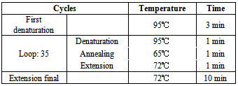
The primers sequences were designed to amplify the complete sequence of ampC gene. The sequences of forward and reverse primers is shown in table 2. Master Mix for PCR contained 2 μl PCR buffer, 0.7 μl of each dNTPs, 0.9μl MgCl2, 1 μl of each primers (forward and reverse), 0.3 μl Taq DNA polymerase (Sinna Gen, Iran) and 2 μl DNA template with 20 μl final volume in each tube. Setup condition for DNA amplification is shown in table 3. PCR products were analyzed by horizontal gel electrophoresis with 1.5% agarose and ethidium bromide in TBE buffer and running at 100 V for 1 hour. DNA molecular size marker was included in all gels (100-bp DNA ladder, Sinna Gen, Iran).
Table 2. Primer pair used for AmpC β-lactamase screening
     
Table 3. Setup condition for PCR reaction
     

4. Results

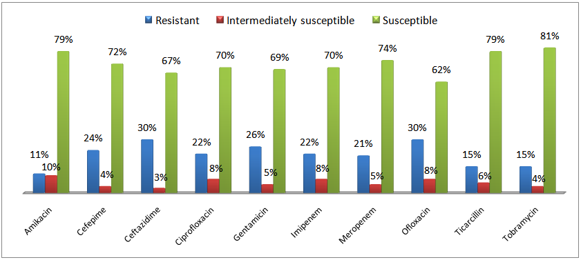
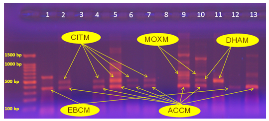
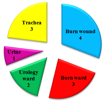
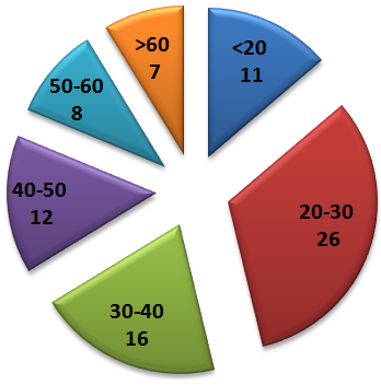
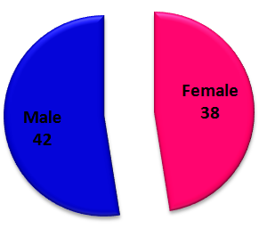
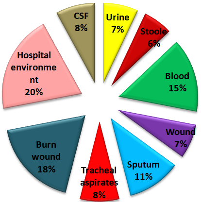
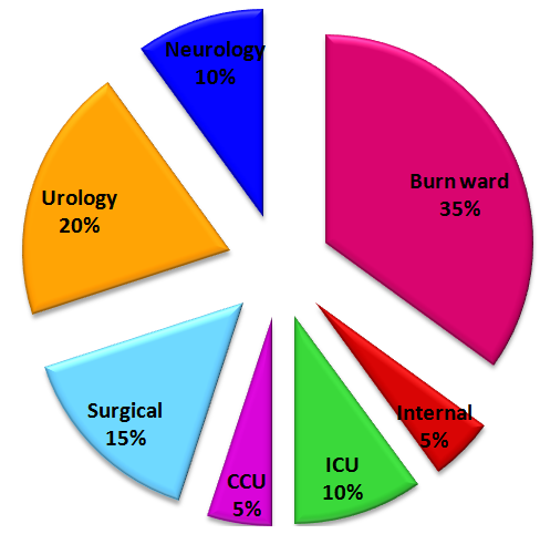
The antimicrobial susceptibility testing results is shown in chart 1. the highest resistance was seen for Ceftazidime and Ofloxacin (30%) and the least resistance was seen for Amikacin (11%). 11 (11%) isolates were MDR and were resistant to more than three classes of antibiotics. 13% (13 isolates) of them were found to be AmpC producing strains in phenotypic tests and 11 of them (85%) carried AmpC genes from different six families so they shows polymorphism (figure 1). Among this Ampc producing strains, 4 of them were from burn wound, 1 from urine, 3 from trachea and 5 from hospital environment (3 from burn ward and 2 from urology ward) (chart 2). Based on the age and sex, most of the pseudomonas aeruginosa samples were detected from 20-30 age group (32%) (Chart 3) and males (42%) (chart 4), and based on the samples, most of the pseudomonas aeruginosa samples were detected from burn wound (18%) in clinical samples (chart 5) and from burn ward (35%) in environmental samples (chart 6). Statistical Analyses were done by SPSS V.16 and the relationship between variables were determining. Based on the results, there is not significant relationship between AmpC-producing strains and Sex, Age & Kind of samples (P value > 0.05).
Chart 1. Antibiotic susceptibility pattern of isolated P. aeruginosas
Figure 1. PCR Amplification of AmpC Gene in AmpC positive phenotypic tests Pseudomonas aeruginosas
Chart 2. Frequency of the Ampc producing strains based on kind of samples
Chart 3. Frequency of pseudomonas aeruginosa based on Age
Chart 4. Frequency of pseudomonas aeruginosa based on Sex
Chart 5. Frequency of pseudomonas aeruginosa based on samples
Chart 6. Frequency of pseudomonas aeruginosa based on sampling wards

5. Discussion

The emergence of AmpC β-lactamases-producing P. aeruginosa propose a serious challenge to antimicrobial chemotherapy because of continued increases immunosuppressed / compromised patient populations and the ability of the bacteria to rapidly mutate and adapt to antibacterial agent [3,34]. In recent decades, a large number of molecular techniques have been developed for detection of serious microbial pathogens and their resistance or pathogenic markers. While many have found practical application in routine microbiological diagnostics, others are currently used in research only. If these modern methods are introduced into diagnostics, they often help in rapid and accurate detection of certain microorganisms or their resistance and pathogenic determinants, so for control of this pathogenic bacteria, the use of treatments based on microbiological and pharmacological data, should be priorities. Based on this study, in comparison with recent researches in Iran, the rate of resistance for most of the antibiotics used in this study were decreased but considering the ever-increasing prevalence of resistant in this bacteria, rapid identification and choose suitable antimicrobial treatment are needed to prevent of further resistant.

Note

1. Double disc plus combined discs

References

[1]  Shacheraghi F, Shakibaie MR, and Noveiri H. Molecular Identification of ESBL Genes blaGES-1, blaVEB-1, blaCTX-M blaOXA-1, blaOXA-4, blaOXA-10 and blaPER-1 in Pseudomonas aeruginosa Strains Isolated from Burn Patients by PCR, RFLP and Sequencing Techniques. World Academy of Science, Engineering and Technology. 2010; 37: 1189-1193.
[2]  Juan C, Macia MD, Gutierrez O, Vidal C, Perez JL, and Oliver A. Molecular Mechanisms of ß-Lactam Resistance Mediated by AmpC Hyperproduction in Pseudomonas aeruginosa Clinical Strains. Antimicrobial agents and chemotherapy. 2005; 49(11): 4733–4738.
[3]  Zavascki A.P, Carvalhaes CG, Picao RC, and Gales A.C. Multidrug-resistant Pseudomonas aeruginosa and Acinetobacterbaumannii: resistance mechanisms and implications for therapy. Expert Review of Anti-Infective Therapy. 2012; 8(1): 71-93.
[4]  Maatallah M, Cheriaa J, Backhrouf A, Iversen A, Grundmann H, et al. Population Structure of Pseudomonas aeruginosa from Five Mediterranean Countries: Evidence for Frequent Recombination and Epidemic Occurrence of CC235. 2011; 6(10): e25617.
[5]  McGowan JE. Resistance in nonfermenting Gram-negative bacteria: multidrug resistance to the maximum. The American Journal of Medicine. 2006; 119(6): S29–S36.
[6]  Strateva T, Ouzounova-Raykova V, Markova B, Todorova A, Marteva-Proevska Y, and Mitov I. Problematic clinical isolates of Pseudomonas aeruginosa from the university hospitals in Sofia, Bulgaria: current status of antimicrobial resistance and prevailing resistance mechanisms. Journal of Medical Microbiology. 2007; 56: 956–963.
[7]  Magiorakos AP, Srinivasan A, Carey RB, Carmeli Y, Falagas ME, et al. Multidrug-resistant, extensively drug-resistant and pandrug-resistant bacteria: an international expert proposal for interim standard definitions for acquired resistance. Clinical Microbiology and Infection. 2011; 18(3): 268–281.
[8]  Bonomo RA, Scabo D. Mechanisms of multidrug resistance in Acinetobacter species and Pseudomonas aeruginosa. Clinical Infectious Diseases. 2006; 43(2): 49-56.
[9]  Livermore DM. Multiple mechanisms of antimicrobial resistance in Pseudomonas aeruginosa: Our Worst Nightmare? Clinical Infectious Diseases. 2002; 34(5): 634–640.
[10]  Woodford N, Ellington MJ. The emergence of antibiotic resistance by mutation. Clinical Microbiology and Infection. 2007; 13: 5–18.
[11]  Philippon A, Arlet G, and Jacoby GA. Plasmid-Determined AmpC-Type ß-Lactamases .Antimicrobial agents and chemotherapy. 2002; 46(1): 1–11.
[12]  Lodge JM, Minchin SD, Piddock LJV, and Busby SJW. Cloning, sequencing and analysis of the structural gene and regulatory region of the Pseudomonas aeruginosa chromosomal ampC ß-lactamase. Biochem. J. 1990; 272: 627-631.
[13]  Bennett PM. Plasmid encoded antibiotic resistance: acquisition and transfer of antibiotic resistance genes in bacteria. British Journal of Pharmacology. 2008; 153(S1): S347–S357.
[14]  Chroma M, Kolar M. Genetic methods for detection of antibiotic resistance: focus on extended-spectrum β-lactamases. 2010; 154(4): 289–296.
[15]  Livermore DM, Woodford N. The ß-lactamase threat in Enterobacteriaceae, Pseudomonas and Acinetobacter. Trends Microbial. 2006; 14(9):413-420.
[16]  Ambler RP. The structure of ß-lactamases. Philosophical transaction, the Royal society, Lond B. biological sciences. 1980; 289(1036): 321-331.
[17]  Bush K, Jacoby GA, and Medeiros AA. A functional classification scheme for ß-lactamases and its correlation with molecular structure. Antimicrob. Agents Chemother. 1995; 39(6): 1211–1233.
[18]  Sanders CC. Chromosomal cephalosporinases responsible for multiple resistance to newer ß-lactam antibiotics. Annual Review of Microbiology. 1987; 41: 573–593.
[19]  Giwercman B, Lambert PA, Rosdahl VT, Shand GT, and Hoiby N. Rapid emergence of resistance in Pseudomonas aeruginosa in cystic fibrosis patients due to in-vivo selection of stable partially derepressed ß-lactamase producing strains. Antimicrobial agents and chemotherapy. 1990; 26: 247–259.
[20]  Livermore DM. Clinical significance of beta-lactamase induction and stable derepression in gram-negative rods. Eur. J. Clin. Microbiol. 1987; 6: 439–445.
[21]  Livermore DM. ß-Lactamases in laboratory and clinical resistance. Clin. Microbiol. Rev. 1995; 8: 557–584.
[22]  Jacoby GA .AmpC ß –lactamases. Clinical Microbiology. Rev. 2009; 22(1): 161-182.
[23]  Dumas JL, Delden CV, Perron K, and Kohler T. Analysis of antibiotic resistance gene expression in Pseudomonas aeruginosa by quantitative real-time-PCR. Federation of European Microbiological Societies Microbiol Lett. 2006; 254: 217–225.
[24]  Langaee TY, Gagnon L, and HuletskyA. Inactivation of the ampD Gene in Pseudomonas aeruginosa Leads to Moderate- Basal-Level and Hyperinducible AmpC ß-Lactamase Expression. Antimicrobial Agents and Chemotherapy. 2000; 44(3): 583–589.
[25]  Hancock REW, Speert DP. Antibiotic resistance in Pseudomonas aeruginosa: mechanisms and impact on treatment. Drug Resistance Updates. 2000; 3: 247–255.
[26]  Jacobs C, Frere JM, and Normark S. Cytosolic intermediates for cell wall biosynthesis and degradation control inducible ß-lactam resistance in gram-negative bacteria. Cell Press. 1997; 88(6): 823–832.
[27]  Wiedemann B, Dietz H, and Pfeifle D. Induction of ß-lactamase in Enterobacter cloacae. Clinical Infectious Diseases. 1988; 27(1): S42–S47.
[28]  Langaee TY, Dargis M, and HuletskyA. An ampD gene in Pseudomonas aeruginosa encodes a negative regulator of AmpC ß-lactamase expression. Antimicrob. Agents Chemother. 1998; 42:3296–3300.
[29]  Lodge J, Busby S, and Piddock L. Investigation of the Pseudomonas aeruginosaampR gene and its role at the chromosomal ampC promoter. Federation of European Microbiological Societies Microbiol Lett. 1993; 111: 315–320.
[30]  Manoharan A, Sugumar M, Kumar A, Jose H, and Mathai D. Phenotypic & molecular characterization of AmpC β-lactamases among Escherichia coli, Klebsiella spp. & Enterobacter spp. from five Indian Medical Centers. Indian J Med Res. 2012; 135: 359-364.
[31]  Mansouri S, Chitsaz M, Haji-hosseini R, Mirzaee M, and Ghini MH. Determining resistance of E.coli clinical isolates producing AmpC betalactamase based on their phenotypic and genotypic properties. Shahed university J. 1388; 19(80): 1-11.
[32]  Magiorakos AP, Srinivasan A, Carey RB, Carmeli Y, Falagas ME, et al. Multidrug-resistant, extensively drug-resistant and pandrug-resistant bacteria: an international expert proposal for interim standard definitions for acquired resistance. Clinical Microbiology and Infection. 2011; 18(3): 268-281.
[33]  Jiang X, Zhang Z, Li M, Zhou D, Ruan F, and Lu Y. Detection of Extended-Spectrum ß-Lactamases in Clinical Isolates of Pseudomonas aeruginosa. Antimicrobial Agents and Chemotherapy. 2006; 50(9): 2990–2995.
[34]  Tassios PT, Gennimata V, L. Spaliara-Kalogeropoulou, D. Kairis, C. Koutsia, A. C. Vatopoulos, and N. J. Legakis. 1997. Multiresistant Pseudomonas aeruginosaserogroup O: 11 outbreak in an intensive care unit. Clinical Microbiology and Infection. 3(6): 621–628.